Neuropilin-1 (Npn-1, previously known as Neuropilin) and Npn-2 (previously known as Npn-1-related molecule) are type I transmembrane proteins that bind members of the class III secreted semaphorin subfamily which are implicated in repulsive axon guidance. The extracellular domain of these proteins is composed of two N-terminal CUB (complement-binding) domains (domains a1 and a2), two domains with homology to coagulation factors V and VIII (domains b1 and b2) and a MAM (meprin) domain (domain c). In the absence of ligands, neuropilins can form homo- and hetero-oligomers via homophilic interactions of their MAM domains. At the amino acid sequence level, Npn-1and Npn-2 share 44% identity. Npn-1 and Npn-2 show different binding specificities for different members of the semaphorin family. The expression patterns of Npn-1 and Npn-2 in developing neurons of the central and peripheral nervous systems are largely, though not completely nonoverlapping. Npn‑1 and Npn-2 are also expressed by endothelial and tumor cells and have been shown to be isoform-specific receptors for VEGF165. Npn‑1 was also reported to bind PlGF-2 and the VEGF-like protein from of virus NZ2.

Key Product Details
Species Reactivity
Validated:
Cited:
Applications
Validated:
Cited:
Label
Antibody Source
Product Specifications
Immunogen
Gln23-Asp857 (Val809-Asp825 del)
Accession # O35276
Specificity
Clonality
Host
Isotype
Endotoxin Level
Scientific Data Images for Neuropilin-2 Antibody
Detection of Mouse and Rat Neuropilin‑2 by Western Blot.
Western blot shows lysates of C6 rat glioma cell line, LL/2 mouse Lewis lung carcinoma cell line, and bEnd.3 mouse endothelioma cell line. PVDF membrane was probed with 0.5 µg/mL of Goat Anti-Mouse/Rat Neuropilin-2 Antigen Affinity-purified Polyclonal Antibody (Catalog # AF567) followed by HRP-conjugated Anti-Goat IgG Secondary Antibody (Catalog # HAF017). A specific band was detected for Neuropilin-2 at approximately 110 kDa (as indicated). This experiment was conducted under reducing conditions and using Immunoblot Buffer Group 1.
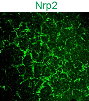
Neuropilin‑2 in Rat Brain.
Neuropilin-2 was detected in perfusion fixed frozen sections of rat brain using Goat Anti-Mouse/Rat Neuropilin-2 Antigen Affinity-purified Polyclonal Antibody (Catalog # AF567) at 15 µg/mL overnight at 4 °C. Tissue was stained using the NorthernLights™ 557-conjugated Anti-Goat IgG Secondary Antibody (red; Catalog # NL001) and counterstained with DAPI (blue). Specific staining was localized to cytoplasm in neurons. View our protocol for Fluorescent IHC Staining of Frozen Tissue Sections.
Detection of Mouse and Rat Neuropilin‑2 by Simple WesternTM.
Simple Western lane view shows lysates of C6 rat glioma cell line, LL/2 mouse Lewis lung carcinoma cell line, and bEnd.3 mouse endothelioma cell line, loaded at 0.2 mg/mL. A specific band was detected for Neuropilin-2 at approximately 140 kDa (as indicated) using 5 µg/mL of Goat Anti-Mouse/Rat Neuropilin-2 Antigen Affinity-purified Polyclonal Antibody (Catalog # AF567) followed by 1:50 dilution of HRP-conjugated Anti-Goat IgG Secondary Antibody (Catalog # HAF109). This experiment was conducted under reducing conditions and using the 12-230 kDa separation system.Applications for Neuropilin-2 Antibody
Blockade of Receptor-ligand Interaction
Immunohistochemistry
Sample: Perfusion fixed frozen sections of rat brain
Simple Western
Sample: C6 rat glioma cell line, LL/2 mouse Lewis lung carcinoma cell line, and bEnd.3 mouse endothelioma cell line
Western Blot
Sample: C6 rat glioma cell line, LL/2 mouse Lewis lung carcinoma cell line, and bEnd.3 mouse endothelioma cell line
Reviewed Applications
Read 7 reviews rated 4.4 using AF567 in the following applications:
Formulation, Preparation, and Storage
Purification
Reconstitution
Reconstitute at 0.2 mg/mL in sterile PBS. For liquid material, refer to CoA for concentration.
Formulation
Shipping
Stability & Storage
- 12 months from date of receipt, -20 to -70 °C as supplied.
- 1 month, 2 to 8 °C under sterile conditions after reconstitution.
- 6 months, -20 to -70 °C under sterile conditions after reconstitution.
Calculators
Background: Neuropilin-2
References
- Fujisawa, H. and T. Kitsukawa (1998) Curr. Opin. Neurobiol. 8:587.
- Neufeld, G. et al. (1999) FASEB J. 13:9.
- Poltorak, Z. et al. (2000) J. Biol. Chem. 275:18040.
Alternate Names
Gene Symbol
UniProt
Additional Neuropilin-2 Products
Product Documents for Neuropilin-2 Antibody
Certificate of Analysis
To download a Certificate of Analysis, please enter a lot or batch number in the search box below.
Note: Certificate of Analysis not available for kit components.
Product Specific Notices for Neuropilin-2 Antibody
This product or the use of this product is covered by U.S. Patents owned by The Regents of the University of California. This product is for research use only and is not to be used for commercial purposes. Use of this product to produce products for sale or for diagnostic, therapeutic or drug discovery purposes is prohibited. In order to obtain a license to use this product for such purposes, contact The Regents of the University of California.
U.S. Patent # 6,054,293, 6,623,738, and other U.S. and international patents pending.
For research use only
Citations for Neuropilin-2 Antibody
 Powered by Bioz
Customer Reviews for Neuropilin-2 Antibody (7)
Have you used Neuropilin-2 Antibody?
Submit a review and receive an Amazon gift card!
$25/€18/£15/$25CAN/¥2500 Yen for a review with an image
$10/€7/£6/$10CAN/¥1110 Yen for a review without an image
Submit a review
Customer Images
-
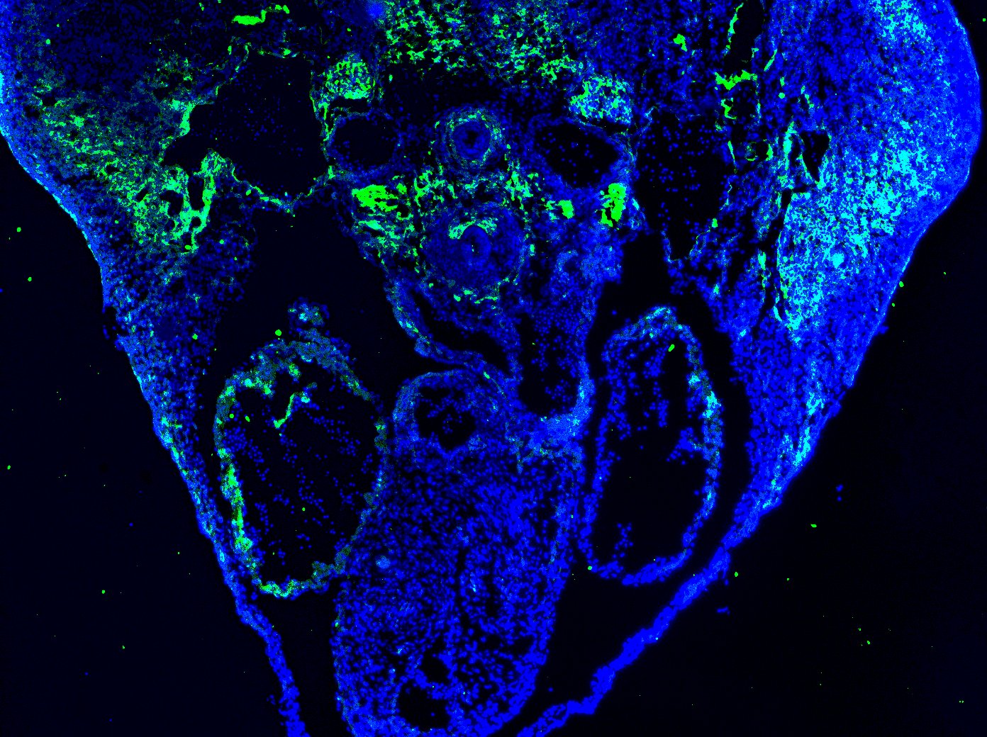
Application: Immunocytochemistry/ImmunofluorescenceSample Tested: E12.5 mouse embryo fixed in 4% PFASpecies: MouseVerified Customer | Posted 12/03/2020Antibody was stained on E12.5 mouse sections (attached picture) as well as E9.5 mouse sections. Worked well. Note the positive stain in the veins and the negative signal in the adjacent arteries. Dilution used - 1:20

-
Application: ImmunohistochemistrySample Tested: Embryonic tissueSpecies: MouseVerified Customer | Posted 11/08/2016
-
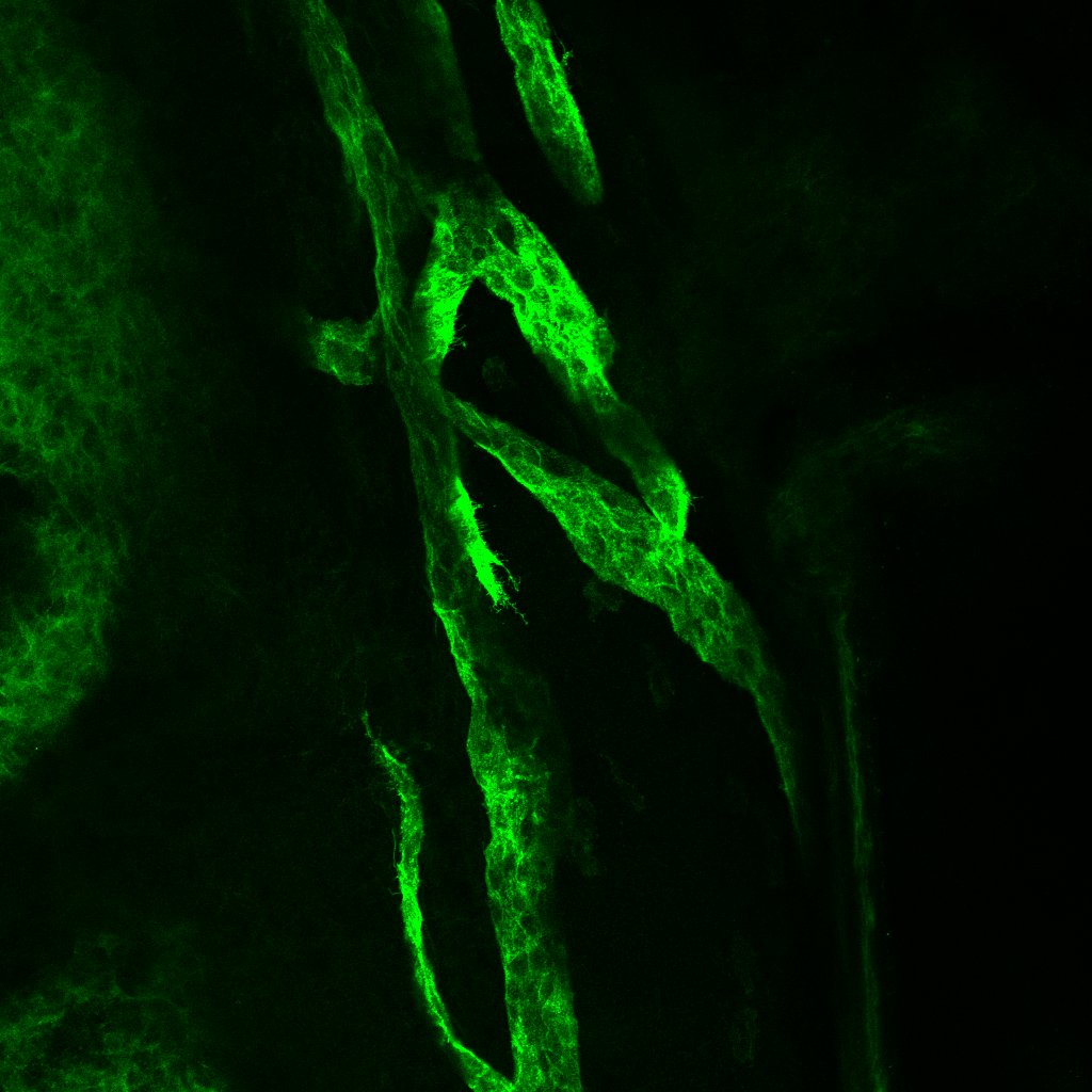
Application: Whole mount immunofluorescenceSample Tested: Whole mount eye tissueSpecies: MouseVerified Customer | Posted 09/07/2016Whole mouse eye tissue was fixed in 2% PFA, blocked and incubated O/N in AF567 diluted 1:250. Signal was detected using an alexafluor 488-labeled donkey anti goat secondary
-
Application: ImmunofluorescenceSample Tested: Hippocampal neuronsSpecies: MouseVerified Customer | Posted 12/19/2014
-
Application: ImmunofluorescenceSample Tested: Hippocampal neurons and astrocytesSpecies: MouseVerified Customer | Posted 12/19/2014
-
Application: Western BlotSample Tested: RN22 cellsSpecies: RatVerified Customer | Posted 12/19/2014
-
Application: Western BlotSample Tested: Primary hippocampal neuronsSpecies: MouseVerified Customer | Posted 12/19/2014
There are no reviews that match your criteria.
Protocols
Find general support by application which include: protocols, troubleshooting, illustrated assays, videos and webinars.
- Antigen Retrieval Protocol (PIER)
- Antigen Retrieval for Frozen Sections Protocol
- Appropriate Fixation of IHC/ICC Samples
- Cellular Response to Hypoxia Protocols
- Chromogenic IHC Staining of Formalin-Fixed Paraffin-Embedded (FFPE) Tissue Protocol
- Chromogenic Immunohistochemistry Staining of Frozen Tissue
- ClariTSA™ Fluorophore Kits
- Detection & Visualization of Antibody Binding
- Fluorescent IHC Staining of Frozen Tissue Protocol
- Graphic Protocol for Heat-induced Epitope Retrieval
- Graphic Protocol for the Preparation and Fluorescent IHC Staining of Frozen Tissue Sections
- Graphic Protocol for the Preparation and Fluorescent IHC Staining of Paraffin-embedded Tissue Sections
- Graphic Protocol for the Preparation of Gelatin-coated Slides for Histological Tissue Sections
- IHC Sample Preparation (Frozen sections vs Paraffin)
- Immunofluorescent IHC Staining of Formalin-Fixed Paraffin-Embedded (FFPE) Tissue Protocol
- Immunohistochemistry (IHC) and Immunocytochemistry (ICC) Protocols
- Immunohistochemistry Frozen Troubleshooting
- Immunohistochemistry Paraffin Troubleshooting
- Preparing Samples for IHC/ICC Experiments
- Preventing Non-Specific Staining (Non-Specific Binding)
- Primary Antibody Selection & Optimization
- Protocol for Heat-Induced Epitope Retrieval (HIER)
- Protocol for Making a 4% Formaldehyde Solution in PBS
- Protocol for VisUCyte™ HRP Polymer Detection Reagent
- Protocol for the Preparation & Fixation of Cells on Coverslips
- Protocol for the Preparation and Chromogenic IHC Staining of Frozen Tissue Sections
- Protocol for the Preparation and Chromogenic IHC Staining of Frozen Tissue Sections - Graphic
- Protocol for the Preparation and Chromogenic IHC Staining of Paraffin-embedded Tissue Sections
- Protocol for the Preparation and Chromogenic IHC Staining of Paraffin-embedded Tissue Sections - Graphic
- Protocol for the Preparation and Fluorescent IHC Staining of Frozen Tissue Sections
- Protocol for the Preparation and Fluorescent IHC Staining of Paraffin-embedded Tissue Sections
- Protocol for the Preparation of Gelatin-coated Slides for Histological Tissue Sections
- R&D Systems Quality Control Western Blot Protocol
- TUNEL and Active Caspase-3 Detection by IHC/ICC Protocol
- The Importance of IHC/ICC Controls
- Troubleshooting Guide: Immunohistochemistry
- Troubleshooting Guide: Western Blot Figures
- Western Blot Conditions
- Western Blot Protocol
- Western Blot Protocol for Cell Lysates
- Western Blot Troubleshooting
- Western Blot Troubleshooting Guide
- View all Protocols, Troubleshooting, Illustrated assays and Webinars
Associated Pathways
