NanoLuc® (Nluc) Luciferase Antibody (965853)
R&D Systems | Catalog # MAB100261

Key Product Details
Species Reactivity
Validated:
Cited:
Applications
Validated:
Cited:
Label
Antibody Source
Product Specifications
Immunogen
Specificity
Clonality
Host
Isotype
Scientific Data Images for NanoLuc® (Nluc) Luciferase Antibody (965853)
Detection of NanoLuc® Luciferase by Western Blot.
Western blot shows Recombinant NanoLuc®. PVDF membrane was probed with 2 µg/mL of Mouse Anti-Human NanoLuc® (Nluc) Luciferase Monoclonal Antibody (Catalog # MAB100261) followed by HRP-conjugated Anti-Mouse IgG Secondary Antibody (Catalog # HAF018). A specific band was detected for NanoLuc® at approximately 20 kDa (as indicated). This experiment was conducted under reducing conditions and using Immunoblot Buffer Group 1. Luciferase by Western Blot")
Detection of NanoLuc® (Nluc) Luciferase by Western Blot
Luciferase assay for quantifying the release of GPC4 from astrocytes. A, Nluc is inserted at the N terminus, after the endogenous signal peptide, to preserve GPC4 trafficking. B, Primary astrocytes were nucleofected with Nluc-GPC4 and treated with and without PI-PLC. Western botting with alpha -Nluc antibody showed the expected 20 kDa size shift in the N-terminal fragment. PI-PLC treatment facilitates the release of Nluc-GPC4, confirming the GPI-anchorage of the construct. C, Representative trace of one experiment showing the linear kinetics of GPC4 release from astrocyte culture (R2 = 0.998). Error bars indicate the standard error of the mean. D, Astrocytes expressing Nluc-GPC4 were incubated in fresh media with and without PI-PLC for 3 h, and Nluc signal was measured in the cell lysate and media. Nluc signal was normalized to untreated lysate conditions for each biological replicate. PI-PLC treatment resulted in the decrease in the luciferase activity of the cell lysate and the corresponding increase in the activity in the media. These data show Nluc-GPC4 is quantitative in measuring released versus surface pools of GPC4. Error bars indicate 95% CI of the mean here and in following graphs. The requirement of the GPI-anchorage for PI-PLC-dependent release of GPC4 is shown in Extended Data Figure 2-1. E, The release rate (media over lysate activity) of Nluc-GPC4 and Nluc-Prion was normalized to Nluc-GPC4 release. Nluc-GPC4 is released ∼2-fold more than Nluc-Prion (t test p < 0.0001, Cohen’s d = 3.59); ***p < 0.001. Image collected and cropped by CiteAb from the following open publication (https://pubmed.ncbi.nlm.nih.gov/34301723), licensed under a CC-BY license. Not internally tested by R&D Systems. Luciferase by Western Blot")
Detection of NanoLuc® (Nluc) Luciferase by Western Blot
Iterative optimization of the SEPLuc and Antares bioluminescence signal ratios. (A) cyOFP1 was inserted into the existing membrane-anchored SEPLuc fusion via the 5-amino acid linker (n = 2). (B) cyOFP1 was fused to another Nanoluc sequence, then linked downstream of SEPLuc via E2A (n = 2). (C) Antares was cloned downstream of SEPLuc and IRES (n = 2). (D) The Antares-SEPLuc construct. Antares is followed by T2A and puromycin (colored purple), then by IRES and SEPLuc. (n = 3). (E) Comparison of the Antares-SEPLuc constructs utilizing wildtype IRES and the mutant variants IRESv11 and IRESv24 (n = 3). (F) Comparison of the Antares-SEPLuc IRESv24 constructs utilizing two weaker Nanoluc precursors, C1A4E and C1A4E+6, transiently expressed in SW982 cells (n = 2). (G) Western blot of whole cell lysates from SW982 transfected with vector control, Nanoluc, SEPLuc, Antares, Antares-SEPLuc, Antares-SEPLuc IRESv24, and pHLuc. All constructs were transfected into a model cell line, HEK293T for initial assessment of their expression, unless stated otherwise. Downward arrows on spectral scan values indicate the wavelengths at which peaks are expected (450 nm for Nanoluc, 510 nm for SEP, and 580 nm for Antares). Luciferase assay values were normalized to the Nanoluc emission at 450 nm. Error bars are SD. Image collected and cropped by CiteAb from the following open publication (https://pubmed.ncbi.nlm.nih.gov/32457886), licensed under a CC-BY license. Not internally tested by R&D Systems.Applications for NanoLuc® (Nluc) Luciferase Antibody (965853)
Western Blot
Sample: Recombinant NanoLuc®
Reviewed Applications
Read 1 review rated 5 using MAB100261 in the following applications:
Formulation, Preparation, and Storage
Purification
Reconstitution
Reconstitute at 0.5 mg/mL in sterile PBS. For liquid material, refer to CoA for concentration.
Formulation
Shipping
Stability & Storage
- 12 months from date of receipt, -20 to -70 °C as supplied.
- 1 month, 2 to 8 °C under sterile conditions after reconstitution.
- 6 months, -20 to -70 °C under sterile conditions after reconstitution.
Calculators
Background: Luciferase
Alternate Names
Additional Luciferase Products
Product Documents for NanoLuc® (Nluc) Luciferase Antibody (965853)
Certificate of Analysis
To download a Certificate of Analysis, please enter a lot or batch number in the search box below.
Note: Certificate of Analysis not available for kit components.
Product Specific Notices for NanoLuc® (Nluc) Luciferase Antibody (965853)
NanoLuc and NanoBiT are registered trademarks of Promega Corporation.
For research use only
Citations for NanoLuc® (Nluc) Luciferase Antibody (965853)
 Powered by Bioz
Customer Reviews for NanoLuc® (Nluc) Luciferase Antibody (965853) (1)
Have you used NanoLuc® (Nluc) Luciferase Antibody (965853)?
Submit a review and receive an Amazon gift card!
$25/€18/£15/$25CAN/¥2500 Yen for a review with an image
$10/€7/£6/$10CAN/¥1110 Yen for a review without an image
Submit a review
Customer Images
-
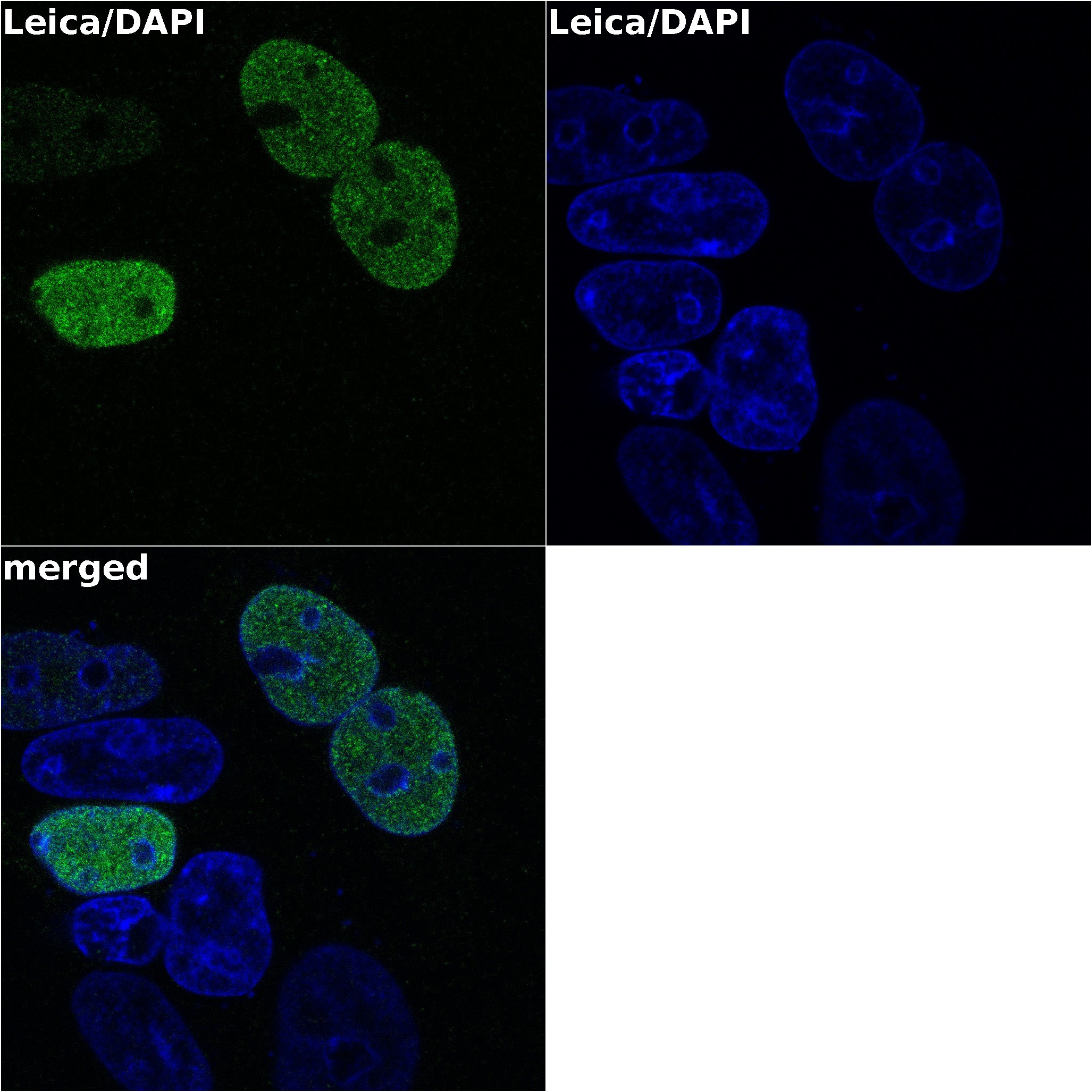
Application: Immunocytochemistry/ImmunofluorescenceSample Tested: HEK293 human embryonic kidney cell lineSpecies: HumanVerified Customer | Posted 06/08/2020Really nice antibody to work with. It gives you really bright pictures.

There are no reviews that match your criteria.
Protocols
Find general support by application which include: protocols, troubleshooting, illustrated assays, videos and webinars.
- Cellular Response to Hypoxia Protocols
- R&D Systems Quality Control Western Blot Protocol
- Troubleshooting Guide: Western Blot Figures
- Western Blot Conditions
- Western Blot Protocol
- Western Blot Protocol for Cell Lysates
- Western Blot Troubleshooting
- Western Blot Troubleshooting Guide
- View all Protocols, Troubleshooting, Illustrated assays and Webinars
FAQs for NanoLuc® (Nluc) Luciferase Antibody (965853)
-
Q: What is the immunogen for the NanoLuc® (Nluc) Luciferase Antibody, Catalog # MAB100261? Does this antibody recognize LgBit or SmBit?
A: The immunoogen for MAB100261 is a Synthetic peptide of NanoLuc® (Nluc) Luciferase. For more details on the compound, Promega Corporation should be contacted. NanoBit (LgBit+SmBit) is a derivative of the original product NanoLuc, where portion of the protein is split to get two complementary peptides. MAB100261 detects an epitope on NanoLuc that is not part of LgBit or SmBit. SmBit is only 11 amino acids in length.