TrkA, the product of the proto-oncogene trk, is a member of the neurotrophic tyrosine kinase receptor family that has three members. TrkA, TrkB, and TrkC preferentially bind NGF, NT-4, and BDNF and NT-3, respectively. All Trk family proteins share a conserved complex subdomain organization consisting of a signal peptide, two cysteine-rich domains, a cluster of three leucine-rich motifs, and two immunoglobulin-like domains in the extracellular region, as well as an intracellular region that contains the tyrosine kinase domain. Two distinct rat TrkA isoforms (TrkA-I and Trk-A-II) that differ by a 6-amino acid insertion in their extracellular domain have been identified. The longer TrkA isoform is the only isoform expressed within neuronal tissues whereas the shorter TrkA-I is expressed mainly in non-neuronal tissues. NGF binds to TrkA with low affinity and activates its cytoplasmic kinase, initiating a signaling cascade that mediates neuronal survival and differentiation. Higher affinity binding of NGF requires the co-expression of TrkA with the p75 NGF receptor (NGF R), a member of the tumor necrosis factor receptor superfamily. NGF R binds all neurotrophins with low affinity and modulates Trk activity as well as alters the specificity of Trk receptors for their ligands. NGF R can also mediate cell death when expressed independent of Trk.

Key Product Details
Species Reactivity
Validated:
Rat
Cited:
Human, Mouse, Rat, Chicken, Transgenic Mouse, Xenograft
Applications
Validated:
Immunohistochemistry, Western Blot
Cited:
Immunohistochemistry, Immunohistochemistry-Frozen, Western Blot, Neutralization, Flow Cytometry, Immunocytochemistry, ELISA Development (Capture), IHC-F
Label
Unconjugated
Antibody Source
Polyclonal Goat IgG
Loading...
Product Specifications
Immunogen
Mouse myeloma cell line NS0-derived recombinant rat TrkA
Ala33-Pro418
Accession # P35739
Ala33-Pro418
Accession # P35739
Specificity
Detects rat TrkA in direct ELISAs and Western blots. In direct ELISAs and Western blots, approximately 5% cross-reactivity with recombinant human TrkA and less than 1% cross-reactivity with recombinant mouse (rm) TrkB and rmTrkC is observed.
Clonality
Polyclonal
Host
Goat
Isotype
IgG
Scientific Data Images for Rat TrkA Antibody
Detection of Rat TrkA by Western Blot.
Western blot shows lysate of rat striatum tissue. PVDF membrane was probed with 1 µg/mL of Goat Anti-Rat TrkA Antigen Affinity-purified Polyclonal Antibody (Catalog # AF1056) followed by HRP-conjugated Anti-Goat IgG Secondary Antibody (Catalog # HAF017). A specific band was detected for TrkA at approximately 140 kDa (as indicated). This experiment was conducted under reducing conditions and using Immunoblot Buffer Group 1.
TrkA in Rat Dorsal Root Ganglion.
TrkA was detected in perfusion fixed frozen sections of rat dorsal root ganglion using 15 µg/mL Rat TrkA Antigen Affinity-purified Polyclonal Antibody (Catalog # AF1056) overnight at 4 °C. Tissue was stained (red). View our protocol for Fluorescent IHC Staining of Frozen Tissue Sections.
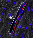
Detection of Porcine TrkA by Immunohistochemistry
Immunohistochemical characterization of ChR2-expressing DRG neurons. A, Immunolabeling of ChR2+ neurons (Venus; green) with either NF200, TrkC, CGRP, TrkA, TRPV1, or IB4 (red) in the L4 DRG contralateral and ipsilateral to PNI on day 14. B, Percentage of colocalization of each marker in ChR2+ DRG neurons [n = 4 rats (two to three slices per rat)]. Size-frequency histogram (inset) illustrating the distribution of the cross-sectional areas of ChR2+ NF200+ DRG neurons in the L4 DRG ipsilateral (371 cells) and contralateral (316 cells) to PNI on day 14. Values represent mean ± SEM. Scale bar: 100 μm. Image collected and cropped by CiteAb from the following open publication (https://pubmed.ncbi.nlm.nih.gov/29468190), licensed under a CC-BY license. Not internally tested by R&D Systems.
Detection of Rat TrkA by Immunohistochemistry
Immunohistochemical characterization of ChR2-expressing DRG neurons. A, Immunolabeling of ChR2+ neurons (Venus; green) with either NF200, TrkC, CGRP, TrkA, TRPV1, or IB4 (red) in the L4 DRG contralateral and ipsilateral to PNI on day 14. B, Percentage of colocalization of each marker in ChR2+ DRG neurons [n = 4 rats (two to three slices per rat)]. Size-frequency histogram (inset) illustrating the distribution of the cross-sectional areas of ChR2+ NF200+ DRG neurons in the L4 DRG ipsilateral (371 cells) and contralateral (316 cells) to PNI on day 14. Values represent mean ± SEM. Scale bar: 100 μm. Image collected and cropped by CiteAb from the following open publication (https://pubmed.ncbi.nlm.nih.gov/29468190), licensed under a CC-BY license. Not internally tested by R&D Systems.Applications for Rat TrkA Antibody
Application
Recommended Usage
Immunohistochemistry
5-15 µg/mL
Sample: Perfusion fixed frozen sections of rat dorsal root ganglion
Sample: Perfusion fixed frozen sections of rat dorsal root ganglion
Western Blot
1 µg/mL
Sample: Rat striatum
Sample: Rat striatum
Reviewed Applications
Read 2 reviews rated 4.5 using AF1056 in the following applications:
Formulation, Preparation, and Storage
Purification
Antigen Affinity-purified
Reconstitution
Reconstitute at 0.2 mg/mL in sterile PBS. For liquid material, refer to CoA for concentration.
Loading...
Formulation
Lyophilized from a 0.2 μm filtered solution in PBS with Trehalose. *Small pack size (SP) is supplied either lyophilized or as a 0.2 µm filtered solution in PBS.
Shipping
Lyophilized product is shipped at ambient temperature. Liquid small pack size (-SP) is shipped with polar packs. Upon receipt, store immediately at the temperature recommended below.
Stability & Storage
Use a manual defrost freezer and avoid repeated freeze-thaw cycles.
- 12 months from date of receipt, -20 to -70 °C as supplied.
- 1 month, 2 to 8 °C under sterile conditions after reconstitution.
- 6 months, -20 to -70 °C under sterile conditions after reconstitution.
Calculators
Background: TrkA
References
- Esposito, D. et al. (2001) J. Biol. Chem. 276:32687.
- Sofroniew, M.V. et al. (2001) Annu. Rev. Neurosci. 24:1217.
Long Name
Neurotrophic Tyrosine Kinase Receptor A
Alternate Names
NTRK-1, NTRK1
Gene Symbol
NTRK1
UniProt
Additional TrkA Products
Product Documents for Rat TrkA Antibody
Certificate of Analysis
To download a Certificate of Analysis, please enter a lot or batch number in the search box below.
Note: Certificate of Analysis not available for kit components.
Product Specific Notices for Rat TrkA Antibody
For research use only
Related Research Areas
Citations for Rat TrkA Antibody
 Powered by Bioz
Customer Reviews for Rat TrkA Antibody (2)
4.5 out of 5
2 Customer Ratings
Have you used Rat TrkA Antibody?
Submit a review and receive an Amazon gift card!
$25/€18/£15/$25CAN/¥2500 Yen for a review with an image
$10/€7/£6/$10CAN/¥1110 Yen for a review without an image
Submit a review
Customer Images
Showing
1
-
2 of
2 reviews
Showing All
Filter By:
-
Application: Immunocytochemistry/ImmunofluorescenceSample Tested: Bone ExtractsSpecies: MouseVerified Customer | Posted 10/31/2020

-
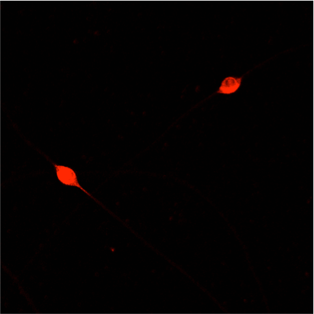
Application: Immunocytochemistry/ImmunofluorescenceSample Tested: E12 DRG neuronsSpecies: MouseVerified Customer | Posted 11/22/2019E12 mouse DRG neurons grown overnight with 10ng/ml NGF, then fixed in 4% PFA, blocked for 1 h with 5% BSA, and incubated overnight in 1% BSA containing AF1056 (1:300). A fluorophore-conjugated secondary antibody was used to visualise reactivity.
There are no reviews that match your criteria.
Protocols
Find general support by application which include: protocols, troubleshooting, illustrated assays, videos and webinars.
- Antigen Retrieval Protocol (PIER)
- Antigen Retrieval for Frozen Sections Protocol
- Appropriate Fixation of IHC/ICC Samples
- Cellular Response to Hypoxia Protocols
- Chromogenic IHC Staining of Formalin-Fixed Paraffin-Embedded (FFPE) Tissue Protocol
- Chromogenic Immunohistochemistry Staining of Frozen Tissue
- ClariTSA™ Fluorophore Kits
- Detection & Visualization of Antibody Binding
- Fluorescent IHC Staining of Frozen Tissue Protocol
- Graphic Protocol for Heat-induced Epitope Retrieval
- Graphic Protocol for the Preparation and Fluorescent IHC Staining of Frozen Tissue Sections
- Graphic Protocol for the Preparation and Fluorescent IHC Staining of Paraffin-embedded Tissue Sections
- Graphic Protocol for the Preparation of Gelatin-coated Slides for Histological Tissue Sections
- IHC Sample Preparation (Frozen sections vs Paraffin)
- Immunofluorescent IHC Staining of Formalin-Fixed Paraffin-Embedded (FFPE) Tissue Protocol
- Immunohistochemistry (IHC) and Immunocytochemistry (ICC) Protocols
- Immunohistochemistry Frozen Troubleshooting
- Immunohistochemistry Paraffin Troubleshooting
- Preparing Samples for IHC/ICC Experiments
- Preventing Non-Specific Staining (Non-Specific Binding)
- Primary Antibody Selection & Optimization
- Protocol for Heat-Induced Epitope Retrieval (HIER)
- Protocol for Making a 4% Formaldehyde Solution in PBS
- Protocol for VisUCyte™ HRP Polymer Detection Reagent
- Protocol for the Preparation & Fixation of Cells on Coverslips
- Protocol for the Preparation and Chromogenic IHC Staining of Frozen Tissue Sections
- Protocol for the Preparation and Chromogenic IHC Staining of Frozen Tissue Sections - Graphic
- Protocol for the Preparation and Chromogenic IHC Staining of Paraffin-embedded Tissue Sections
- Protocol for the Preparation and Chromogenic IHC Staining of Paraffin-embedded Tissue Sections - Graphic
- Protocol for the Preparation and Fluorescent IHC Staining of Frozen Tissue Sections
- Protocol for the Preparation and Fluorescent IHC Staining of Paraffin-embedded Tissue Sections
- Protocol for the Preparation of Gelatin-coated Slides for Histological Tissue Sections
- R&D Systems Quality Control Western Blot Protocol
- TUNEL and Active Caspase-3 Detection by IHC/ICC Protocol
- The Importance of IHC/ICC Controls
- Troubleshooting Guide: Immunohistochemistry
- Troubleshooting Guide: Western Blot Figures
- Western Blot Conditions
- Western Blot Protocol
- Western Blot Protocol for Cell Lysates
- Western Blot Troubleshooting
- Western Blot Troubleshooting Guide
- View all Protocols, Troubleshooting, Illustrated assays and Webinars
Loading...
Associated Pathways

