
Interleukin 8 (IL-8), also named monocyte-derived neutrophil chemotactic factor (MDNCF), neutrophil-activating protein 1 (NAP-1), neutrophil-activating factor (NAF) and granulocyte chemotactic peptide (GCP), belongs to the Glu-Leu-Arg motif containing (ELR+) CXC chemokine family and has been designated CXCL8. IL-8 is a potent neutrophil chemoattractant that recruits neutrophils to sites of inflammation. IL-8 also activates neutrophil functions and through a poorly understood mechanism, promotes angiogenesis. The biological activites of IL-8 is mediated by two types of G protein-coupled chemokine receptors, CXCR1 and CXCR2. In normal tissues, IL-8 expression and secretion is barely detectable. Upon stimulation by a wide range of pro-inflammatory signals including exposure to IL-1, TNF, bacterial or viral products, IL-8 production is rapidly induced in many different cell types. Secreted IL-8 is not glycosylated but has N‑terminal sequence heterogenecity due to proteolytic processing. In human, two major forms, the 72 amino acid (aa) monocyte-derived IL-8 and the 77 aa endothelial IL-8 have been identified. Whereas the 72 aa isoform is a more potent chemoattractant, only the 77 aa isoform can induce apoptosis in leukemic cells. The N‑terminal pentapeptide in the 77 aa isoform has been identified as the active site for the IL-8 apoptotic activity. Canine IL-8 encodes a 101 aa precursor protein with a putative 22 aa signal peptide. It shares 77% and 87% aa sequence identity with human and porcine IL-8, respectively. Similar to human IL-8, recombinant canine IL-8 also undergoes
N‑terminal processing. Two major peptides (the 79 aa and 74 aa variants that differ by an analogous N‑terminal pentapeptide) are present in the recombinant canine IL-8 preparations.

Scientific images may be resized, cropped, or adjusted for brightness or contrast for presentation purposes. These updates are limited to presentation and do not affect the underlying scientific interpretation of the data. Where available, additional source data may be provided upon request.
Key Product Details
Validated by
Species Reactivity
Applications
Label
Antibody Source
Product Specifications
Immunogen
Ala23-Pro101 & & Val28-Pro101
Accession # P41324
Specificity
Clonality
Host
Isotype
Endotoxin Level
Scientific Data Images for Canine IL-8/CXCL8 Antibody
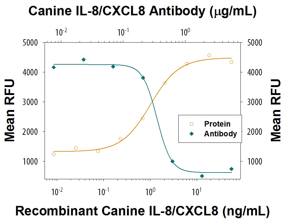
Chemotaxis Induced by IL-8/CXCL8 and Neutralization by Canine IL-8/CXCL8 Antibody.
Recombinant Canine IL-8/CXCL8 (Catalog # 1608-CL) chemoattracts the BaF3 mouse pro-B cell line transfected with human CXCR2 in a dose-dependent manner (orange line). The amount of cells that migrated through to the lower chemotaxis chamber was measured by Resazurin (Catalog # AR002). Chemotaxis elicited by Recombinant Canine IL-8/CXCL8 (10 ng/mL) is neutralized (green line) by increasing concentrations of Goat Anti-Canine IL-8/CXCL8 Antigen Affinity-purified Polyclonal Antibody (Catalog # AF1608). The ND50 is typically 0.1-0.5 µg/mL.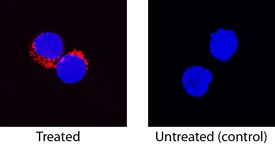
IL-8/CXCL8 in Canine PBMCs.
IL-8/CXCL8 was detected in immersion fixed canine peripheral blood mononuclear cells (PBMCs) treated with Concanavalin A using Goat Anti-Canine IL-8/CXCL8 Antigen Affinity-purified Polyclonal Antibody (Catalog # AF1608) at 15 µg/mL for 3 hours at room temperature. Cells were stained using the NorthernLights™ 557-conjugated Anti-Goat IgG Secondary Antibody (red; Catalog # NL001) and counterstained with DAPI (blue). Specific staining was localized to cytoplasm. View our protocol for Fluorescent ICC Staining of Non-adherent Cells.Applications for Canine IL-8/CXCL8 Antibody
Immunocytochemistry
Sample: Immersion fixed canine peripheral blood mononuclear cells treated with Concanavalin A
Western Blot
Sample: Recombinant Canine IL-8/CXCL8 (Catalog # 1608-CL)
Neutralization
Formulation, Preparation, and Storage
Purification
Reconstitution
Reconstitute at 0.2 mg/mL in sterile PBS. For liquid material, refer to CoA for concentration.
Formulation
Shipping
Stability & Storage
- 12 months from date of receipt, -20 to -70 °C as supplied.
- 1 month, 2 to 8 °C under sterile conditions after reconstitution.
- 6 months, -20 to -70 °C under sterile conditions after reconstitution.
Calculators
Background: IL-8/CXCL8
References
- Van Damme, J. et al. (1998) in The Cytokine Handbook, A.W. Thomson, ed., Academic Press, New York., p. 271.
- Terui, Y. et al. (1998) Blood, 92:2672.
- Terui, Y. et al. (1999) Cancer Research 59:5651.
Long Name
Alternate Names
Gene Symbol
UniProt
Additional IL-8/CXCL8 Products
Product Documents for Canine IL-8/CXCL8 Antibody
Certificate of Analysis
To download a Certificate of Analysis, please enter a lot or batch number in the search box below.
Note: Certificate of Analysis not available for kit components.
Product Specific Notices for Canine IL-8/CXCL8 Antibody
For research use only
Customer Reviews for Canine IL-8/CXCL8 Antibody
There are currently no reviews for this product. Be the first to review Canine IL-8/CXCL8 Antibody and earn rewards!
Have you used Canine IL-8/CXCL8 Antibody?
Submit a review and receive an Amazon gift card!
$25/€18/£15/$25CAN/¥2500 for a review with an image
$10/€7/£6/$10CAN/¥1110 for a review without an image
Submit a review
Protocols
Find general support by application which include: protocols, troubleshooting, illustrated assays, videos and webinars.
- Appropriate Fixation of IHC/ICC Samples
- Cellular Response to Hypoxia Protocols
- ClariTSA™ Fluorophore Kits
- Detection & Visualization of Antibody Binding
- ICC Cell Smear Protocol for Suspension Cells
- ICC Immunocytochemistry Protocol Videos
- ICC for Adherent Cells
- Immunocytochemistry (ICC) Protocol
- Immunocytochemistry Troubleshooting
- Immunofluorescence of Organoids Embedded in Cultrex Basement Membrane Extract
- Immunohistochemistry (IHC) and Immunocytochemistry (ICC) Protocols
- Preparing Samples for IHC/ICC Experiments
- Preventing Non-Specific Staining (Non-Specific Binding)
- Primary Antibody Selection & Optimization
- Protocol for VisUCyte™ HRP Polymer Detection Reagent
- Protocol for the Fluorescent ICC Staining of Cell Smears - Graphic
- Protocol for the Fluorescent ICC Staining of Cultured Cells on Coverslips - Graphic
- Protocol for the Preparation and Fluorescent ICC Staining of Cells on Coverslips
- Protocol for the Preparation and Fluorescent ICC Staining of Non-adherent Cells
- Protocol for the Preparation and Fluorescent ICC Staining of Stem Cells on Coverslips
- Protocol for the Preparation of a Cell Smear for Non-adherent Cell ICC - Graphic
- R&D Systems Quality Control Western Blot Protocol
- TUNEL and Active Caspase-3 Detection by IHC/ICC Protocol
- The Importance of IHC/ICC Controls
- Troubleshooting Guide: Western Blot Figures
- Western Blot Conditions
- Western Blot Protocol
- Western Blot Protocol for Cell Lysates
- Western Blot Troubleshooting
- Western Blot Troubleshooting Guide
- View all Protocols, Troubleshooting, Illustrated assays and Webinars


