GSK-3 beta Antibody (3D10) - BSA Free
Novus Biologicals | Catalog # NBP1-47470
![Western Blot: GSK-3 beta Antibody (3D10)BSA Free [NBP1-47470]](https://resources.rndsystems.com/images/products/GSK-3-beta-Antibody-3D10-Western-Blot-NBP1-47470-img0012.jpg "Western Blot: GSK-3 beta Antibody (3D10)BSA Free [NBP1-47470]")
Key Product Details
Validated by
Species Reactivity
Validated:
Cited:
Applications
Validated:
Cited:
Label
Antibody Source
Format
Product Specifications
Immunogen
Clonality
Host
Isotype
Theoretical MW
Disclaimer note: The observed molecular weight of the protein may vary from the listed predicted molecular weight due to post translational modifications, post translation cleavages, relative charges, and other experimental factors.
Scientific Data Images for GSK-3 beta Antibody (3D10) - BSA Free
Western Blot: GSK-3 beta Antibody (3D10)BSA Free [NBP1-47470]
Western Blot: GSK-3 beta Antibody (3D10) [NBP1-47470] - Analysis of GSK-3 beta in mouse beta cell line (betaTC3) using anti-GSK-3 beta antibody. Image from verified customer review.![Immunocytochemistry/ Immunofluorescence: GSK-3 beta Antibody (3D10) - BSA Free [NBP1-47470]](https://resources.rndsystems.com/images/products/GSK-3-beta-Antibody-3D10-Immunocytochemistry-Immunofluorescence-NBP1-47470-img0006.jpg "Immunocytochemistry/ Immunofluorescence: GSK-3 beta Antibody (3D10) - BSA Free [NBP1-47470]")
Immunocytochemistry/ Immunofluorescence: GSK-3 beta Antibody (3D10) - BSA Free [NBP1-47470]
Immunocytochemistry/Immunofluorescence: GSK-3 beta Antibody (3D10) [NBP1-47470] - Analysis of NIH/3T3 (left) and U251 (right) cells using GSK3 beta mouse mAb (green). Blue: DRAQ5 fluorescent DNA dye. Red: Actin filaments have been labeled with Alexa Fluor-555 phalloidin.![Immunohistochemistry: GSK-3 beta Antibody (3D10) - BSA Free [NBP1-47470]](https://resources.rndsystems.com/images/products/GSK-3-beta-Antibody-3D10-Immunohistochemistry-NBP1-47470-img0014.jpg "Immunohistochemistry: GSK-3 beta Antibody (3D10) - BSA Free [NBP1-47470]")
![Flow Cytometry: GSK-3 beta Antibody (3D10) - BSA Free [NBP1-47470]](https://resources.rndsystems.com/images/products/GSK-3-beta-Antibody-3D10-Flow-Cytometry-NBP1-47470-img0013.jpg "Flow Cytometry: GSK-3 beta Antibody (3D10) - BSA Free [NBP1-47470]")
Flow Cytometry: GSK-3 beta Antibody (3D10) - BSA Free [NBP1-47470]
Flow Cytometry: GSK-3 beta Antibody (3D10) [NBP1-47470] - An intracellular stain was performed on HeLa cells with GSK-3 beta (3D10) antibody NBP1-47470PE (blue) and a matched isotype control (orange). Cells were fixed with 4% PFA and then permeablized with 0.1% saponin. Cells were incubated in an antibody dilution of 2.5 ug/mL for 30 minutes at room temperature. Both antibodies were conjugated to Phycoerythrin.![Western Blot: GSK-3 beta Antibody (3D10)BSA Free [NBP1-47470]](https://resources.rndsystems.com/images/products/GSK-3-beta-Antibody-3D10-Western-Blot-NBP1-47470-img0008.jpg "Western Blot: GSK-3 beta Antibody (3D10)BSA Free [NBP1-47470]")
Western Blot: GSK-3 beta Antibody (3D10)BSA Free [NBP1-47470]
Western Blot: GSK-3 beta Antibody (3D10) [NBP1-47470] - Analysis using GSK3 beta mouse mAb against A549 (1), K562 (2), PC-12 (3), NIH/3T3 (4), and HEK293 (5) cell lysates.![Immunohistochemistry-Paraffin: GSK-3 beta Antibody (3D10) - BSA Free [NBP1-47470]](https://resources.rndsystems.com/images/products/GSK-3-beta-Antibody-3D10-Immunohistochemistry-Paraffin-NBP1-47470-img0007.jpg "Immunohistochemistry-Paraffin: GSK-3 beta Antibody (3D10) - BSA Free [NBP1-47470]")
Immunohistochemistry-Paraffin: GSK-3 beta Antibody (3D10) - BSA Free [NBP1-47470]
Immunohistochemistry-Paraffin: GSK-3 beta Antibody (3D10) [NBP1-47470] - Analysis of paraffin-embedded human lung cancer (left) and breast cancer tissues (right) using GSK3 beta mouse mAb with DAB staining.![Flow Cytometry: GSK-3 beta Antibody (3D10) - BSA Free [NBP1-47470]](https://resources.rndsystems.com/images/products/GSK-3-beta-Antibody-3D10-Flow-Cytometry-NBP1-47470-img0005.jpg "Flow Cytometry: GSK-3 beta Antibody (3D10) - BSA Free [NBP1-47470]")
Flow Cytometry: GSK-3 beta Antibody (3D10) - BSA Free [NBP1-47470]
Flow Cytometry: GSK-3 beta Antibody (3D10) [NBP1-47470] - Flow cytometric analysis of Hela cells using GSK3 beta mouse mAb (green) and negative control (purple).![Simple Western: GSK-3 beta Antibody (3D10)BSA Free [NBP1-47470]](https://resources.rndsystems.com/images/products/GSK-3-beta-Antibody-3D10-Simple-Western-NBP1-47470-img0009.jpg "Simple Western: GSK-3 beta Antibody (3D10)BSA Free [NBP1-47470]")
Simple Western: GSK-3 beta Antibody (3D10)BSA Free [NBP1-47470]
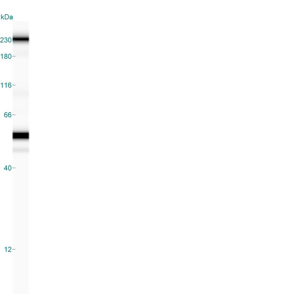
Simple Western: GSK-3 beta Antibody (3D10) [NBP1-47470] - Simple Western lane view shows a specific band for GSK-3 Beta in 0.5 mg/ml of Hek293 lysate. This experiment was performed under reducing conditions using the 12-230 kDa separation system.![Simple Western: GSK-3 beta Antibody (3D10)BSA Free [NBP1-47470]](https://resources.rndsystems.com/images/products/GSK-3-beta-Antibody-3D10-Simple-Western-NBP1-47470-img0010.jpg "Simple Western: GSK-3 beta Antibody (3D10)BSA Free [NBP1-47470]")
Simple Western: GSK-3 beta Antibody (3D10)BSA Free [NBP1-47470]
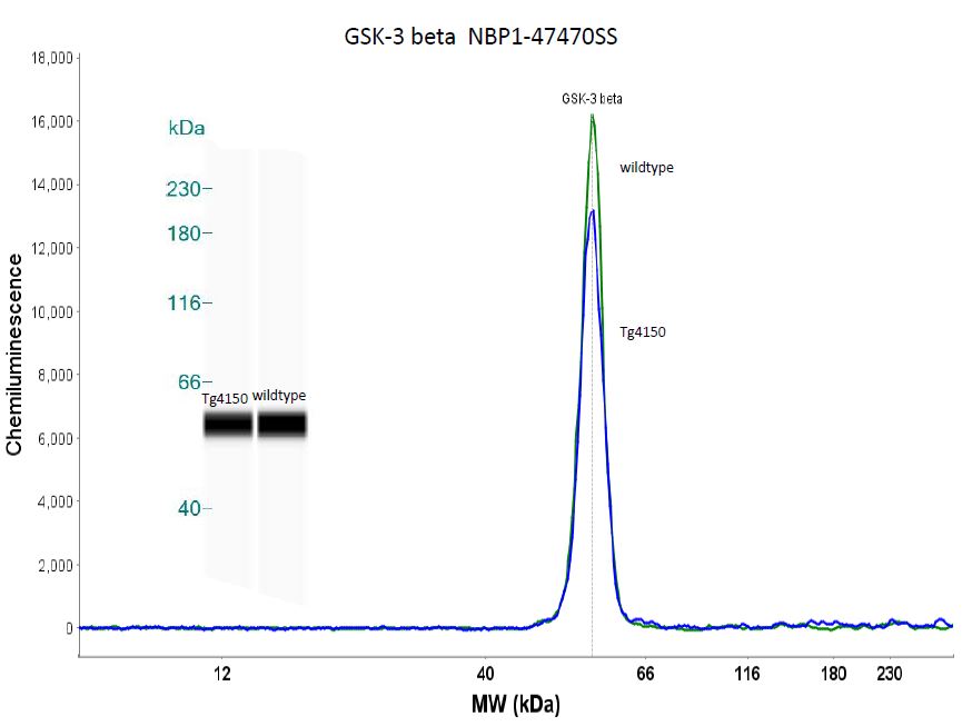
Simple Western: GSK-3 beta Antibody (3D10) [NBP1-47470] - Simple western analysis of mouse brain tissue (striatum) from 4 month old Tg4150 and wildtype mice. Image courtesy of Dr. Brandi Wasek-Patterson at Baylor Research Institute, Institute of Metabolic Disease. - BSA Free [NBP1-47470] -")
Immunocytochemistry/ Immunofluorescence: GSK-3 beta Antibody (3D10) - BSA Free [NBP1-47470] -
Immunocytochemistry/ Immunofluorescence: GSK-3 beta Antibody (3D10) - BSA Free [NBP1-47470] - Fap1-inhibition with SLV peptide increases Fas & Gsk3 beta phosphorylation in CD133+ cells in a murine xenograft modelSW620 cells were injected in the flanks of athymic Nude mice & tumor volume was determination biweekly. Mice were treated weekly with oxaliplatin (days 0, 7 & 14) & injected daily with Fap1 blocking SLV peptide or VLS control peptide, or treated with SLV or VLS peptide alone (n=12 per cohort). Tumors were simultaneously harvested from cohorts of mice when control tumors were >2,000 mm3. (A) SLV peptide increases Fas phosphorylation in CD133+ xenograft tumors with or without oxaliplatin. Immunofluorescent detection of phospho-Fas or CD133 was performed with DAPI staining of nuclei. (B) SLV peptide increases Gsk3 beta phosphorylation in CD133+ xenograft tumors with or without oxaliplatin. Immunofluorescent detection of phospho-Gsk3 beta or CD133 was performed with DAPI staining of nuclei. Image collected & cropped by CiteAb from the following publication (https://pubmed.ncbi.nlm.nih.gov/29899829), licensed under a CC-BY license. Not internally tested by Novus Biologicals. - BSA Free [NBP1-47470] -")
Immunocytochemistry/ Immunofluorescence: GSK-3 beta Antibody (3D10) - BSA Free [NBP1-47470] -
Immunocytochemistry/ Immunofluorescence: GSK-3 beta Antibody (3D10) - BSA Free [NBP1-47470] - Fap1-inhibition with SLV peptide increases phosphorylation of Fap1-substrates Fas & Gsk3 beta in a murine xenograft modelSW620 cells were injected in the flanks of athymic Nude mice & tumor volume was determination biweekly. Mice were treated weekly with oxaliplatin (days 0, 7 & 14) & injected daily with Fap1 blocking SLV peptide or VLS control peptide, or treated with SLV or VLS peptide alone (n=12 per cohort). Tumors were simultaneously harvested from cohorts of mice when control tumors were >2,000 mm3. (A) SLV peptide increases gland formation in xenograft tumors with or without oxaliplatin. Histology was analyzed by hematoxylin/ eosin staining. Fap1 expression was determined by immunofluorescence. Relative fluorescent intensity (RFI) of Fap1 staining is indicated below relevant panels. (B) SLV peptide increases Fas-phosphorylation in xenograft tumors with or without by oxaliplatin. Immunofluorescent detection of total versus phospho-Fas was performed with DAPI staining of nuclei. Areas without gland formation were selected for this study. (C) SLV peptide increases Gsk3 beta -phosphorylation with or without oxaliplatin. Immunofluorescent detection of total versus phospho- Gsk3 beta was performed with DAPI staining of nuclei. Areas without gland formation were selected for this study. Image collected & cropped by CiteAb from the following publication (https://pubmed.ncbi.nlm.nih.gov/29899829), licensed under a CC-BY license. Not internally tested by Novus Biologicals.Applications for GSK-3 beta Antibody (3D10) - BSA Free
ELISA
Flow Cytometry
Immunocytochemistry/ Immunofluorescence
Immunohistochemistry
Immunohistochemistry-Paraffin
Simple Western
Western Blot
In Simple Western only 10 - 15 uL of the recommended dilution is used per data point.
See Simple Western Antibody Database for Simple Western validation: Tested in Mesenchymal stem cells, Hek293 lysate 0.5 mg/mL, separated by Size, antibody dilution of 1:50, apparent MW was 56 kDa. Separated by Size-Wes, Sally Sue/Peggy Sue.
Reviewed Applications
Read 4 reviews rated 4.8 using NBP1-47470 in the following applications:
Flow Cytometry Panel Builder
Bio-Techne Knows Flow Cytometry
Save time and reduce costly mistakes by quickly finding compatible reagents using the Panel Builder Tool.
Advanced Features
- Spectra Viewer - Custom analysis of spectra from multiple fluorochromes
- Spillover Popups - Visualize the spectra of individual fluorochromes
- Antigen Density Selector - Match fluorochrome brightness with antigen density

Formulation, Preparation, and Storage
Purification
Formulation
Format
Preservative
Concentration
Shipping
Stability & Storage
Background: GSK-3 beta
Long Name
Alternate Names
Entrez Gene IDs
Gene Symbol
UniProt
Additional GSK-3 beta Products
Product Documents for GSK-3 beta Antibody (3D10) - BSA Free
Certificate of Analysis
To download a Certificate of Analysis, please enter a lot or batch number in the search box below.
Product Specific Notices for GSK-3 beta Antibody (3D10) - BSA Free
This product is for research use only and is not approved for use in humans or in clinical diagnosis. Primary Antibodies are guaranteed for 1 year from date of receipt.
Related Research Areas
Citations for GSK-3 beta Antibody (3D10) - BSA Free
 Powered by Bioz
Customer Reviews for GSK-3 beta Antibody (3D10) - BSA Free (4)
Have you used GSK-3 beta Antibody (3D10) - BSA Free?
Submit a review and receive an Amazon gift card!
$25/€18/£15/$25CAN/¥2500 Yen for a review with an image
$10/€7/£6/$10CAN/¥1110 Yen for a review without an image
Submit a review
Customer Images
-
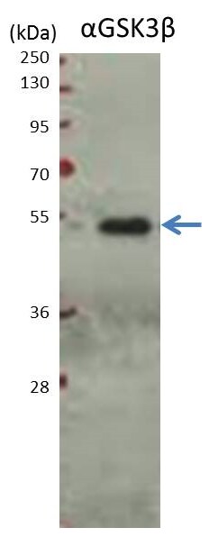
Application: Western BlotSample Tested: MM1S (multiple myeloma cell line)Species: HumanVerified Customer | Posted 11/30/2016Western blot analysis of GSK-3 beta antibody NBP1-47470 against MM1S cell lysate.Conditions: - MM1S cells lysed with NP40 lysis buffer -protein quantified with BCA assay, 10 ug total protein loaded per lane -protein resolved by SDS-PAGE on 7.5% gel -transferred onto PVDF membrane and blocked with 5% BSA in TBST for 2hrs at room temperature -incubated with antibody (1:10,000) in TBST (+1% BSA) overnight at 4 deg with shaking -secondary incubation with HRP-conjugated anti-mouse antibody (1:10,000) for 1hr at room temperature -visualized with ECL chemilumnescence, imaged with film, exposure for 2 mins

-
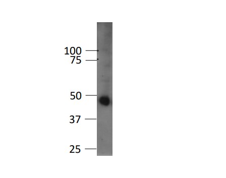
Application: Western BlotSample Tested: mouse beta cell lineSpecies: MouseVerified Customer | Posted 11/10/2016western blot of Gsk3beta (1:1000) in mouse beta cell line (betaTC3) nuclear extract.
-
Application: Simple WesternSample Tested:Species: MouseVerified Customer | Posted 08/05/2015Simple western analysis of mouse brain tissue (striatum) from 4 month old Tg4150 and wildtype mice.
-
Application: Simple WesternSample Tested: Human mesenchymal stem cell whole cell lysateSpecies: HumanVerified Customer | Posted 03/24/2015Human mesenchymal stem cell whole cell lysate
There are no reviews that match your criteria.
Protocols
Find general support by application which include: protocols, troubleshooting, illustrated assays, videos and webinars.
- 7-Amino Actinomycin D (7-AAD) Cell Viability Flow Cytometry Protocol
- Antigen Retrieval Protocol (PIER)
- Antigen Retrieval for Frozen Sections Protocol
- Appropriate Fixation of IHC/ICC Samples
- Cellular Response to Hypoxia Protocols
- Chromogenic IHC Staining of Formalin-Fixed Paraffin-Embedded (FFPE) Tissue Protocol
- Chromogenic Immunohistochemistry Staining of Frozen Tissue
- ClariTSA™ Fluorophore Kits
- Detection & Visualization of Antibody Binding
- ELISA Sample Preparation & Collection Guide
- ELISA Troubleshooting Guide
- Extracellular Membrane Flow Cytometry Protocol
- Flow Cytometry Protocol for Cell Surface Markers
- Flow Cytometry Protocol for Staining Membrane Associated Proteins
- Flow Cytometry Staining Protocols
- Flow Cytometry Troubleshooting Guide
- Fluorescent IHC Staining of Frozen Tissue Protocol
- Graphic Protocol for Heat-induced Epitope Retrieval
- Graphic Protocol for the Preparation and Fluorescent IHC Staining of Frozen Tissue Sections
- Graphic Protocol for the Preparation and Fluorescent IHC Staining of Paraffin-embedded Tissue Sections
- Graphic Protocol for the Preparation of Gelatin-coated Slides for Histological Tissue Sections
- How to Run an R&D Systems DuoSet ELISA
- How to Run an R&D Systems Quantikine ELISA
- How to Run an R&D Systems Quantikine™ QuicKit™ ELISA
- ICC Cell Smear Protocol for Suspension Cells
- ICC Immunocytochemistry Protocol Videos
- ICC for Adherent Cells
- IHC Sample Preparation (Frozen sections vs Paraffin)
- Immunocytochemistry (ICC) Protocol
- Immunocytochemistry Troubleshooting
- Immunofluorescence of Organoids Embedded in Cultrex Basement Membrane Extract
- Immunofluorescent IHC Staining of Formalin-Fixed Paraffin-Embedded (FFPE) Tissue Protocol
- Immunohistochemistry (IHC) and Immunocytochemistry (ICC) Protocols
- Immunohistochemistry Frozen Troubleshooting
- Immunohistochemistry Paraffin Troubleshooting
- Intracellular Flow Cytometry Protocol Using Alcohol (Methanol)
- Intracellular Flow Cytometry Protocol Using Detergents
- Intracellular Nuclear Staining Flow Cytometry Protocol Using Detergents
- Intracellular Staining Flow Cytometry Protocol Using Alcohol Permeabilization
- Intracellular Staining Flow Cytometry Protocol Using Detergents to Permeabilize Cells
- Preparing Samples for IHC/ICC Experiments
- Preventing Non-Specific Staining (Non-Specific Binding)
- Primary Antibody Selection & Optimization
- Propidium Iodide Cell Viability Flow Cytometry Protocol
- Protocol for Heat-Induced Epitope Retrieval (HIER)
- Protocol for Liperfluo
- Protocol for Making a 4% Formaldehyde Solution in PBS
- Protocol for VisUCyte™ HRP Polymer Detection Reagent
- Protocol for the Characterization of Human Th22 Cells
- Protocol for the Characterization of Human Th9 Cells
- Protocol for the Fluorescent ICC Staining of Cell Smears - Graphic
- Protocol for the Fluorescent ICC Staining of Cultured Cells on Coverslips - Graphic
- Protocol for the Preparation & Fixation of Cells on Coverslips
- Protocol for the Preparation and Chromogenic IHC Staining of Frozen Tissue Sections
- Protocol for the Preparation and Chromogenic IHC Staining of Frozen Tissue Sections - Graphic
- Protocol for the Preparation and Chromogenic IHC Staining of Paraffin-embedded Tissue Sections
- Protocol for the Preparation and Chromogenic IHC Staining of Paraffin-embedded Tissue Sections - Graphic
- Protocol for the Preparation and Fluorescent ICC Staining of Cells on Coverslips
- Protocol for the Preparation and Fluorescent ICC Staining of Non-adherent Cells
- Protocol for the Preparation and Fluorescent ICC Staining of Stem Cells on Coverslips
- Protocol for the Preparation and Fluorescent IHC Staining of Frozen Tissue Sections
- Protocol for the Preparation and Fluorescent IHC Staining of Paraffin-embedded Tissue Sections
- Protocol for the Preparation of Gelatin-coated Slides for Histological Tissue Sections
- Protocol for the Preparation of a Cell Smear for Non-adherent Cell ICC - Graphic
- Protocol: Annexin V and PI Staining by Flow Cytometry
- Protocol: Annexin V and PI Staining for Apoptosis by Flow Cytometry
- Quantikine HS ELISA Kit Assay Principle, Alkaline Phosphatase
- Quantikine HS ELISA Kit Principle, Streptavidin-HRP Polymer
- R&D Systems Quality Control Western Blot Protocol
- Sandwich ELISA (Colorimetric) – Biotin/Streptavidin Detection Protocol
- Sandwich ELISA (Colorimetric) – Direct Detection Protocol
- TUNEL and Active Caspase-3 Detection by IHC/ICC Protocol
- The Importance of IHC/ICC Controls
- Troubleshooting Guide: ELISA
- Troubleshooting Guide: Fluorokine Flow Cytometry Kits
- Troubleshooting Guide: Immunohistochemistry
- Troubleshooting Guide: Western Blot Figures
- Western Blot Conditions
- Western Blot Protocol
- Western Blot Protocol for Cell Lysates
- Western Blot Troubleshooting
- Western Blot Troubleshooting Guide
- View all Protocols, Troubleshooting, Illustrated assays and Webinars
FAQs for GSK-3 beta Antibody (3D10) - BSA Free
-
Q: I received this antibody in February 2012 and stored the unopened package immediately at 4C. Today (4/5/2012), I opened the package, placed the vial and centrifuged it briefly. I opened the top and found some thick beige-colored liquid which measured around 50 ul. I don't think I can use the antibody. Could you help me with the problem, please?
A: NBP1-47470 is supplied as ascitic fluid, which frequently has a beige-like color, and this could be what you're seeing in your vial. Otherwise, I'm concerned that the antibody may have been damaged by storing it a 4 C for such a long time. There is a very low concentration of preservative in this antibody, which is why we recommend storage at -20 C long term.
-
Q: Our customer would like to know whether NBP1-47470, GSK3 beta Antibody can be used in IF. It shows on the website that this antibody can be used to detect the target protein through ICC/IF. But his sampels are paraffin-embedded tissues, no cultured cells. Thus he is not sure. Please give us some advice and help.
A: Our GSK3 beta antibody with catalogue number NBP1-47470 has been validated for IHC with paraffin-embedded tissues, and has also been validated for immunocytochemistry. Our immunocytochemistry data can be seen in image 3 on our website, whilst our IHC-P data can be seen in image 4. This antibody is covered by our 100% quality guarantee for your customer's application of IHC with paraffin-embedded tissues. Currently tested species are human, mouse, rat and primate.
-
Q: I received this antibody in February 2012 and stored the unopened package immediately at 4C. Today (4/5/2012), I opened the package, placed the vial and centrifuged it briefly. I opened the top and found some thick beige-colored liquid which measured around 50 ul. I don't think I can use the antibody. Could you help me with the problem, please?
A: NBP1-47470 is supplied as ascitic fluid, which frequently has a beige-like color, and this could be what you're seeing in your vial. Otherwise, I'm concerned that the antibody may have been damaged by storing it a 4 C for such a long time. There is a very low concentration of preservative in this antibody, which is why we recommend storage at -20 C long term.
-
Q: Our customer would like to know whether NBP1-47470, GSK3 beta Antibody can be used in IF. It shows on the website that this antibody can be used to detect the target protein through ICC/IF. But his sampels are paraffin-embedded tissues, no cultured cells. Thus he is not sure. Please give us some advice and help.
A: Our GSK3 beta antibody with catalogue number NBP1-47470 has been validated for IHC with paraffin-embedded tissues, and has also been validated for immunocytochemistry. Our immunocytochemistry data can be seen in image 3 on our website, whilst our IHC-P data can be seen in image 4. This antibody is covered by our 100% quality guarantee for your customer's application of IHC with paraffin-embedded tissues. Currently tested species are human, mouse, rat and primate.
Associated Pathways


