
Key Product Details
Validated by
Knockout/Knockdown
Species Reactivity
Validated:
Human
Cited:
Human
Applications
Validated:
Knockout Validated, Western Blot, Flow Cytometry, CyTOF-ready
Cited:
Immunohistochemistry, Western Blot, Flow Cytometry, Immunoprecipitation
Label
Unconjugated
Antibody Source
Monoclonal Mouse IgG1 Clone # 185504
Loading...
Product Specifications
Immunogen
Mouse myeloma cell line NS0-derived recombinant human B7‑H3
Leu29-Pro245
Accession # NP_079516
Leu29-Pro245
Accession # NP_079516
Specificity
Detects human B7-H3 in direct ELISAs and Western blots. In Western blots, no cross-reactivity with recombinant human (rh) B7‑H1, rhB7‑H2, rhB7‑1, rhB7‑2, or recombinant mouse PD-L2 is observed.
Clonality
Monoclonal
Host
Mouse
Isotype
IgG1
Scientific Data Images for Human B7-H3 Antibody (185504)
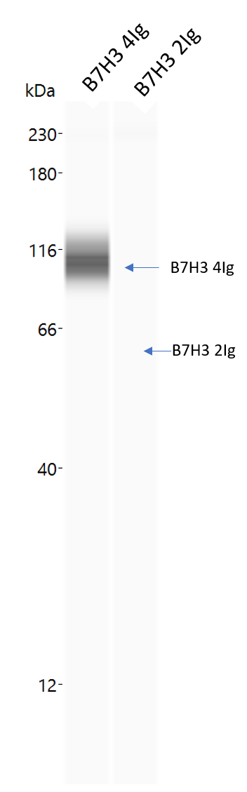
Detection of Human B7‑H3 by Western Blot.
Western blot shows lysates of LNCaP human prostate cancer cell line and U2OS human osteosarcoma cell line. PVDF membrane was probed with 2 µg/mL of Mouse Anti-Human B7-H3 Monoclonal Antibody (Catalog # MAB1027) followed by HRP-conjugated Anti-Mouse IgG Secondary Antibody (Catalog # HAF018). A specific band was detected for B7-H3 at approximately 90-110 kDa (as indicated). This experiment was conducted under reducing conditions and using Immunoblot Buffer Group 1.
Detection of B7-H3 in PC‑3 Human Cell Line by Flow Cytometry.
PC-3 human prostate cancer cell line was stained with Mouse Anti-Human B7-H3 Monoclonal Antibody (Catalog # MAB1027, filled histogram) or isotype control antibody (Catalog # MAB002, open histogram), followed by Phycoerythrin-conjugated Anti-Mouse IgG F(ab')2Secondary Antibody (Catalog # F0102B).
Detection of B7‑H3 in Human Dendritic Cells by Flow Cytometry.
Human monocyte-derived dendritic cells were stained with Mouse Anti-Human B7-H3 Monoclonal Antibody (Catalog # MAB1027, filled histogram) or isotype control antibody (Catalog # MAB002, open histogram), followed by Allophycocyanin-conjugated Anti-Mouse IgG Secondary Antibody (Catalog # F0101B).
Western Blot Shows Human B7‑H3 Specificity by Using Knockout Cell Line.
Western blot shows lysates of U2OS human osteosarcoma parental cell line and B7-H3 knockout U2OS cell line (KO). PVDF membrane was probed with 2 µg/mL of Mouse Anti-Human B7-H3 Monoclonal Antibody (Catalog # MAB1027) followed by HRP-conjugated Anti-Mouse IgG Secondary Antibody (Catalog # HAF018). A specific band was detected for B7-H3 at approximately 95 kDa (as indicated) in the parental U2OS cell line, but is not detectable in knockout U2OS cell line. GAPDH (Catalog # MAB5718) is shown as a loading control. This experiment was conducted under reducing conditions and using Immunoblot Buffer Group 1.Applications for Human B7-H3 Antibody (185504)
Application
Recommended Usage
CyTOF-ready
Ready to be labeled using established conjugation methods. No BSA or other carrier proteins that could interfere with conjugation.
Flow Cytometry
0.25 µg/106 cells
Sample: Human monocyte-derived dendritic cells and PC‑3 human prostate cancer cell line
Sample: Human monocyte-derived dendritic cells and PC‑3 human prostate cancer cell line
Knockout Validated
B7‑H3
is specifically detected in U2OS human osteosarcoma parental cell line but is not detectable in
B7‑H3 knockout U2OS cell line.
Western Blot
2 µg/mL
Sample: LNCaP human prostate cancer cell line and U2OS human osteosarcoma cell line
Sample: LNCaP human prostate cancer cell line and U2OS human osteosarcoma cell line
Reviewed Applications
Read 4 reviews rated 4.5 using MAB1027 in the following applications:
Flow Cytometry Panel Builder
Bio-Techne Knows Flow Cytometry
Save time and reduce costly mistakes by quickly finding compatible reagents using the Panel Builder Tool.
Advanced Features
- Spectra Viewer - Custom analysis of spectra from multiple fluorochromes
- Spillover Popups - Visualize the spectra of individual fluorochromes
- Antigen Density Selector - Match fluorochrome brightness with antigen density

Formulation, Preparation, and Storage
Purification
Protein A or G purified from hybridoma culture supernatant
Reconstitution
Reconstitute at 0.5 mg/mL in sterile PBS. For liquid material, refer to CoA for concentration.
Loading...
Formulation
Lyophilized from a 0.2 μm filtered solution in PBS with Trehalose. *Small pack size (SP) is supplied either lyophilized or as a 0.2 µm filtered solution in PBS.
Shipping
Lyophilized product is shipped at ambient temperature. Liquid small pack size (-SP) is shipped with polar packs. Upon receipt, store immediately at the temperature recommended below.
Stability & Storage
Use a manual defrost freezer and avoid repeated freeze-thaw cycles.
- 12 months from date of receipt, -20 to -70 °C as supplied.
- 1 month, 2 to 8 °C under sterile conditions after reconstitution.
- 6 months, -20 to -70 °C under sterile conditions after reconstitution.
Calculators
Background: B7-H3
References
- Chapoval, A.I. et al. (2001) Nat. Immunol. 2:269.
- Sharpe, A.H. and G.J. Freeman (2002) Nat. Rev. Immunol. 2:116.
- Coyle, A. and J. Gutierrez-Ramos (2001) Nat. Immunol. 2:203.
Long Name
B7 Homolog 3
Alternate Names
B7H3, CD276
Gene Symbol
CD276
UniProt
Additional B7-H3 Products
Product Documents for Human B7-H3 Antibody (185504)
Certificate of Analysis
To download a Certificate of Analysis, please enter a lot or batch number in the search box below.
Note: Certificate of Analysis not available for kit components.
Product Specific Notices for Human B7-H3 Antibody (185504)
For research use only
Citations for Human B7-H3 Antibody (185504)
 Powered by Bioz
Customer Reviews for Human B7-H3 Antibody (185504) (4)
4.5 out of 5
4 Customer Ratings
Have you used Human B7-H3 Antibody (185504)?
Submit a review and receive an Amazon gift card!
$25/€18/£15/$25CAN/¥2500 Yen for a review with an image
$10/€7/£6/$10CAN/¥1110 Yen for a review without an image
Submit a review
Customer Images
Showing
1
-
4 of
4 reviews
Showing All
Filter By:
-
Application: Western BlotSample Tested: Osteosarcoma U2OS cell lineSpecies: HumanVerified Customer | Posted 06/22/2022

-
Application: Simple WesternSample Tested: Purified proteinSpecies: HumanVerified Customer | Posted 05/01/2020
-
Application: Functional AssaySample Tested: 3T3-L1 mouse embryonic fibroblast adipose-like cell lineSpecies: MouseVerified Customer | Posted 05/07/2019
-
Application: Simple WesternSample Tested: Purified proteinSpecies: HumanVerified Customer | Posted 11/02/2018
There are no reviews that match your criteria.
Protocols
Find general support by application which include: protocols, troubleshooting, illustrated assays, videos and webinars.
- 7-Amino Actinomycin D (7-AAD) Cell Viability Flow Cytometry Protocol
- Cellular Response to Hypoxia Protocols
- Extracellular Membrane Flow Cytometry Protocol
- Flow Cytometry Protocol for Cell Surface Markers
- Flow Cytometry Protocol for Staining Membrane Associated Proteins
- Flow Cytometry Staining Protocols
- Flow Cytometry Troubleshooting Guide
- Intracellular Flow Cytometry Protocol Using Alcohol (Methanol)
- Intracellular Flow Cytometry Protocol Using Detergents
- Intracellular Nuclear Staining Flow Cytometry Protocol Using Detergents
- Intracellular Staining Flow Cytometry Protocol Using Alcohol Permeabilization
- Intracellular Staining Flow Cytometry Protocol Using Detergents to Permeabilize Cells
- Propidium Iodide Cell Viability Flow Cytometry Protocol
- Protocol for Liperfluo
- Protocol for the Characterization of Human Th22 Cells
- Protocol for the Characterization of Human Th9 Cells
- Protocol: Annexin V and PI Staining by Flow Cytometry
- Protocol: Annexin V and PI Staining for Apoptosis by Flow Cytometry
- R&D Systems Quality Control Western Blot Protocol
- Troubleshooting Guide: Fluorokine Flow Cytometry Kits
- Troubleshooting Guide: Western Blot Figures
- Western Blot Conditions
- Western Blot Protocol
- Western Blot Protocol for Cell Lysates
- Western Blot Troubleshooting
- Western Blot Troubleshooting Guide
- View all Protocols, Troubleshooting, Illustrated assays and Webinars
Loading...