
Key Product Details
Species Reactivity
Validated:
Human, Canine
Cited:
Human, Mouse
Applications
Validated:
Immunohistochemistry, Western Blot, Flow Cytometry, Immunocytochemistry, CyTOF-ready
Cited:
Immunohistochemistry, Immunohistochemistry-Paraffin, Immunohistochemistry-Frozen, Flow Cytometry, Immunocytochemistry
Label
Unconjugated
Antibody Source
Monoclonal Mouse IgG1 Clone # 74902
Loading...
Product Specifications
Immunogen
S. frugiperda insect ovarian cell line Sf 21-derived recombinant human NGF R
Lys29-Asn250
Accession # P08138
Lys29-Asn250
Accession # P08138
Specificity
Detects human NGF R in direct ELISAs and Western blots. In direct ELISAs, no crossreactivity with recombinant human (rh) 4-1BB, rhCD27, rhCD40, rhBAFF R, rhCD30, rhDR3, rhDR6, rhEDAR, rhFas, rhHVEM, rhGITR, rhLTR B, recominant mouse (rm) NGF R, rhOPG, rmOX40, rhRANK, rhTAJ, rhTNF RI or rhTNF RII is observed.
Clonality
Monoclonal
Host
Mouse
Isotype
IgG1
Scientific Data Images for NGFR/TNFRSF16 Antibody (74902)
Detection of Human NGF R/TNFRSF16 by Western Blot.
Western blot shows lysates of SW480 human colorectal adenocarcinoma cell line. PVDF membrane was probed with 2 µg/mL of Mouse Anti-Human/Canine NGF R/TNFRSF16 Monoclonal Antibody (Catalog # MAB367) followed by HRP-conjugated Anti-Mouse IgG Secondary Antibody (Catalog # HAF018). A specific band was detected for NGF R/TNFRSF16 at approximately 65 kDa (as indicated). GAPDH (Catalog # MAB5718) is shown as a loading control. This experiment was conducted under non-reducing conditions and using Immunoblot Buffer Group 1.
Detection of NGF R/TNFRSF16 in SH‑SY5Y Human Cell Line by Flow Cytometry.
SH-SY5Y human neuroblastoma cell line was stained with Mouse Anti-Human NGF R/TNFRSF16 Monoclonal Antibody (Catalog # MAB367, filled histogram) or isotype control antibody (Catalog # MAB002, open histogram) followed by anti-Mouse IgG PE-conjugated Secondary Antibody (Catalog # F0102B). View our protocol for Staining Membrane-associated Proteins.
NGF R/TNFRSF16 in Canine Mesenchymal Stem Cells.
NGF R/TNFRSF16 was detected in immersion fixed canine mesenchymal stem cells using Mouse Anti-Human/Canine NGF R/TNFRSF16 Monoclonal Antibody (Catalog # MAB367) at 10 µg/mL for 3 hours at room temperature. Cells were stained using the NorthernLights™ 557-conjugated Anti-Goat IgG Secondary Antibody (red; Catalog # NL001) and counterstained with DAPI (blue). Specific staining was localized to cell surfaces. View our protocol for Fluorescent ICC Staining of Stem Cells on Coverslips.
NGF R/TNFRSF16 in Human Mesenchymal Stem Cells.
NGF R/TNFRSF16 was detected in immersion fixed human mesenchymal stem cells using Mouse Anti-Human/Canine NGF R/TNFRSF16 Monoclonal Antibody (Catalog # MAB367) at 10 µg/mL for 3 hours at room temperature. Cells were stained using the NorthernLights™ 557-conjugated Anti-Goat IgG Secondary Antibody (red; Catalog # NL001) and counterstained with DAPI (blue). Specific staining was localized to cell surfaces. View our protocol for Fluorescent ICC Staining of Stem Cells on Coverslips.
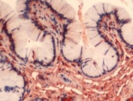
NGF R/TNFRSF16 in Human Brain.
NGF R/TNFRSF16 was detected in immersion fixed paraffin-embedded sections of human brain (cortex) using 25 µg/mL Mouse Anti-Human/Canine NGF R/TNFRSF16 Monoclonal Antibody (Catalog # MAB367) overnight at 4 °C. Tissue was stained with the Anti-Mouse HRP-DAB Cell & Tissue Staining Kit (brown; Catalog # CTS002) and counterstained with hematoxylin (blue). View our protocol for Chromogenic IHC Staining of Paraffin-embedded Tissue Sections.
Detection of NGFR/TNFRSF16 by Western Blot
HPV16 E6-E7 increased p75NTR expression through PI3K/Akt signaling pathway in TE-1 cellsA. Proteins involved in PI3K/Akt signaling pathway and p75NTR protein were analysed by western blotting. TE-1 cells were treated by negative control RNA, GAPDH RNA, PI3K siRNA, respectively. Equal protein loading was evaluated by beta -actin. “-” means HPV16 E6-E7 negative which represents TE-1-control cells, “+” means HPV16 E6-E7 positive which represents TE-1-psb cells. B. Proteins involved in PI3K/Akt signaling pathway and p75NTR protein were analysed by western blotting. TE-1 cells were treated by negative control RNA, GAPDH RNA, Akt siRNA, respectively. Equal protein loading was evaluated by beta -actin. “-” means HPV16 E6-E7 negative which represents TE-1-control cells, “+” means HPV16 E6-E7 positive which represents TE-1-psb cells. C. Densitometric of western blotting bands in Figure A were analyzed and expressed relative to the loading control, beta -actin. Data are typical of three experiments and the histogram values are mean ± S.D. *P<0.05,**P<0.01,***P<0.001, relative to control cells in control siRNA group. ###P<0.001, relative to TE-1-psb cell in control siRNA group. D. Densitometric of western blotting bands in Figure B were analyzed and expressed relative to the loading control, beta -actin. Data are typical of three experiments and the histogram values are mean ± S.D. *P<0.05,**P<0.01,***P<0.001, relative to control cells in control siRNA group or Akt siRNA group. ###P<0.001, relative to TE-1-psb cell in control siRNA group. Image collected and cropped by CiteAb from the following open publication (https://pubmed.ncbi.nlm.nih.gov/27489353), licensed under a CC-BY license. Not internally tested by R&D Systems.
Detection of NGFR/TNFRSF16 by Western Blot
HPV16 E6-E7 increased p75NTR expression through PI3K/Akt signaling pathway in Eca109 cellsA. Proteins involved in PI3K/Akt signaling pathway and p75NTR protein were analysed by western blotting. Eca109 cells were treated by negative control RNA, GAPDH RNA, PI3K siRNA, respectively. Equal protein loading was evaluated by beta -actin. “-” means HPV16 E6-E7 negative which represents Eca109-control cells, “+”means HPV16 E6-E7 positive which represents Eca109-psb cells. B. Proteins involved in PI3K/Akt signaling pathway and p75NTR protein were analysed by western blotting. Eca109 cells were treated by negative control RNA, GAPDH RNA, Akt siRNA, respectively. Equal protein loading was evaluated by beta -actin. “-”means HPV16 E6-E7 negative which represents Eca109-control cells, “+”means HPV16 E6-E7 positive which represents Eca109-psb cells. C. Densitometric of western blotting bands in Figure A were analyzed and expressed relative to the loading control, beta -actin. Data are typical of three experiments and the histogram values are mean ± S.D. *P<0.05,**P<0.01,***P<0.001, relative to control cells in control siRNA group. ###P<0.001, relative to Eca109-psb cell in control siRNA group. D. Densitometric of western blotting bands in Figure B were analyzed and expressed relative to the loading control, beta -actin. Data are typical of three experiments and the histogram values are mean ± S.D. *P<0.05,**P<0.01,***P<0.001, relative to control cells in control siRNA group or Akt siRNA group. ###P<0.001, relative to Eca109-psb cell in control siRNA group. Image collected and cropped by CiteAb from the following open publication (https://pubmed.ncbi.nlm.nih.gov/27489353), licensed under a CC-BY license. Not internally tested by R&D Systems.
Detection of NGFR/TNFRSF16 by Western Blot
HPV16 E6-E7 increased p75NTR expression through PI3K/Akt signaling pathway in TE-1 cellsA. Proteins involved in PI3K/Akt signaling pathway and p75NTR protein were analysed by western blotting. TE-1 cells were treated by negative control RNA, GAPDH RNA, PI3K siRNA, respectively. Equal protein loading was evaluated by beta -actin. “-” means HPV16 E6-E7 negative which represents TE-1-control cells, “+” means HPV16 E6-E7 positive which represents TE-1-psb cells. B. Proteins involved in PI3K/Akt signaling pathway and p75NTR protein were analysed by western blotting. TE-1 cells were treated by negative control RNA, GAPDH RNA, Akt siRNA, respectively. Equal protein loading was evaluated by beta -actin. “-” means HPV16 E6-E7 negative which represents TE-1-control cells, “+” means HPV16 E6-E7 positive which represents TE-1-psb cells. C. Densitometric of western blotting bands in Figure A were analyzed and expressed relative to the loading control, beta -actin. Data are typical of three experiments and the histogram values are mean ± S.D. *P<0.05,**P<0.01,***P<0.001, relative to control cells in control siRNA group. ###P<0.001, relative to TE-1-psb cell in control siRNA group. D. Densitometric of western blotting bands in Figure B were analyzed and expressed relative to the loading control, beta -actin. Data are typical of three experiments and the histogram values are mean ± S.D. *P<0.05,**P<0.01,***P<0.001, relative to control cells in control siRNA group or Akt siRNA group. ###P<0.001, relative to TE-1-psb cell in control siRNA group. Image collected and cropped by CiteAb from the following open publication (https://pubmed.ncbi.nlm.nih.gov/27489353), licensed under a CC-BY license. Not internally tested by R&D Systems.
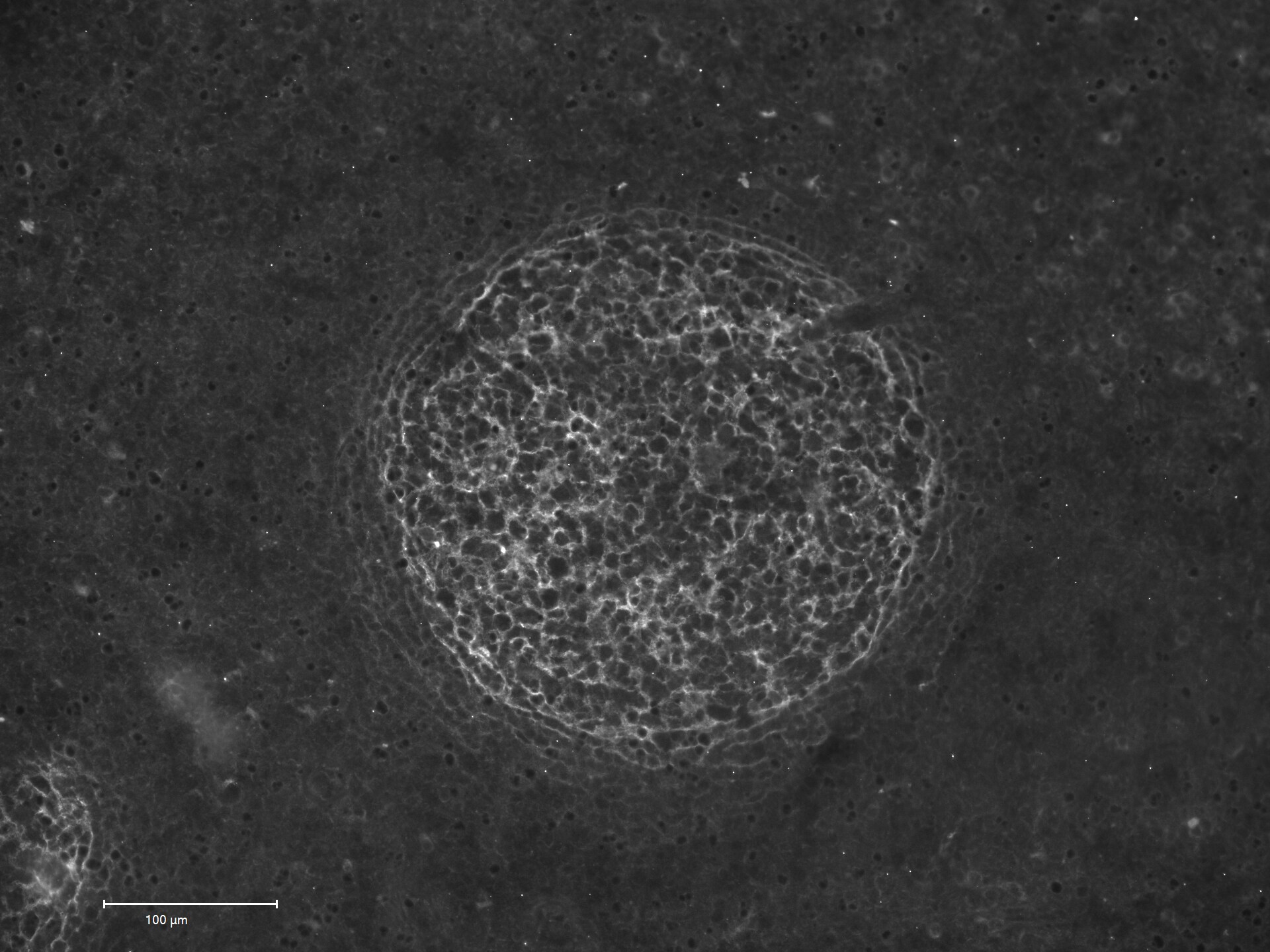
Detection of NGFR/TNFRSF16 by Western Blot
PI3K/Akt signaling pathway activation and increased p75NTR expression induced by HPV16 E6-E7 were inhibited by LY294002 in ESCC cellsA. Proteins involved in PI3K/Akt signaling pathway and p75NTR protein were analysed by western blotting. Cells were treated with LY294002 (10μmol/L) for 24h. Equal protein loading was evaluated by beta -actin. B. Densitometric of western blotting bands in Figure A were analyzed and expressed relative to the loading control, beta -actin. Data are typical of three experiments and the histogram values are mean ± S.D. **P<0.01,***P<0.001, relative to control cells without LY294002 treatment. ###P<0.001, relative to psb cells without LY294002 treatment. C. p-Akt, PI3K and p75NTR proteins of ESCC cells were detected by immunofluorescence. D. PI3K and p75NTR proteins of spheres formed from ESCC cells were detected by immunofluorescence. Image collected and cropped by CiteAb from the following open publication (https://pubmed.ncbi.nlm.nih.gov/27489353), licensed under a CC-BY license. Not internally tested by R&D Systems.
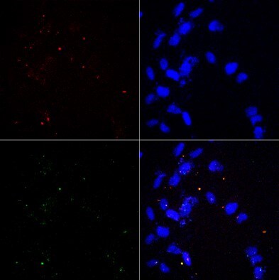
Detection of NGFR/TNFRSF16 by Immunohistochemistry
Visualization of CD271 and CD81 expression in bone marrow vascular regions by confocal microscopy.(A) Confocal scan of vascular region in BM biopsies with 3D orthographic cross-section view, co-stained with mouse anti-CD271, rabbit anti-CD81, and DAPI. (B) Single channel data for the florescent markers in A. (C) Intensity profile for all channels in A across a cell of interest in a representative z plane. Scale bars represent 50 μm. Yellow dashed lines indicated vessel surface. Image collected and cropped by CiteAb from the following open publication (https://pubmed.ncbi.nlm.nih.gov/36876630), licensed under a CC-BY license. Not internally tested by R&D Systems.
Detection of NGFR/TNFRSF16 by Immunohistochemistry
Visualization of CD271 and CD81 expression in bone marrow vascular regions by confocal microscopy.(A) Confocal scan of vascular region in BM biopsies with 3D orthographic cross-section view, co-stained with mouse anti-CD271, rabbit anti-CD81, and DAPI. (B) Single channel data for the florescent markers in A. (C) Intensity profile for all channels in A across a cell of interest in a representative z plane. Scale bars represent 50 μm. Yellow dashed lines indicated vessel surface. Image collected and cropped by CiteAb from the following open publication (https://pubmed.ncbi.nlm.nih.gov/36876630), licensed under a CC-BY license. Not internally tested by R&D Systems.Applications for NGFR/TNFRSF16 Antibody (74902)
Application
Recommended Usage
CyTOF-ready
Ready to be labeled using established conjugation methods. No BSA or other carrier proteins that could interfere with conjugation.
Flow Cytometry
0.25 µg/106 cells
Sample: SHSY-5Y human cell line
Sample: SHSY-5Y human cell line
Immunocytochemistry
8-25 µg/mL
Sample: Immersion fixed canine and human mesenchymal stem cells
Sample: Immersion fixed canine and human mesenchymal stem cells
Immunohistochemistry
8-25 µg/mL
Sample: Immersion fixed paraffin-embedded sections of human brain (cortex)
Sample: Immersion fixed paraffin-embedded sections of human brain (cortex)
Western Blot
2 µg/mL
Sample: SW480 human colorectal adenocarcinoma cell line
under non-reducing conditions only
Sample: SW480 human colorectal adenocarcinoma cell line
under non-reducing conditions only
Reviewed Applications
Read 3 reviews rated 4.7 using MAB367 in the following applications:
Flow Cytometry Panel Builder
Bio-Techne Knows Flow Cytometry
Save time and reduce costly mistakes by quickly finding compatible reagents using the Panel Builder Tool.
Advanced Features
- Spectra Viewer - Custom analysis of spectra from multiple fluorochromes
- Spillover Popups - Visualize the spectra of individual fluorochromes
- Antigen Density Selector - Match fluorochrome brightness with antigen density

Formulation, Preparation, and Storage
Purification
Protein A or G purified from hybridoma culture supernatant
Reconstitution
Reconstitute at 0.5 mg/mL in sterile PBS. For liquid material, refer to CoA for concentration.
Loading...
Formulation
Lyophilized from a 0.2 μm filtered solution in PBS with Trehalose. *Small pack size (SP) is supplied either lyophilized or as a 0.2 µm filtered solution in PBS.
Shipping
Lyophilized product is shipped at ambient temperature. Liquid small pack size (-SP) is shipped with polar packs. Upon receipt, store immediately at the temperature recommended below.
Stability & Storage
Use a manual defrost freezer and avoid repeated freeze-thaw cycles.
- 12 months from date of receipt, -20 to -70 °C as supplied.
- 1 month, 2 to 8 °C under sterile conditions after reconstitution.
- 6 months, -20 to -70 °C under sterile conditions after reconstitution.
Calculators
Background: NGFR/TNFRSF16
References
- Barker, P.A. et al. (1992) Mol. Cell Biochem. 110:1.
- Stemple, D.L. et al. (1992) Cell 71:973.
- Morrison, S.J. et al. (1999) Cell 96:737.
- Mujtaba, T. et al. (1998) Dev. Biol. 200:1.
- Campagnolo, L. et al. (2001) Biol. Reprod. 64:464.
- Cassiman, D. et al. (2001) Hepatology 33:148.
Long Name
Nerve Growth Factor Receptor
Alternate Names
CD271, NGF R, p75 NTR, TNFRSF16
Gene Symbol
NGFR
UniProt
Additional NGFR/TNFRSF16 Products
Product Documents for NGFR/TNFRSF16 Antibody (74902)
Certificate of Analysis
To download a Certificate of Analysis, please enter a lot or batch number in the search box below.
Note: Certificate of Analysis not available for kit components.
Product Specific Notices for NGFR/TNFRSF16 Antibody (74902)
For research use only
Citations for NGFR/TNFRSF16 Antibody (74902)
 Powered by Bioz
Customer Reviews for NGFR/TNFRSF16 Antibody (74902) (3)
4.7 out of 5
3 Customer Ratings
Have you used NGFR/TNFRSF16 Antibody (74902)?
Submit a review and receive an Amazon gift card!
$25/€18/£15/$25CAN/¥2500 Yen for a review with an image
$10/€7/£6/$10CAN/¥1110 Yen for a review without an image
Submit a review
Customer Images
Showing
1
-
3 of
3 reviews
Showing All
Filter By:
-
Application: ImmunocytochemistrySample Tested: primary astrocytesSpecies: HumanVerified Customer | Posted 02/25/2022Primary astrocytes co-stained for cilia (Arl13b, red, top left), P75NTR (green, bottom left) and DAPI (blue, top right) and merged image (bottom right).

-
Application: ImmunohistochemistrySample Tested: Uterus tissueSpecies: HumanVerified Customer | Posted 09/04/2021
-
Application: Immunohistochemistry-FrozenSample Tested: human tonsilSpecies: HumanVerified Customer | Posted 02/29/2020NGFR on human tonsil, dilution 1:100pH 9 epitope retrieval
There are no reviews that match your criteria.
Protocols
Find general support by application which include: protocols, troubleshooting, illustrated assays, videos and webinars.
- 7-Amino Actinomycin D (7-AAD) Cell Viability Flow Cytometry Protocol
- Antigen Retrieval Protocol (PIER)
- Antigen Retrieval for Frozen Sections Protocol
- Appropriate Fixation of IHC/ICC Samples
- Cellular Response to Hypoxia Protocols
- Chromogenic IHC Staining of Formalin-Fixed Paraffin-Embedded (FFPE) Tissue Protocol
- Chromogenic Immunohistochemistry Staining of Frozen Tissue
- ClariTSA™ Fluorophore Kits
- Detection & Visualization of Antibody Binding
- Extracellular Membrane Flow Cytometry Protocol
- Flow Cytometry Protocol for Cell Surface Markers
- Flow Cytometry Protocol for Staining Membrane Associated Proteins
- Flow Cytometry Staining Protocols
- Flow Cytometry Troubleshooting Guide
- Fluorescent IHC Staining of Frozen Tissue Protocol
- Graphic Protocol for Heat-induced Epitope Retrieval
- Graphic Protocol for the Preparation and Fluorescent IHC Staining of Frozen Tissue Sections
- Graphic Protocol for the Preparation and Fluorescent IHC Staining of Paraffin-embedded Tissue Sections
- Graphic Protocol for the Preparation of Gelatin-coated Slides for Histological Tissue Sections
- ICC Cell Smear Protocol for Suspension Cells
- ICC Immunocytochemistry Protocol Videos
- ICC for Adherent Cells
- IHC Sample Preparation (Frozen sections vs Paraffin)
- Immunocytochemistry (ICC) Protocol
- Immunocytochemistry Troubleshooting
- Immunofluorescence of Organoids Embedded in Cultrex Basement Membrane Extract
- Immunofluorescent IHC Staining of Formalin-Fixed Paraffin-Embedded (FFPE) Tissue Protocol
- Immunohistochemistry (IHC) and Immunocytochemistry (ICC) Protocols
- Immunohistochemistry Frozen Troubleshooting
- Immunohistochemistry Paraffin Troubleshooting
- Intracellular Flow Cytometry Protocol Using Alcohol (Methanol)
- Intracellular Flow Cytometry Protocol Using Detergents
- Intracellular Nuclear Staining Flow Cytometry Protocol Using Detergents
- Intracellular Staining Flow Cytometry Protocol Using Alcohol Permeabilization
- Intracellular Staining Flow Cytometry Protocol Using Detergents to Permeabilize Cells
- Preparing Samples for IHC/ICC Experiments
- Preventing Non-Specific Staining (Non-Specific Binding)
- Primary Antibody Selection & Optimization
- Propidium Iodide Cell Viability Flow Cytometry Protocol
- Protocol for Heat-Induced Epitope Retrieval (HIER)
- Protocol for Liperfluo
- Protocol for Making a 4% Formaldehyde Solution in PBS
- Protocol for VisUCyte™ HRP Polymer Detection Reagent
- Protocol for the Characterization of Human Th22 Cells
- Protocol for the Characterization of Human Th9 Cells
- Protocol for the Fluorescent ICC Staining of Cell Smears - Graphic
- Protocol for the Fluorescent ICC Staining of Cultured Cells on Coverslips - Graphic
- Protocol for the Preparation & Fixation of Cells on Coverslips
- Protocol for the Preparation and Chromogenic IHC Staining of Frozen Tissue Sections
- Protocol for the Preparation and Chromogenic IHC Staining of Frozen Tissue Sections - Graphic
- Protocol for the Preparation and Chromogenic IHC Staining of Paraffin-embedded Tissue Sections
- Protocol for the Preparation and Chromogenic IHC Staining of Paraffin-embedded Tissue Sections - Graphic
- Protocol for the Preparation and Fluorescent ICC Staining of Cells on Coverslips
- Protocol for the Preparation and Fluorescent ICC Staining of Non-adherent Cells
- Protocol for the Preparation and Fluorescent ICC Staining of Stem Cells on Coverslips
- Protocol for the Preparation and Fluorescent IHC Staining of Frozen Tissue Sections
- Protocol for the Preparation and Fluorescent IHC Staining of Paraffin-embedded Tissue Sections
- Protocol for the Preparation of Gelatin-coated Slides for Histological Tissue Sections
- Protocol for the Preparation of a Cell Smear for Non-adherent Cell ICC - Graphic
- Protocol: Annexin V and PI Staining by Flow Cytometry
- Protocol: Annexin V and PI Staining for Apoptosis by Flow Cytometry
- R&D Systems Quality Control Western Blot Protocol
- TUNEL and Active Caspase-3 Detection by IHC/ICC Protocol
- The Importance of IHC/ICC Controls
- Troubleshooting Guide: Fluorokine Flow Cytometry Kits
- Troubleshooting Guide: Immunohistochemistry
- Troubleshooting Guide: Western Blot Figures
- Western Blot Conditions
- Western Blot Protocol
- Western Blot Protocol for Cell Lysates
- Western Blot Troubleshooting
- Western Blot Troubleshooting Guide
- View all Protocols, Troubleshooting, Illustrated assays and Webinars


