CD5L (CD5 antigen-like), also known as Sp alpha and AIM, is a 50 kDa secreted glycoprotein that belongs to the SRCR (scavenger receptor cysteine rich) group B family of proteins. Group B proteins are distinguished by SRCR domains that are encoded by a single exon (1 - 3). The human CD5L cDNA encodes a 347 amino acid (aa) precursor that includes a 19 aa signal sequence and three SRCR domains (4, 5). Among group B proteins, CD5L is most closely related to CD5 and CD6, with which it shares 18% and 31% aa sequence identity, respectively. CD5L is up-regulated in macrophages at inflammatory sites. It sustains inflammatory reactions by both increasing the phagocytic capacity of macrophages and impeding the apoptosis of local macrophages, NK cells, and T cells (6, 7). Agonists of the LXR and RXR nuclear hormone receptors induce CD5L upregulation in macrophages and reduce macrophage apoptosis (8, 9). Oxidized LDL (which acts through LXR/RXR) is taken up by macrophages, promoting their development into foam cells. The increased level of CD5L protects foam cells from apoptosis but permits more rapid cellular accumulation and atherosclerotic plaque formation (9). In activated B cells, however, the combination of CD5L and TGF-beta inhibits proliferation. The binding of CD5L to splenic B cells is increased following TGF-beta exposure, suggesting that TGF-beta increases the expression or availability of an unidentified CD5L receptor (5, 10). CD5L also functions as a pattern recognition molecule by binding both lipoteichoic acid on Gram positive and lipopolysaccharide on Gram negative bacteria (11). In the thymic cortex, CD5L protects cortical CD4+CD8+ thymocytes from apoptosis (12). CD5L circulates in the serum in complex with IgM (13).

Key Product Details
Species Reactivity
Validated:
Human
Cited:
Human
Applications
Validated:
Immunohistochemistry, Western Blot
Cited:
ELISA Development
Label
Unconjugated
Antibody Source
Polyclonal Goat IgG
Loading...
Product Specifications
Immunogen
Mouse myeloma cell line NS0-derived recombinant human CD5L
Ser20-Gly347
Accession # O43866
Ser20-Gly347
Accession # O43866
Specificity
Detects human CD5L in direct ELISAs and Western blots. In these formats, less than 5% cross‑reactivity with recombinant mouse CD5L and less than 1% cross-reactivity with recombinant human CD5 is observed.
Clonality
Polyclonal
Host
Goat
Isotype
IgG
Scientific Data Images for Human CD5L Antibody
CD5L in Human Spleen.
CD5L was detected in immersion fixed paraffin-embedded sections of human spleen using Goat Anti-Human CD5L Antigen Affinity-purified Polyclonal Antibody (Catalog # AF2797) at 5 µg/mL for 1 hour at room temperature followed by incubation with the Anti-Goat IgG VisUCyte™ HRP Polymer Antibody (VC004). Before incubation with the primary antibody, tissue was subjected to heat-induced epitope retrieval using Antigen Retrieval Reagent-Basic (CTS013). Tissue was stained using DAB (brown) and counterstained with hematoxylin (blue). Specific staining was localized to lymphocytes. Staining was performed using our protocol for IHC Staining with VisUCyte HRP Polymer Detection Reagents.Applications for Human CD5L Antibody
Application
Recommended Usage
Immunohistochemistry
5-15 µg/mL
Sample: Immersion fixed paraffin-embedded sections of human spleen
Sample: Immersion fixed paraffin-embedded sections of human spleen
Western Blot
0.1 µg/mL
Sample: Recombinant Human CD5L (Catalog # 2797-CL)
Sample: Recombinant Human CD5L (Catalog # 2797-CL)
Reviewed Applications
Read 4 reviews rated 4.3 using AF2797 in the following applications:
Formulation, Preparation, and Storage
Purification
Antigen Affinity-purified
Reconstitution
Reconstitute at 0.2 mg/mL in sterile PBS. For liquid material, refer to CoA for concentration.
Loading...
Formulation
Lyophilized from a 0.2 μm filtered solution in PBS with Trehalose. *Small pack size (SP) is supplied either lyophilized or as a 0.2 µm filtered solution in PBS.
Shipping
Lyophilized product is shipped at ambient temperature. Liquid small pack size (-SP) is shipped with polar packs. Upon receipt, store immediately at the temperature recommended below.
Stability & Storage
Use a manual defrost freezer and avoid repeated freeze-thaw cycles.
- 12 months from date of receipt, -20 to -70 °C as supplied.
- 1 month, 2 to 8 °C under sterile conditions after reconstitution.
- 6 months, -20 to -70 °C under sterile conditions after reconstitution.
Calculators
Background: CD5L
References
- Sarrias, M.R. et al. (2004) Crit. Rev. Immunol. 24:1.
- Resnick, D. et al. (1994) Trends Biochem. Sci. 19:5.
- Mukhopadhyay, S. and S. Gordon (2004) Immunobiology 209:39.
- Gebe, J.A. et al. (1997) J. Biol. Chem. 272:6151.
- Gebe, J.A. et al. (2000) Immunol. 99:78.
- Haruta, I. et al. (2001) J. Biol. Chem. 276:22910.
- Kuwata, K. et al. (2003) Am. J. Pathol. 162:837.
- Valledor, A.F. et al. (2004) Proc. Natl. Acad. Sci. 101:17813.
- Arai, S. et al. (2005) Cell Metab. 1:201.
- Yusa, S. et al. (1999) Eur. J. Immunol. 29:1086.
- Sarrias, M-R. et al. (2005) J. Biol. Chem. 280:35391.
- Miyazaki, T. et al. (1999) J. Exp. Med. 189:413.
- Tissot, J.D. et al. (2002) Electrophoresis 23:1203.
Long Name
CD5 Antigen-like
Alternate Names
AIM, API6, CD5L, CT-2, SP-alpha
Gene Symbol
CD5L
UniProt
Additional CD5L Products
Product Documents for Human CD5L Antibody
Certificate of Analysis
To download a Certificate of Analysis, please enter a lot or batch number in the search box below.
Note: Certificate of Analysis not available for kit components.
Product Specific Notices for Human CD5L Antibody
For research use only
Related Research Areas
Citations for Human CD5L Antibody
 Powered by Bioz
Customer Reviews for Human CD5L Antibody (4)
4.3 out of 5
4 Customer Ratings
Have you used Human CD5L Antibody?
Submit a review and receive an Amazon gift card!
$25/€18/£15/$25CAN/¥2500 Yen for a review with an image
$10/€7/£6/$10CAN/¥1110 Yen for a review without an image
Submit a review
Customer Images
Showing
1
-
4 of
4 reviews
Showing All
Filter By:
-
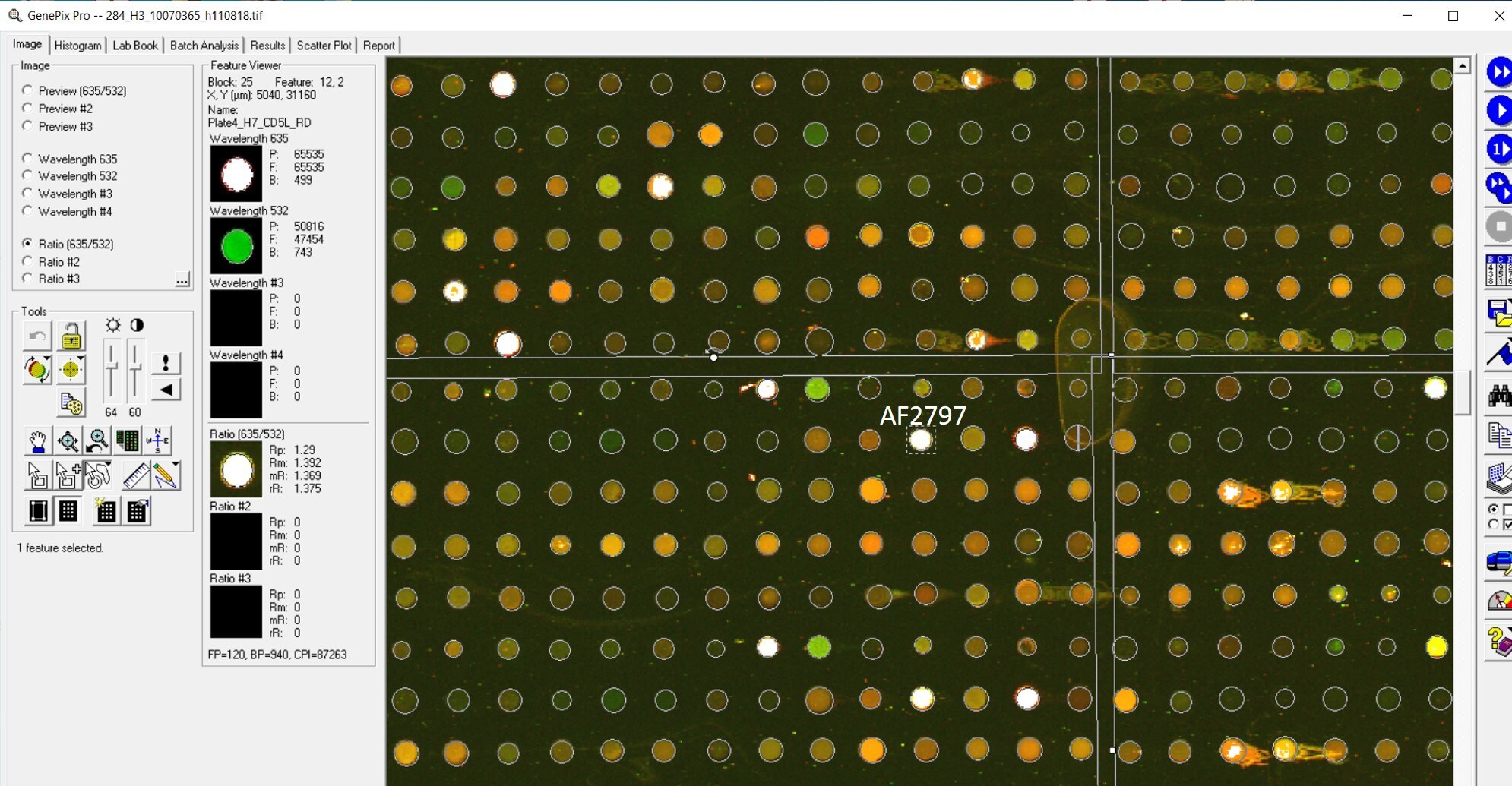
Application: MicroarraysSample Tested: EDTA PlasmaSpecies: HumanVerified Customer | Posted 01/14/2021

-
Application: MicroarraysSample Tested: EDTA PlasmaSpecies: HumanVerified Customer | Posted 11/07/2018
-
Application: ELISASample Tested: Plasma and SerumSpecies: Human and MouseVerified Customer | Posted 11/07/2018
-
Application: MicroarraySample Tested: EDTA PlasmaSpecies: HumanVerified Customer | Posted 11/02/2018
There are no reviews that match your criteria.
Protocols
Find general support by application which include: protocols, troubleshooting, illustrated assays, videos and webinars.
- Antigen Retrieval Protocol (PIER)
- Antigen Retrieval for Frozen Sections Protocol
- Appropriate Fixation of IHC/ICC Samples
- Cellular Response to Hypoxia Protocols
- Chromogenic IHC Staining of Formalin-Fixed Paraffin-Embedded (FFPE) Tissue Protocol
- Chromogenic Immunohistochemistry Staining of Frozen Tissue
- ClariTSA™ Fluorophore Kits
- Detection & Visualization of Antibody Binding
- Fluorescent IHC Staining of Frozen Tissue Protocol
- Graphic Protocol for Heat-induced Epitope Retrieval
- Graphic Protocol for the Preparation and Fluorescent IHC Staining of Frozen Tissue Sections
- Graphic Protocol for the Preparation and Fluorescent IHC Staining of Paraffin-embedded Tissue Sections
- Graphic Protocol for the Preparation of Gelatin-coated Slides for Histological Tissue Sections
- IHC Sample Preparation (Frozen sections vs Paraffin)
- Immunofluorescent IHC Staining of Formalin-Fixed Paraffin-Embedded (FFPE) Tissue Protocol
- Immunohistochemistry (IHC) and Immunocytochemistry (ICC) Protocols
- Immunohistochemistry Frozen Troubleshooting
- Immunohistochemistry Paraffin Troubleshooting
- Preparing Samples for IHC/ICC Experiments
- Preventing Non-Specific Staining (Non-Specific Binding)
- Primary Antibody Selection & Optimization
- Protocol for Heat-Induced Epitope Retrieval (HIER)
- Protocol for Making a 4% Formaldehyde Solution in PBS
- Protocol for VisUCyte™ HRP Polymer Detection Reagent
- Protocol for the Preparation & Fixation of Cells on Coverslips
- Protocol for the Preparation and Chromogenic IHC Staining of Frozen Tissue Sections
- Protocol for the Preparation and Chromogenic IHC Staining of Frozen Tissue Sections - Graphic
- Protocol for the Preparation and Chromogenic IHC Staining of Paraffin-embedded Tissue Sections
- Protocol for the Preparation and Chromogenic IHC Staining of Paraffin-embedded Tissue Sections - Graphic
- Protocol for the Preparation and Fluorescent IHC Staining of Frozen Tissue Sections
- Protocol for the Preparation and Fluorescent IHC Staining of Paraffin-embedded Tissue Sections
- Protocol for the Preparation of Gelatin-coated Slides for Histological Tissue Sections
- R&D Systems Quality Control Western Blot Protocol
- TUNEL and Active Caspase-3 Detection by IHC/ICC Protocol
- The Importance of IHC/ICC Controls
- Troubleshooting Guide: Immunohistochemistry
- Troubleshooting Guide: Western Blot Figures
- Western Blot Conditions
- Western Blot Protocol
- Western Blot Protocol for Cell Lysates
- Western Blot Troubleshooting
- Western Blot Troubleshooting Guide
- View all Protocols, Troubleshooting, Illustrated assays and Webinars
Loading...