Human CEACAM-1/CD66a Antibody (283324)
R&D Systems | Catalog # MAB22441

Key Product Details
Species Reactivity
Validated:
Human
Cited:
Human
Applications
Validated:
Immunohistochemistry, Western Blot, ELISA Capture (Matched Antibody Pair)
Cited:
Immunohistochemistry-Paraffin, Western Blot, Microarray
Label
Unconjugated
Antibody Source
Monoclonal Mouse IgG1 Clone # 283324
Loading...
Product Specifications
Immunogen
Mouse myeloma cell line NS0-derived recombinant human CEACAM-1
Gln35-Gly428
Accession # P13688
Gln35-Gly428
Accession # P13688
Specificity
Detects human CEACAM-1 in ELISAs and Western blots. In ELISAs and Western blots, no cross-reactivity with recombinant human (rh) CD31, rhICAM-1, -2, -3, recombinant mouse MAdCAM-1, or rhVCAM-1 was observed. In sandwich ELISAs, no cross-reactivity with rhCEACAM‑3, rhCEACAM-5, or rhCEACAM-6 was observed.
Clonality
Monoclonal
Host
Mouse
Isotype
IgG1
Scientific Data Images for Human CEACAM-1/CD66a Antibody (283324)
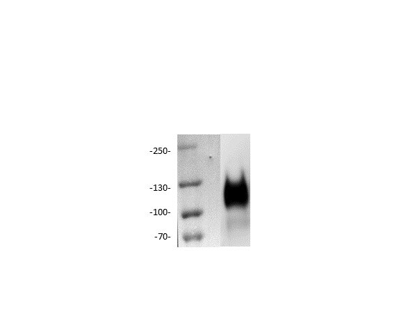
Detection of Human CEACAM‑1/CD66a by Western Blot.
Western blot shows lysates of human liver tissue and HepG2 human hepatocellular carcinoma cell line. PVDF membrane was probed with 2 µg/mL of Mouse Anti-Human CEACAM-1/CD66a Monoclonal Antibody (Catalog # MAB22441) followed by HRP-conjugated Anti-Mouse IgG Secondary Antibody (Catalog # HAF018). Specific bands were detected for CEACAM-1/CD66a at approximately 100-150 kDa (as indicated). This experiment was conducted under reducing conditions and using Immunoblot Buffer Group 1.
CEACAM‑1/CD66a in Human Colon.
CEACAM‑1/CD66a was detected in immersion fixed paraffin-embedded sections of human colon using Mouse Anti-Human CEACAM‑1/CD66a Monoclonal Antibody (Catalog # MAB22441) at 25 µg/mL overnight at 4 °C. Tissue was stained using the Anti-Mouse HRP-DAB Cell & Tissue Staining Kit (brown; Catalog # CTS002) and counterstained with hematoxylin (blue). Specific labeling was localized to stromal cells. View our protocol for Chromogenic IHC Staining of Paraffin-embedded Tissue Sections.
CEACAM‑1/CD66a in Human Colon Cancer Tissue.
CEACAM-1/CD66a was detected in immersion fixed paraffin-embedded sections of human colon cancer tissue using Mouse Anti-Human CEACAM-1/CD66a Monoclonal Antibody (Catalog # MAB22441) at 25 µg/mL over-night at 4 °C. Tissue was stained using the Anti-Mouse HRP-DAB Cell & Tissue Staining Kit (brown; Catalog # CTS002) and counterstained with hematoxylin (blue). View our protocol for Chromogenic IHC Staining of Paraffin-embedded Tissue Sections.Applications for Human CEACAM-1/CD66a Antibody (283324)
Application
Recommended Usage
Immunohistochemistry
8-25 µg/mL
Sample: Immersion fixed paraffin-embedded sections of human colon and human colon cancer tissue
Sample: Immersion fixed paraffin-embedded sections of human colon and human colon cancer tissue
Western Blot
2 µg/mL
Sample: Human liver tissue and HepG2 human hepatocellular carcinoma cell line
Sample: Human liver tissue and HepG2 human hepatocellular carcinoma cell line
Human CEACAM-1/CD66a Sandwich Immunoassay
Please Note: Optimal dilutions of this antibody should be experimentally determined.
Reviewed Applications
Read 1 review rated 4 using MAB22441 in the following applications:
Formulation, Preparation, and Storage
Purification
Protein A or G purified from hybridoma culture supernatant
Reconstitution
Reconstitute at 0.5 mg/mL in sterile PBS. For liquid material, refer to CoA for concentration.
Loading...
Formulation
Lyophilized from a 0.2 μm filtered solution in PBS with Trehalose. *Small pack size (SP) is supplied either lyophilized or as a 0.2 µm filtered solution in PBS.
Shipping
Lyophilized product is shipped at ambient temperature. Liquid small pack size (-SP) is shipped with polar packs. Upon receipt, store immediately at the temperature recommended below.
Stability & Storage
Use a manual defrost freezer and avoid repeated freeze-thaw cycles.
- 12 months from date of receipt, -20 to -70 °C as supplied.
- 1 month, 2 to 8 °C under sterile conditions after reconstitution.
- 6 months, -20 to -70 °C under sterile conditions after reconstitution.
Calculators
Background: CEACAM-1/CD66a
References
- Beauchemin, N. et al. (1999) Exp. Cell Res. 252:243.
- Thompson, J. et al. (1992) Genomics 12:761.
- Waggener, C. and S. Ergun (2000) Exp. Cell Res. 261:19.
- Barnett, T.R. et al. (1989) J. Cell Biol. 108:267.
- Hinoda, Y. et al. (1988) Proc. Natl. Acad. Sci. USA 85:6959.
- Kuroki, M. et al. (1991) Biochem. Biophys. Res. Commun. 176:578.
- Barnett, T.R. et al. (1993) Mol. Cell. Biol. 13:1273.
- Watt, S.M. et al. (1994) Blood 84:200.
- Oikawa, S. et al. (1992) Biochem. Biophys. Res. Commun. 186:881.
- Klaas, P.J.M. et al. (2005) FEBS Lett. 579:6159.
- Bogoevska, V. et al. (2005) Glycobiology 16:197.
Long Name
Carcinoembryonic Antigen-related Cell Adhesion Molecule 1
Alternate Names
BGP1, Biliary Glycoprotein 1, CD66a, Cea-1, CEACAM1, Hv-1, Hv-2, MHVR
Gene Symbol
CEACAM1
UniProt
Additional CEACAM-1/CD66a Products
Product Documents for Human CEACAM-1/CD66a Antibody (283324)
Certificate of Analysis
To download a Certificate of Analysis, please enter a lot or batch number in the search box below.
Note: Certificate of Analysis not available for kit components.
Product Specific Notices for Human CEACAM-1/CD66a Antibody (283324)
For research use only
Citations for Human CEACAM-1/CD66a Antibody (283324)
 Powered by Bioz
Customer Reviews for Human CEACAM-1/CD66a Antibody (283324) (1)
4 out of 5
1 Customer Rating
Have you used Human CEACAM-1/CD66a Antibody (283324)?
Submit a review and receive an Amazon gift card!
$25/€18/£15/$25CAN/¥2500 Yen for a review with an image
$10/€7/£6/$10CAN/¥1110 Yen for a review without an image
Submit a review
Customer Images
Showing
1
-
1 of
1 review
Showing All
Filter By:
-
Application: Western BlotSample Tested: A549 human lung carcinoma cell lineSpecies: HumanVerified Customer | Posted 04/08/2022

There are no reviews that match your criteria.
Protocols
Find general support by application which include: protocols, troubleshooting, illustrated assays, videos and webinars.
- Antigen Retrieval Protocol (PIER)
- Antigen Retrieval for Frozen Sections Protocol
- Appropriate Fixation of IHC/ICC Samples
- Cellular Response to Hypoxia Protocols
- Chromogenic IHC Staining of Formalin-Fixed Paraffin-Embedded (FFPE) Tissue Protocol
- Chromogenic Immunohistochemistry Staining of Frozen Tissue
- ClariTSA™ Fluorophore Kits
- Detection & Visualization of Antibody Binding
- Fluorescent IHC Staining of Frozen Tissue Protocol
- Graphic Protocol for Heat-induced Epitope Retrieval
- Graphic Protocol for the Preparation and Fluorescent IHC Staining of Frozen Tissue Sections
- Graphic Protocol for the Preparation and Fluorescent IHC Staining of Paraffin-embedded Tissue Sections
- Graphic Protocol for the Preparation of Gelatin-coated Slides for Histological Tissue Sections
- IHC Sample Preparation (Frozen sections vs Paraffin)
- Immunofluorescent IHC Staining of Formalin-Fixed Paraffin-Embedded (FFPE) Tissue Protocol
- Immunohistochemistry (IHC) and Immunocytochemistry (ICC) Protocols
- Immunohistochemistry Frozen Troubleshooting
- Immunohistochemistry Paraffin Troubleshooting
- Preparing Samples for IHC/ICC Experiments
- Preventing Non-Specific Staining (Non-Specific Binding)
- Primary Antibody Selection & Optimization
- Protocol for Heat-Induced Epitope Retrieval (HIER)
- Protocol for Making a 4% Formaldehyde Solution in PBS
- Protocol for VisUCyte™ HRP Polymer Detection Reagent
- Protocol for the Preparation & Fixation of Cells on Coverslips
- Protocol for the Preparation and Chromogenic IHC Staining of Frozen Tissue Sections
- Protocol for the Preparation and Chromogenic IHC Staining of Frozen Tissue Sections - Graphic
- Protocol for the Preparation and Chromogenic IHC Staining of Paraffin-embedded Tissue Sections
- Protocol for the Preparation and Chromogenic IHC Staining of Paraffin-embedded Tissue Sections - Graphic
- Protocol for the Preparation and Fluorescent IHC Staining of Frozen Tissue Sections
- Protocol for the Preparation and Fluorescent IHC Staining of Paraffin-embedded Tissue Sections
- Protocol for the Preparation of Gelatin-coated Slides for Histological Tissue Sections
- R&D Systems Quality Control Western Blot Protocol
- TUNEL and Active Caspase-3 Detection by IHC/ICC Protocol
- The Importance of IHC/ICC Controls
- Troubleshooting Guide: Immunohistochemistry
- Troubleshooting Guide: Western Blot Figures
- Western Blot Conditions
- Western Blot Protocol
- Western Blot Protocol for Cell Lysates
- Western Blot Troubleshooting
- Western Blot Troubleshooting Guide
- View all Protocols, Troubleshooting, Illustrated assays and Webinars
Loading...
Associated Pathways
