
CXCL11, also known as I-TAC, SCYB9B, H174 and beta -R1, is a non-ELR CXC chemokine. CXCL11 cDNA encodes a 94 amino acid (aa) residue precursor protein with a 21 aa residue putative signal sequence, which is cleaved to form the mature 73 aa residue protein. CXCL11 shares 36% and 37% amino acid sequence homology with IP-10 and MIG (two other known human non-ELR CXC chemokines), respectively. CXCL11 is expressed at low levels in normal tissues including thymus, spleen and pancreas. The expression of CXCL11 mRNA is radically up regulated in IFN-gamma and IL-1 stimulated astrocytes. Moderate increase in expression is also observed in stimulated monocytes. CXCL11 has potent chemoattractant activity for IL-2 activated T cells and transfected cell lines expressing CXCR3, but not freshly isolated T cells, neutrophils, or monocytes. The gene encoding CXCL11 has been mapped to chromosome 4.

Scientific images may be resized, cropped, or adjusted for brightness or contrast for presentation purposes. These updates are limited to presentation and do not affect the underlying scientific interpretation of the data. Where available, additional source data may be provided upon request.
Key Product Details
Species Reactivity
Validated:
Cited:
Applications
Validated:
Cited:
Label
Antibody Source
Product Specifications
Immunogen
Phe22-Phe94
Accession # O14625
Specificity
Clonality
Host
Isotype
Endotoxin Level
Scientific Data Images for Human CXCL11/I-TAC Antibody
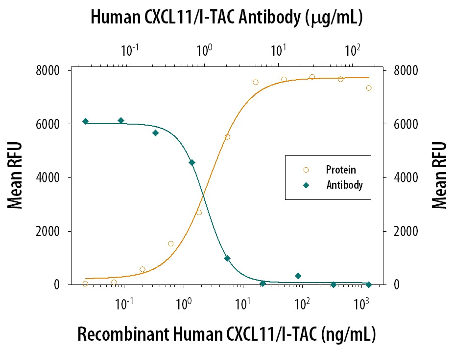
Chemotaxis Induced by CXCL11/I‑TAC and Neutralization by Human CXCL11/I‑TAC Antibody.
Recombinant Human CXCL11/I-TAC (Catalog # 672-IT) chemoattracts the BaF3 mouse pro-B cell line transfected with human CXCR3 in a dose-dependent manner (orange line). The amount of cells that migrated through to the lower chemotaxis chamber was measured by Resazurin (Catalog # AR002). Chemotaxis elicited by Recombinant Human CXCL11/I-TAC (0.05 µg/mL) is neutralized (green line) by increasing concentrations of Goat Anti-Human CXCL11/I-TAC Antigen Affinity-purified Polyclonal Antibody (Catalog # AF260). The ND50 is typically 0.5-1.5 µg/mL.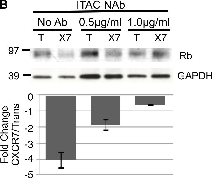
Detection of Human CXCL11/I-TAC by Block/Neutralize
CXCR7-mediated Rb degradation requires ligand.pLEC cultures were infected with Trans only or Trans+CXCR7. At 6 hours post-infection, media was replaced with media only or media containing (A) SDF-1/CXCL12 neutralizing antibody at 1.5, or 3.0 µg/ml or (B) ITAC/CXCL11 neutralizing antibody at 0.5 or 1.0 µg/ml. At 20 hours post infection, cells were lysed and analyzed by western blot for total Rb levels. Densitometry analysis was performed and fold change values were calculated from GAPDH-normalized optical density ratios as described for Figure 3. Bar graphs (bottom) represent the average fold change at each condition for two independent experiments. Image collected and cropped by CiteAb from the following open publication (https://dx.plos.org/10.1371/journal.pone.0069828), licensed under a CC-BY license. Not internally tested by R&D Systems.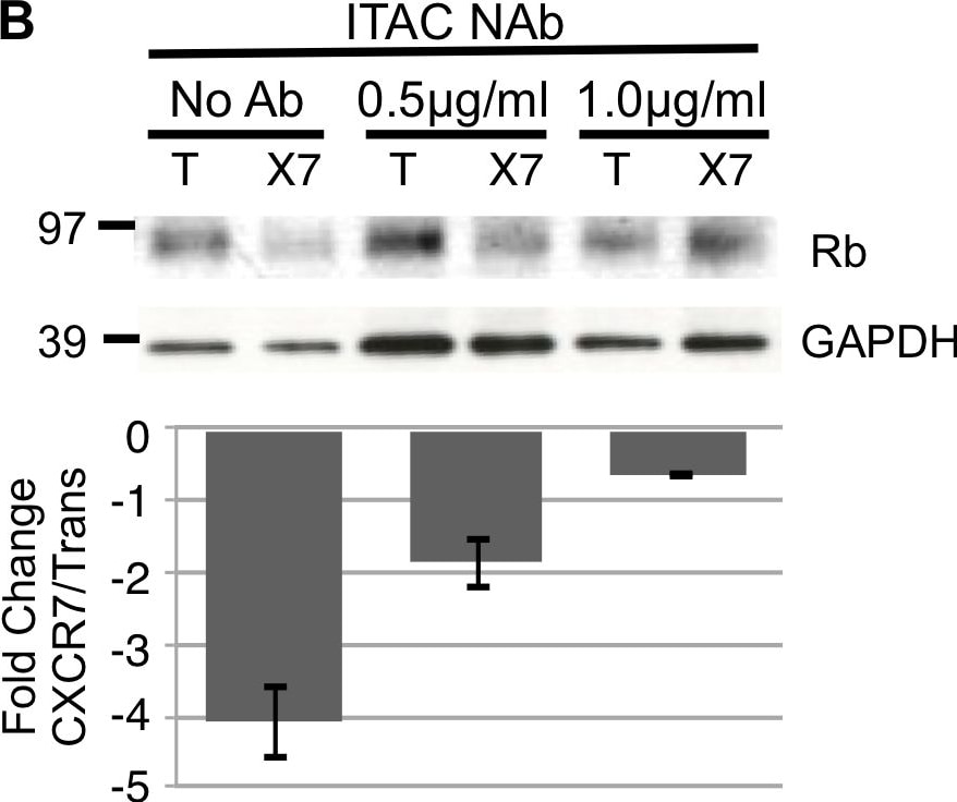
Detection of Human CXCL11/I-TAC by Block/Neutralize
CXCR7-mediated Rb degradation requires ligand.pLEC cultures were infected with Trans only or Trans+CXCR7. At 6 hours post-infection, media was replaced with media only or media containing (A) SDF-1/CXCL12 neutralizing antibody at 1.5, or 3.0 µg/ml or (B) ITAC/CXCL11 neutralizing antibody at 0.5 or 1.0 µg/ml. At 20 hours post infection, cells were lysed and analyzed by western blot for total Rb levels. Densitometry analysis was performed and fold change values were calculated from GAPDH-normalized optical density ratios as described for Figure 3. Bar graphs (bottom) represent the average fold change at each condition for two independent experiments. Image collected and cropped by CiteAb from the following open publication (https://dx.plos.org/10.1371/journal.pone.0069828), licensed under a CC-BY license. Not internally tested by R&D Systems.
Human CXCL11 / I-TAC ELISA Standard Curve
Recombinant Human CXCL11/I‑TAC (Catalog # 672-IT) was serially diluted and captured by Mouse Anti-Human CXCL11/I‑TAC Monoclonal Antibody (Catalog # MAB672) coated on a Clear Polystyrene Microplate (Catalog # DY990). Goat Anti-Human CXCL11/I‑TAC Antigen Affinity-purified Polyclonal Antibody (Catalog # AF260) was biotinylated and incubated with the protein captured on the plate. Detection of the standard curve was achieved by incubating Streptavidin-HRP (Catalog # DY998)Applications for Human CXCL11/I-TAC Antibody
Western Blot
Sample: Recombinant Human CXCL11/I-TAC (Catalog # 672-IT)
Neutralization
Reviewed Applications
Read 1 review rated 5 using AF260 in the following applications:
Formulation, Preparation, and Storage
Purification
Reconstitution
Reconstitute at 0.2 mg/mL in sterile PBS. For liquid material, refer to CoA for concentration.
Formulation
Shipping
Stability & Storage
- 12 months from date of receipt, -20 to -70 °C as supplied.
- 1 month, 2 to 8 °C under sterile conditions after reconstitution.
- 6 months, -20 to -70 °C under sterile conditions after reconstitution.
Calculators
Background: CXCL11/I-TAC
References
- Cole, K. et al. (1998) J. Exp. Med. 187:2009.
- Sandhya Rani, M. et al. (1996) J. Biol. Chem. 271:22878.
- Lou, Y. et al. (1998) J. Neurovirol. 4:575.
Alternate Names
Gene Symbol
UniProt
Additional CXCL11/I-TAC Products
Product Documents for Human CXCL11/I-TAC Antibody
Certificate of Analysis
To download a Certificate of Analysis, please enter a lot or batch number in the search box below.
Note: Certificate of Analysis not available for kit components.
Product Specific Notices for Human CXCL11/I-TAC Antibody
For research use only
Related Research Areas
Citations for Human CXCL11/I-TAC Antibody
 Powered by Bioz
Customer Reviews for Human CXCL11/I-TAC Antibody (1)
Have you used Human CXCL11/I-TAC Antibody?
Submit a review and receive an Amazon gift card!
$25/€18/£15/$25CAN/¥2500 for a review with an image
$10/€7/£6/$10CAN/¥1110 for a review without an image
Submit a review
-
Application: Western BlotSample Tested: See PMID 19552626Species: HumanVerified Customer | Posted 01/07/2015
There are no reviews that match your criteria.
Protocols
Find general support by application which include: protocols, troubleshooting, illustrated assays, videos and webinars.
- Cellular Response to Hypoxia Protocols
- R&D Systems Quality Control Western Blot Protocol
- Troubleshooting Guide: Western Blot Figures
- Western Blot Conditions
- Western Blot Protocol
- Western Blot Protocol for Cell Lysates
- Western Blot Troubleshooting
- Western Blot Troubleshooting Guide
- View all Protocols, Troubleshooting, Illustrated assays and Webinars
Associated Pathways
