Human CXCL5/ENA-78 Antibody (33170)
R&D Systems | Catalog # MAB654

Key Product Details
Validated by
Biological Validation
Species Reactivity
Validated:
Human
Cited:
Human, Mouse
Applications
Validated:
ELISA Capture (Matched Antibody Pair), Neutralization, Intracellular Staining by Flow Cytometry, CyTOF-ready
Cited:
ELISA Development, Mass Cytometry
Label
Unconjugated
Antibody Source
Monoclonal Mouse IgG1 Clone # 33170
Loading...
Product Specifications
Immunogen
E. coli-derived recombinant human CXCL5/ENA-78
Ala37-Asn114
Accession # P42830
Ala37-Asn114
Accession # P42830
Specificity
Detects human CXCL5/ENA-78 in ELISAs. Does not cross-react with recombinant human CXCL1, 2, or 3.
Clonality
Monoclonal
Host
Mouse
Isotype
IgG1
Endotoxin Level
<0.10 EU per 1 μg of the antibody by the LAL method.
Scientific Data Images for Human CXCL5/ENA-78 Antibody (33170)
Detection of CXCL5/ENA‑78 in Human PBMCs by Flow Cytometry.
Human peripheral blood mononculear cells (PBMCs) either treated with LPS (filled histogram) or untreated (open histogram) were stained with Mouse Anti-Human CXCL5/ENA-78 Monoclonal Antibody (Catalog # MAB654), followed by Allophycocyanin-conjugated Anti-Mouse IgG Secondary Antibody (Catalog # F0101B). To facilitate intracellular staining, cells were fixed with Flow Cytometry Fixation Buffer (Catalog # FC004) and permeabilized with Flow Cytometry Permeabilization/Wash Buffer I (Catalog # FC005). View our protocol for Staining Intracellular Molecules.
Chemotaxis Induced by CXCL5/ENA-78 and Neutralization by Human CXCL5/ENA-78 Antibody.
Recombinant Human CXCL5/ENA-78 (254-XB) chemoattracts the BaF3 mouse pro-B cell line transfected with human CXCR2 in a dose-dependent manner (orange line). The amount of cells that migrated through to the lower chemotaxis chamber was measured by Resazurin (AR002). Chemotaxis elicited by Recombinant Human CXCL5/ ENA-78 (30 ng/mL) is neutralized (green line) by increasing concentrations of Mouse Anti-Human CXCL5/ENA-78 Monoclonal Antibody (Catalog # MAB654). The ND50is typically 0.65-5 µg/mL.
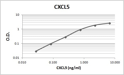
Human CXCL5 / ENA-78 ELISA Standard Curve
Recombinant Human CXCL5/ENA‑78 (Catalog # 254-XB) was serially diluted and captured by Mouse Anti-Human CXCL5/ENA‑78 Monoclonal Antibody (Catalog # MAB654) coated on a Clear Polystyrene Microplate (Catalog # DY990). Goat Anti-Human CXCL5/ENA‑78 Antigen Affinity-purified Polyclonal Antibody (Catalog # AF254) was biotinylated and incubated with the protein captured on the plate. Detection of the standard curve was achieved by incubating Streptavidin-HRP (Catalog # DY998)Applications for Human CXCL5/ENA-78 Antibody (33170)
Application
Recommended Usage
CyTOF-ready
Ready to be labeled using established conjugation methods. No BSA or other carrier proteins that could interfere with conjugation.
Intracellular Staining by Flow Cytometry
0.25 µg/106 cells
Sample: Human peripheral blood mononculear cells (PBMCs) treated with LPS were fixed with Flow Cytometry Fixation Buffer (Catalog # FC004) and permeabilized with Flow Cytometry Permeabilization/Wash Buffer I (Catalog # FC005)
Sample: Human peripheral blood mononculear cells (PBMCs) treated with LPS were fixed with Flow Cytometry Fixation Buffer (Catalog # FC004) and permeabilized with Flow Cytometry Permeabilization/Wash Buffer I (Catalog # FC005)
Neutralization
Measured by its ability to neutralize CXCL5/ENA‑78-induced chemotaxis in the BaF3 mouse pro‑B cell line transfected with human CXCR2. The Neutralization Dose (ND50) is typically 0.65-5 µg/mL in the presence of 30 ng/mL Recombinant Human CXCL5/ENA‑78.
Human CXCL5/ENA-78 Sandwich Immunoassay
Please Note: Optimal dilutions of this antibody should be experimentally determined.
Reviewed Applications
Read 1 review rated 4 using MAB654 in the following applications:
Flow Cytometry Panel Builder
Bio-Techne Knows Flow Cytometry
Save time and reduce costly mistakes by quickly finding compatible reagents using the Panel Builder Tool.
Advanced Features
- Spectra Viewer - Custom analysis of spectra from multiple fluorochromes
- Spillover Popups - Visualize the spectra of individual fluorochromes
- Antigen Density Selector - Match fluorochrome brightness with antigen density

Formulation, Preparation, and Storage
Purification
Protein A or G purified from ascites
Reconstitution
Reconstitute at 0.5 mg/mL in sterile PBS. For liquid material, refer to CoA for concentration.
Loading...
Formulation
Lyophilized from a 0.2 μm filtered solution in PBS with Trehalose. *Small pack size (SP) is supplied either lyophilized or as a 0.2 µm filtered solution in PBS.
Shipping
Lyophilized product is shipped at ambient temperature. Liquid small pack size (-SP) is shipped with polar packs. Upon receipt, store immediately at the temperature recommended below.
Stability & Storage
Use a manual defrost freezer and avoid repeated freeze-thaw cycles.
- 12 months from date of receipt, -20 to -70 °C as supplied.
- 1 month, 2 to 8 °C under sterile conditions after reconstitution.
- 6 months, -20 to -70 °C under sterile conditions after reconstitution.
Calculators
Background: CXCL5/ENA-78
Additional CXCL5/ENA-78 Products
Product Documents for Human CXCL5/ENA-78 Antibody (33170)
Certificate of Analysis
To download a Certificate of Analysis, please enter a lot or batch number in the search box below.
Note: Certificate of Analysis not available for kit components.
Product Specific Notices for Human CXCL5/ENA-78 Antibody (33170)
For research use only
Related Research Areas
Citations for Human CXCL5/ENA-78 Antibody (33170)
 Powered by Bioz
Customer Reviews for Human CXCL5/ENA-78 Antibody (33170) (1)
4 out of 5
1 Customer Rating
Have you used Human CXCL5/ENA-78 Antibody (33170)?
Submit a review and receive an Amazon gift card!
$25/€18/£15/$25CAN/¥2500 Yen for a review with an image
$10/€7/£6/$10CAN/¥1110 Yen for a review without an image
Submit a review
Customer Images
Showing
1
-
1 of
1 review
Showing All
Filter By:
-
Application: ELISASample Tested: Serum and PlasmaSpecies: HumanVerified Customer | Posted 08/23/2018

There are no reviews that match your criteria.
Protocols
Find general support by application which include: protocols, troubleshooting, illustrated assays, videos and webinars.
- 7-Amino Actinomycin D (7-AAD) Cell Viability Flow Cytometry Protocol
- Extracellular Membrane Flow Cytometry Protocol
- Flow Cytometry Protocol for Cell Surface Markers
- Flow Cytometry Protocol for Staining Membrane Associated Proteins
- Flow Cytometry Staining Protocols
- Flow Cytometry Troubleshooting Guide
- Intracellular Flow Cytometry Protocol Using Alcohol (Methanol)
- Intracellular Flow Cytometry Protocol Using Detergents
- Intracellular Nuclear Staining Flow Cytometry Protocol Using Detergents
- Intracellular Staining Flow Cytometry Protocol Using Alcohol Permeabilization
- Intracellular Staining Flow Cytometry Protocol Using Detergents to Permeabilize Cells
- Propidium Iodide Cell Viability Flow Cytometry Protocol
- Protocol for Liperfluo
- Protocol for the Characterization of Human Th22 Cells
- Protocol for the Characterization of Human Th9 Cells
- Protocol: Annexin V and PI Staining by Flow Cytometry
- Protocol: Annexin V and PI Staining for Apoptosis by Flow Cytometry
- Troubleshooting Guide: Fluorokine Flow Cytometry Kits
- View all Protocols, Troubleshooting, Illustrated assays and Webinars
Loading...
Associated Pathways
