CXCL5, also known as epithelial cell-derived neutrophil-activating peptide (ENA-78), is an 8 kDa proinflammatory member of the CXC subfamily of chemokines. Its Glu-Leu-Arg (ELR) motif confers angiogenic properties and distinguishes it from ELR-CXC chemokines which are angiostatic (1‑3). Human CXCL5 shares 57% amino acid (aa) sequence identity with mouse and rat CXCL5. Among other human ELR+ chemokines, it shares 77% aa sequence identity with CXCL6/GCP-2 and
35%‑51% with CXCL1/GRO alpha, CXCL2/GRO beta, CXCL3/GRO gamma, CXCL7/NAP-2, and CXCL8/IL-8. Inflammatory stimulation up-regulates CXCL5 production in multiple hematopoietic cell types, fibroblasts, endothelial cells, and vascular smooth muscle cells. In vivo, CXCL5 is elevated at sites of inflammation and pulmonary fibrosis where it promotes neutrophil infiltration and activation as well as angiogenesis (3-6). Its upregulation contributes to increased vascularization, tumor growth, and metastasis in many cancers (6-9). Full length CXCL5 (78 aa) is trimmed at the N-terminus by cathepsin G and chymotrypsin to ENA-74 (74 aa) and ENA-70 (70 aa), with the shortened forms showing increased potency relative to full length CXCL5 (10, 11). CXCL5 exerts its effects primarily through interactions with CXCR2 (6, 12). It also binds duffy antigen receptor for chemokines (DARC), which can limit CXCR2-mediated responses (13, 14).

Key Product Details
Validated by
Biological Validation
Species Reactivity
Validated:
Human
Cited:
Human, Mouse
Applications
Validated:
Immunohistochemistry, Western Blot, Neutralization
Cited:
Immunohistochemistry, Immunohistochemistry-Paraffin, Immunohistochemistry-Frozen, Neutralization, Flow Cytometry, ELISA Development, In vivo assay
Label
Unconjugated
Antibody Source
Polyclonal Goat IgG
Loading...
Product Specifications
Immunogen
E. coli-derived recombinant human CXCL5/ENA-78
Ala37-Asn114
Accession # P42830
Ala37-Asn114
Accession # P42830
Specificity
Detects human CXCL5/ENA‑78 in direct ELISAs and Western blots.
Clonality
Polyclonal
Host
Goat
Isotype
IgG
Endotoxin Level
<0.10 EU per 1 μg of the antibody by the LAL method.
Scientific Data Images for Human CXCL5/ENA-78 Antibody
Detection of Human CXCL5/ENA‑78 by Western Blot.
Western blot shows conditioned media (CM) and lysates from A549 human lung carcinoma cell line untreated (-) or treated (+) with human IL-1 beta for 24 hours. PVDF membrane was probed with 1 µg/mL of Goat Anti-Human CXCL5/ENA-78 Antigen Affinity-purified Polyclonal Antibody (Catalog # AF254) followed by HRP-conjugated Anti-Goat IgG Secondary Antibody (HAF109). A specific band was detected for CXCL5/ENA-78 at approximately 10 kDa (as indicated). This experiment was conducted under reducing conditions and using Immunoblot Buffer Group 1.
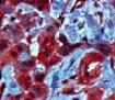
CXCL5/ENA‑78 in Human Breast Cancer Tissue.
CXCL5/ENA-78 was detected in immersion fixed paraffin-embedded sections of human breast cancer tissue using Goat Anti-Human CXCL5/ENA-78 Antigen Affinity-purified Polyclonal Antibody (Catalog # AF254) at 10 µg/mL overnight at 4 °C. Tissue was stained using the Anti-Goat HRP-DAB Cell & Tissue Staining Kit (brown; (CTS008) and counterstained with hematoxylin (blue). Specific staining was localized to cytoplasm. View our protocol for Chromogenic IHC Staining of Paraffin-embedded Tissue Sections.
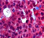
CXCL5/ENA‑78 in Human Breast.
CXCL5/ENA-78 was detected in immersion fixed paraffin-embedded sections of human breast using Goat Anti-Human CXCL5/ENA-78 Antigen Affinity-purified Polyclonal Antibody (Catalog # AF254) at 5 µg/mL overnight at 4 °C. Tissue was stained using the Anti-Goat HRP-DAB Cell & Tissue Staining Kit (brown; (CTS008) and counterstained with hematoxylin (blue). Specific staining was localized to epithelial cells. View our protocol for Chromogenic IHC Staining of Paraffin-embedded Tissue Sections.
Chemotaxis Induced by CXCL5/ENA‑78 and Neutralization by Human CXCL5/ENA‑78 Antibody.
Recombinant Human CXCL5/ENA-78 (254-XB) chemoattracts the BaF3 mouse pro-B cell line transfected with human CXCR2 in a dose-dependent manner (orange line). The amount of cells that migrated through to the lower chemotaxis chamber was measured by Resazurin (AR002). Chemotaxis elicited by Recombinant Human CXCL5/ENA-78 (0.03 µg/mL) is neutralized (green line) by increasing concentrations of Goat Anti-Human CXCL5/ENA-78 Antigen Affinity-purified Polyclonal Antibody (Catalog # AF254). The ND50 is typically 0.2-1.0 µg/mL.
Human CXCL5 / ENA-78 ELISA Standard Curve
Recombinant Human CXCL5/ENA‑78 (Catalog # 254-XB) was serially diluted and captured by Mouse Anti-Human CXCL5/ENA‑78 Monoclonal Antibody (Catalog # MAB654) coated on a Clear Polystyrene Microplate (Catalog # DY990). Goat Anti-Human CXCL5/ENA‑78 Antigen Affinity-purified Polyclonal Antibody (Catalog # AF254) was biotinylated and incubated with the protein captured on the plate. Detection of the standard curve was achieved by incubating Streptavidin-HRP (Catalog # DY998)Applications for Human CXCL5/ENA-78 Antibody
Application
Recommended Usage
Immunohistochemistry
5-15 µg/mL
Sample: Immersion fixed paraffin-embedded sections of human breast and human breast cancer tissue
Sample: Immersion fixed paraffin-embedded sections of human breast and human breast cancer tissue
Western Blot
1 µg/mL
Sample: Conditioned media and lysates from A549 human lung carcinoma cell line treated with human IL-1 beta
Sample: Conditioned media and lysates from A549 human lung carcinoma cell line treated with human IL-1 beta
Neutralization
Measured by its ability to neutralize CXCL5/ENA‑78-induced chemotaxis in the BaF3 mouse pro‑B cell line transfected with human CXCR2. The Neutralization Dose (ND50) is typically 0.2-1.0 µg/mL in the presence of 0.03 µg/mL Recombinant Human CXCL5/ENA‑78.
Reviewed Applications
Read 3 reviews rated 4.7 using AF254 in the following applications:
Formulation, Preparation, and Storage
Purification
Antigen Affinity-purified
Reconstitution
Reconstitute at 0.2 mg/mL in sterile PBS. For liquid material, refer to CoA for concentration.
Loading...
Formulation
Lyophilized from a 0.2 μm filtered solution in PBS with Trehalose. *Small pack size (SP) is supplied either lyophilized or as a 0.2 µm filtered solution in PBS.
Shipping
Lyophilized product is shipped at ambient temperature. Liquid small pack size (-SP) is shipped with polar packs. Upon receipt, store immediately at the temperature recommended below.
Stability & Storage
Use a manual defrost freezer and avoid repeated freeze-thaw cycles.
- 12 months from date of receipt, -20 to -70 °C as supplied.
- 1 month, 2 to 8 °C under sterile conditions after reconstitution.
- 6 months, -20 to -70 °C under sterile conditions after reconstitution.
Calculators
Background: CXCL5/ENA-78
References
- Strieter, R.M. et al. (2005) Cytokine Growth Factor Rev. 16:593.
- Balestrieri, M.L. et al. (2008) Cardiovasc. Res. 78:250.
- Walz, A.. et al. (1991) J. Exp. Med. 174:1355.
- Strieter, R.M. et al. (1992) Immunol. Invest. 21:549.
- Koch, A.E. et al. (1994) J. Clin. Invest. 94:1012.
- Begley, L.A. et al. (2008) Neoplasia 10:244.
- Vandercappellen, J. et al. (2008) Cancer Lett. 267:226.
- Arenberg, D.A. et al. (1998) J. Clin. Invest. 102:465.
- Miyazaki, H. et al. (2006) Cancer Res. 66:4279.
- Wuyts, A. et al. (1999) Eur. J. Biochem. 260:421.
- Nufer, O. et al. (1999) Biochemistry 38:636.
- Ahuja, S.K. and P.M. Murphy (1996) J. Biol. Chem. 271:20545.
- Kashiwazaki, M. et al. (2003) Int. Immunol. 15:1219.
- Horton, L.W. et al. (2007) Cancer Res. 67:9791.
Alternate Names
CXCL5, ENA-78, ENA78
Entrez Gene IDs
6374 (Human)
Gene Symbol
CXCL5
UniProt
Additional CXCL5/ENA-78 Products
Product Documents for Human CXCL5/ENA-78 Antibody
Certificate of Analysis
To download a Certificate of Analysis, please enter a lot or batch number in the search box below.
Note: Certificate of Analysis not available for kit components.
Product Specific Notices for Human CXCL5/ENA-78 Antibody
For research use only
Related Research Areas
Citations for Human CXCL5/ENA-78 Antibody
 Powered by Bioz
Customer Reviews for Human CXCL5/ENA-78 Antibody (3)
4.7 out of 5
3 Customer Ratings
Have you used Human CXCL5/ENA-78 Antibody?
Submit a review and receive an Amazon gift card!
$25/€18/£15/$25CAN/¥2500 Yen for a review with an image
$10/€7/£6/$10CAN/¥1110 Yen for a review without an image
Submit a review
Customer Images

Showing
1
-
3 of
3 reviews
Showing All
Filter By:
-
Application: Immunohistochemistry-ParaffinSample Tested: Breast cancer tissueSpecies: HumanVerified Customer | Posted 04/04/2020

-
Application: Western BlotSample Tested: Hep3B human hepatocellular carcinoma cell line and SK-HEP-1 human liver adenocarcinoma cell lineSpecies: HumanVerified Customer | Posted 02/18/2019
-
Application: ImmunohistochemistrySample Tested: Cancer TissueSpecies: HumanVerified Customer | Posted 10/17/2018
There are no reviews that match your criteria.
Protocols
Find general support by application which include: protocols, troubleshooting, illustrated assays, videos and webinars.
- Antigen Retrieval Protocol (PIER)
- Antigen Retrieval for Frozen Sections Protocol
- Appropriate Fixation of IHC/ICC Samples
- Cellular Response to Hypoxia Protocols
- Chromogenic IHC Staining of Formalin-Fixed Paraffin-Embedded (FFPE) Tissue Protocol
- Chromogenic Immunohistochemistry Staining of Frozen Tissue
- ClariTSA™ Fluorophore Kits
- Detection & Visualization of Antibody Binding
- Fluorescent IHC Staining of Frozen Tissue Protocol
- Graphic Protocol for Heat-induced Epitope Retrieval
- Graphic Protocol for the Preparation and Fluorescent IHC Staining of Frozen Tissue Sections
- Graphic Protocol for the Preparation and Fluorescent IHC Staining of Paraffin-embedded Tissue Sections
- Graphic Protocol for the Preparation of Gelatin-coated Slides for Histological Tissue Sections
- IHC Sample Preparation (Frozen sections vs Paraffin)
- Immunofluorescent IHC Staining of Formalin-Fixed Paraffin-Embedded (FFPE) Tissue Protocol
- Immunohistochemistry (IHC) and Immunocytochemistry (ICC) Protocols
- Immunohistochemistry Frozen Troubleshooting
- Immunohistochemistry Paraffin Troubleshooting
- Preparing Samples for IHC/ICC Experiments
- Preventing Non-Specific Staining (Non-Specific Binding)
- Primary Antibody Selection & Optimization
- Protocol for Heat-Induced Epitope Retrieval (HIER)
- Protocol for Making a 4% Formaldehyde Solution in PBS
- Protocol for VisUCyte™ HRP Polymer Detection Reagent
- Protocol for the Preparation & Fixation of Cells on Coverslips
- Protocol for the Preparation and Chromogenic IHC Staining of Frozen Tissue Sections
- Protocol for the Preparation and Chromogenic IHC Staining of Frozen Tissue Sections - Graphic
- Protocol for the Preparation and Chromogenic IHC Staining of Paraffin-embedded Tissue Sections
- Protocol for the Preparation and Chromogenic IHC Staining of Paraffin-embedded Tissue Sections - Graphic
- Protocol for the Preparation and Fluorescent IHC Staining of Frozen Tissue Sections
- Protocol for the Preparation and Fluorescent IHC Staining of Paraffin-embedded Tissue Sections
- Protocol for the Preparation of Gelatin-coated Slides for Histological Tissue Sections
- R&D Systems Quality Control Western Blot Protocol
- TUNEL and Active Caspase-3 Detection by IHC/ICC Protocol
- The Importance of IHC/ICC Controls
- Troubleshooting Guide: Immunohistochemistry
- Troubleshooting Guide: Western Blot Figures
- Western Blot Conditions
- Western Blot Protocol
- Western Blot Protocol for Cell Lysates
- Western Blot Troubleshooting
- Western Blot Troubleshooting Guide
- View all Protocols, Troubleshooting, Illustrated assays and Webinars
Loading...
Associated Pathways
