Human CXCR1/IL-8RA Antibody (42705)
R&D Systems | Catalog # MAB330
Clone 42705 was used by HLDA to establish CD designation

Key Product Details
Species Reactivity
Validated:
Human
Cited:
Human, Mouse, Rat, Guinea Pig, Rabbit
Applications
Validated:
Neutralization, Flow Cytometry, CyTOF-reported
Cited:
Immunohistochemistry, Immunohistochemistry-Paraffin, Immunohistochemistry-Frozen, Western Blot, Neutralization, Flow Cytometry, Immunocytochemistry, Assay Development, Bioassay, In vivo assay, Functional Assay
Label
Unconjugated
Antibody Source
Monoclonal Mouse IgG2A Clone # 42705
Loading...
Product Specifications
Immunogen
NS0 mouse myeloma cell line transfected with human CXCR1/IL-8 RA
Met1-Leu350
Accession # AAA59159
Met1-Leu350
Accession # AAA59159
Specificity
Detects human CXCR1/IL-8 RA transfectants but not the parental cell line. It does not cross-react with human CXCR2.
Clonality
Monoclonal
Host
Mouse
Isotype
IgG2A
Endotoxin Level
<0.10 EU per 1 μg of the antibody by the LAL method.
Scientific Data Images for Human CXCR1/IL-8RA Antibody (42705)
Detection of CXCR1/IL‑8 RA in Human Blood Granulocytes by Flow Cytometry.
Human peripheral blood granulocytes were stained with Mouse Anti-Human CXCR1/IL-8 RA Monoclonal Antibody (Catalog # MAB330, filled histogram) or isotype control antibody (Catalog # MAB003, open histogram), followed by Phycoerythrin-conjugated Anti-Mouse IgG Secondary Antibody (Catalog # F0102B).
Chemotaxis Induced by CXCL8/IL‑8 and Neutralization by Human CXCR1/IL‑8 RA Antibody.
Recombinant Human CXCL8/IL-8 (Catalog # 208-IL) chemoattracts the BaF3 mouse pro-B cell line transfected with human CXCR1 in a dose-dependent manner (orange line). The amount of cells that migrated through to the lower chemotaxis chamber was measured by Resazurin (Catalog # AR002). Chemotaxis elicited by Recombinant Human CXCL8/IL-8 (1 ng/mL) is neutralized (green line) by increasing concentrations of Mouse Anti-Human CXCR1/IL-8 RA Monoclonal Antibody (Catalog # MAB330). The ND50 is typically 0.4-2.0 µg/mL.
Detection of CXCR1/IL-8RA by Flow Cytometry
IL‐8/CXCR1 contributes to stemness in vitro. (A) Effect on sphere formation of single agent or combination treatment with human recombinant IL‐8, anti‐CXCR1 antibody and/or repertaxin for Caki‐1 (Kruskal–Wallis test, n = 5). (B) Effect on sphere formation of single agent or combination treatment with human recombinant IL‐8, anti‐CXCR1 antibody and repertaxin for 769P cells (Kruskal–Wallis test, n = 5). (C) Flow cytometry analysis of the SP and the CXCR1+ cell compartment upon repertaxin treatment in the spheres derived from Caki‐1. (D) Histograms showing decreased SP and CXCR1+ cells upon repertaxin treatment. The yellow area indicates number of events with verapamil treatment. The red area shows CSC and CXCR1+ populations in the untreated sample. The blue area displays the remaining number of events after repertaxin treatment. Image collected and cropped by CiteAb from the following open publication (https://pubmed.ncbi.nlm.nih.gov/30883740), licensed under a CC-BY license. Not internally tested by R&D Systems.
Detection of CXCR1/IL-8RA by Western Blot
IL-8 as a partial inducer of MMP9 in ESCC cells. (A): ELISA quantification of IL-8 in culture supernatants of ESCC cells after direct co-culture with macrophages. Directly co-cultured ESCC cells secreted more IL-8 than monocultured counterparts. (B): Western blot revealed the expression of known IL-8 receptors, CXCR1 (45 kDa, monomer; 80–90 kDa, dimer and their glycosylated forms) and CXCR2 (41 kDa) in TE-9, TE-10, and TE-11 cell lines. (C,D): Treatment with rhIL-8 (100 ng/mL, for 24 h) upregulated MMP9 mRNA expression in all three ESCC cell lines (C) but only triggered MMP9 secretion from TE-10 (D). Data are expressed as mean ± SEM; * p < 0.05, ** p < 0.01, *** p < 0.001. NS, not significant. Image collected and cropped by CiteAb from the following open publication (https://pubmed.ncbi.nlm.nih.gov/37296952), licensed under a CC-BY license. Not internally tested by R&D Systems.
Detection of CXCR1/IL-8RA by Western Blot
IL-8 as a partial inducer of MMP9 in ESCC cells. (A): ELISA quantification of IL-8 in culture supernatants of ESCC cells after direct co-culture with macrophages. Directly co-cultured ESCC cells secreted more IL-8 than monocultured counterparts. (B): Western blot revealed the expression of known IL-8 receptors, CXCR1 (45 kDa, monomer; 80–90 kDa, dimer and their glycosylated forms) and CXCR2 (41 kDa) in TE-9, TE-10, and TE-11 cell lines. (C,D): Treatment with rhIL-8 (100 ng/mL, for 24 h) upregulated MMP9 mRNA expression in all three ESCC cell lines (C) but only triggered MMP9 secretion from TE-10 (D). Data are expressed as mean ± SEM; * p < 0.05, ** p < 0.01, *** p < 0.001. NS, not significant. Image collected and cropped by CiteAb from the following open publication (https://pubmed.ncbi.nlm.nih.gov/37296952), licensed under a CC-BY license. Not internally tested by R&D Systems.Applications for Human CXCR1/IL-8RA Antibody (42705)
Application
Recommended Usage
CyTOF-reported
Cheng, Y. et al. (2016) J. Immunol. 196: 924. Ready to be labeled using established conjugation methods. No BSA or other carrier proteins that could interfere with conjugation.
Flow Cytometry
0.25 µg/106 cells
Sample: Human whole blood granulocytes
Sample: Human whole blood granulocytes
Neutralization
Measured by its ability to neutralize CXCL8/IL‑8-induced chemotaxis in the BaF3 mouse pro‑B cell line transfected with human CXCR1. The Neutralization Dose (ND50) is typically 0.4-2.0 µg/mL in the presence of 1 ng/mL Recombinant Human CXCL8/IL‑8.
Reviewed Applications
Read 4 reviews rated 4.5 using MAB330 in the following applications:
Flow Cytometry Panel Builder
Bio-Techne Knows Flow Cytometry
Save time and reduce costly mistakes by quickly finding compatible reagents using the Panel Builder Tool.
Advanced Features
- Spectra Viewer - Custom analysis of spectra from multiple fluorochromes
- Spillover Popups - Visualize the spectra of individual fluorochromes
- Antigen Density Selector - Match fluorochrome brightness with antigen density

Formulation, Preparation, and Storage
Purification
Protein A or G purified from hybridoma culture supernatant
Reconstitution
Reconstitute at 0.5 mg/mL in sterile PBS. For liquid material, refer to CoA for concentration.
Loading...
Formulation
Lyophilized from a 0.2 μm filtered solution in PBS with Trehalose. See Certificate of Analysis for details.
*Small pack size (-SP) is supplied either lyophilized or as a 0.2 µm filtered solution in PBS.
*Small pack size (-SP) is supplied either lyophilized or as a 0.2 µm filtered solution in PBS.
Shipping
Lyophilized product is shipped at ambient temperature. Liquid small pack size (-SP) is shipped with polar packs. Upon receipt, store immediately at the temperature recommended below.
Stability & Storage
Use a manual defrost freezer and avoid repeated freeze-thaw cycles.
- 12 months from date of receipt, -20 to -70 °C as supplied.
- 1 month, 2 to 8 °C under sterile conditions after reconstitution.
- 6 months, -20 to -70 °C under sterile conditions after reconstitution.
Calculators
Background: CXCR1/IL-8RA
Long Name
Interleukin 8 Receptor A
Alternate Names
CD181, IL-8 RA, IL8RA
Gene Symbol
CXCR1
UniProt
Additional CXCR1/IL-8RA Products
Product Documents for Human CXCR1/IL-8RA Antibody (42705)
Certificate of Analysis
To download a Certificate of Analysis, please enter a lot or batch number in the search box below.
Note: Certificate of Analysis not available for kit components.
Product Specific Notices for Human CXCR1/IL-8RA Antibody (42705)
For research use only
Citations for Human CXCR1/IL-8RA Antibody (42705)
 Powered by Bioz
Customer Reviews for Human CXCR1/IL-8RA Antibody (42705) (4)
4.5 out of 5
4 Customer Ratings
Have you used Human CXCR1/IL-8RA Antibody (42705)?
Submit a review and receive an Amazon gift card!
$25/€18/£15/$25CAN/¥2500 Yen for a review with an image
$10/€7/£6/$10CAN/¥1110 Yen for a review without an image
Submit a review
Customer Images
Showing
1
-
4 of
4 reviews
Showing All
Filter By:
-
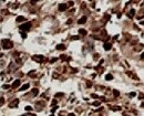
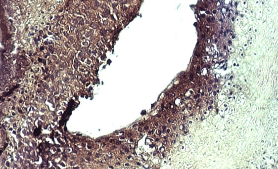
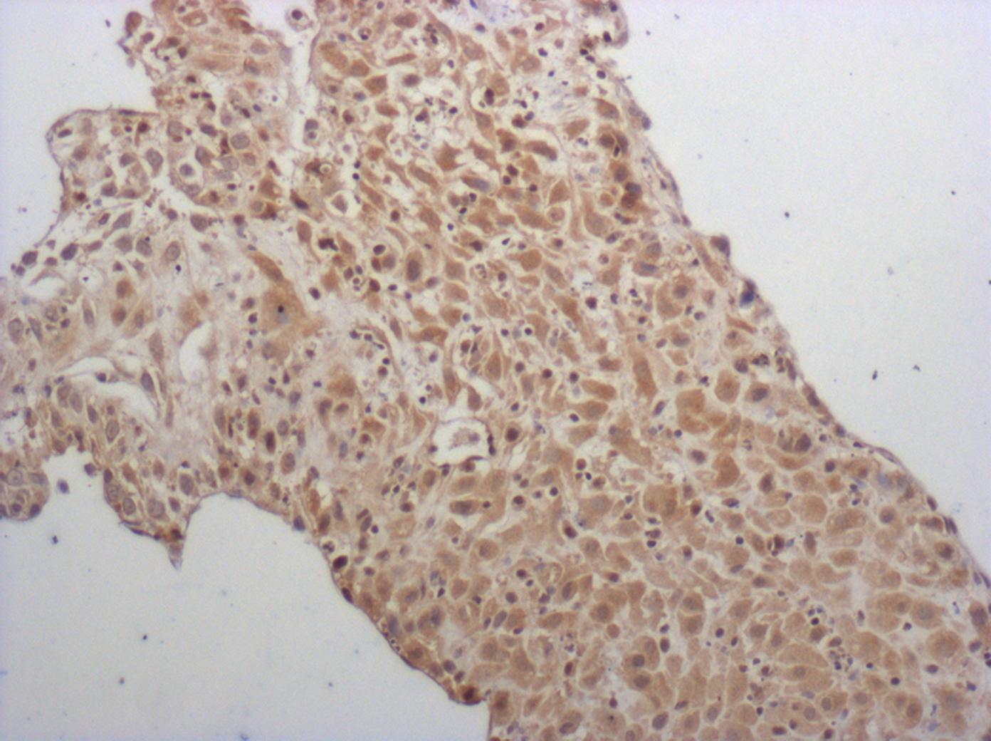
Application: ImmunohistochemistrySample Tested: Cervical cancer tissueSpecies: HumanVerified Customer | Posted 11/15/2021

-
Application: ImmunohistochemistrySample Tested: Pilomatricoma skin tumorSpecies: HumanVerified Customer | Posted 08/09/2021
-
Application: Block/NeutralizeSample Tested: Peripheral blood neutrophilsSpecies: HumanVerified Customer | Posted 04/26/2017
-
Application: ImmunohistochemistrySample Tested: First trimester deciduaSpecies: MouseVerified Customer | Posted 01/23/2017sodium citrate antigen retrieval
There are no reviews that match your criteria.
Protocols
Find general support by application which include: protocols, troubleshooting, illustrated assays, videos and webinars.
- 7-Amino Actinomycin D (7-AAD) Cell Viability Flow Cytometry Protocol
- Extracellular Membrane Flow Cytometry Protocol
- Flow Cytometry Protocol for Cell Surface Markers
- Flow Cytometry Protocol for Staining Membrane Associated Proteins
- Flow Cytometry Staining Protocols
- Flow Cytometry Troubleshooting Guide
- Intracellular Flow Cytometry Protocol Using Alcohol (Methanol)
- Intracellular Flow Cytometry Protocol Using Detergents
- Intracellular Nuclear Staining Flow Cytometry Protocol Using Detergents
- Intracellular Staining Flow Cytometry Protocol Using Alcohol Permeabilization
- Intracellular Staining Flow Cytometry Protocol Using Detergents to Permeabilize Cells
- Propidium Iodide Cell Viability Flow Cytometry Protocol
- Protocol for Liperfluo
- Protocol for the Characterization of Human Th22 Cells
- Protocol for the Characterization of Human Th9 Cells
- Protocol: Annexin V and PI Staining by Flow Cytometry
- Protocol: Annexin V and PI Staining for Apoptosis by Flow Cytometry
- Troubleshooting Guide: Fluorokine Flow Cytometry Kits
- View all Protocols, Troubleshooting, Illustrated assays and Webinars
Loading...
Associated Pathways
