Cyclin E1 (also known as G1/S-specific cyclin-E1 and CCNE1) is a 48-58 kDa member of the cyclin E subfamily, cyclin family of molecules. It associates with Cdk2 kinase and determines substrate specificity for the complex. This complex phosphorylates multiple substrates involved in cell cycle progression and the initiation of DNA replication. Cyclin E1 is required for G1/S phase progression and cell cycle reentry from G0 phase. Its reduced activity during cellular senescence contributes to G1 arrest. Human Cyclin E1 is 410 amino acids (aa) in length. It contains two cyclin box folds (aa 144-234 and 293-363) and is phosphorylated on at least eight Ser/Thr sites. There are at least two alternate splice forms. One is 40-41 kDa in size and utilizes an alternate start site at Met46, while a second is 43-44 kDa in size and shows a deletion of aa 154-196. One other potential start site exists at Met16. There are multiple proteolytic cleavage products that are tumor-associated and show increased destabilizing activity. Cleavage around Gln40 generates 44-45 kDa C-terminal fragments, while cleavage between Ala69Asp70 generates 33-35 kDa C-terminal fragments. Over aa 16-143, human Cyclin E1 shares 67% aa identity with mouse Cyclin E1.

Key Product Details
Species Reactivity
Human
Applications
Immunohistochemistry
Label
Unconjugated
Antibody Source
Monoclonal Mouse IgG2B Clone # 713573
Loading...
Product Specifications
Immunogen
E. coli-derived recombinant human Cyclin E1
Met16-Lys143
Accession # P24864
Met16-Lys143
Accession # P24864
Specificity
Detects human Cyclin E1 in direct ELISAs. In direct ELISAs, approximately 10% cross-reactivity
with recombinant mouse Cyclin E1 is observed.
Clonality
Monoclonal
Host
Mouse
Isotype
IgG2B
Scientific Data Images for Human Cyclin E1 Antibody (713573)
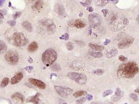
Cyclin E1 in Human Breast.
Cyclin E1 was detected in immersion fixed paraffin-embedded sections of human breast using Mouse Anti-Human Cyclin E1 Monoclonal Antibody (Catalog # MAB6810) at 15 µg/mL overnight at 4 °C. Tissue was stained using the Anti-Mouse HRP-DAB Cell & Tissue Staining Kit (brown; Catalog # CTS002) and counterstained with hematoxylin (blue). Specific staining was localized to epithelial cell nuclei. View our protocol for Chromogenic IHC Staining of Paraffin-embedded Tissue Sections.Applications for Human Cyclin E1 Antibody (713573)
Application
Recommended Usage
Immunohistochemistry
8-25 µg/mL
Sample: Immersion fixed paraffin-embedded sections of human breast
Sample: Immersion fixed paraffin-embedded sections of human breast
Reviewed Applications
Read 1 review rated 5 using MAB6810 in the following applications:
Formulation, Preparation, and Storage
Purification
Protein A or G purified from hybridoma culture supernatant
Reconstitution
Sterile PBS to a final concentration of 0.5 mg/mL. For liquid material, refer to CoA for concentration.
Loading...
Formulation
Lyophilized from a 0.2 μm filtered solution in PBS with Trehalose. *Small pack size (SP) is supplied either lyophilized or as a 0.2 µm filtered solution in PBS.
Shipping
Lyophilized product is shipped at ambient temperature. Liquid small pack size (-SP) is shipped with polar packs. Upon receipt, store immediately at the temperature recommended below.
Stability & Storage
Use a manual defrost freezer and avoid repeated freeze-thaw cycles.
- 12 months from date of receipt, -20 to -70 °C as supplied.
- 1 month, 2 to 8 °C under sterile conditions after reconstitution.
- 6 months, -20 to -70 °C under sterile conditions after reconstitution.
Calculators
Background: Cyclin E1
Alternate Names
CCNE, CCNE1
Gene Symbol
CCNE1
UniProt
Additional Cyclin E1 Products
Product Documents for Human Cyclin E1 Antibody (713573)
Certificate of Analysis
To download a Certificate of Analysis, please enter a lot or batch number in the search box below.
Note: Certificate of Analysis not available for kit components.
Product Specific Notices for Human Cyclin E1 Antibody (713573)
For research use only
Related Research Areas
Customer Reviews for Human Cyclin E1 Antibody (713573) (1)
5 out of 5
1 Customer Rating
Have you used Human Cyclin E1 Antibody (713573)?
Submit a review and receive an Amazon gift card!
$25/€18/£15/$25CAN/¥2500 Yen for a review with an image
$10/€7/£6/$10CAN/¥1110 Yen for a review without an image
Submit a review
Customer Images
Showing
1
-
1 of
1 review
Showing All
Filter By:
-
Application: ImmunohistochemistrySample Tested: laryngeal cancerSpecies: HumanVerified Customer | Posted 03/02/2022

There are no reviews that match your criteria.
Protocols
Find general support by application which include: protocols, troubleshooting, illustrated assays, videos and webinars.
- Antigen Retrieval Protocol (PIER)
- Antigen Retrieval for Frozen Sections Protocol
- Appropriate Fixation of IHC/ICC Samples
- Cellular Response to Hypoxia Protocols
- Chromogenic IHC Staining of Formalin-Fixed Paraffin-Embedded (FFPE) Tissue Protocol
- Chromogenic Immunohistochemistry Staining of Frozen Tissue
- ClariTSA™ Fluorophore Kits
- Detection & Visualization of Antibody Binding
- Fluorescent IHC Staining of Frozen Tissue Protocol
- Graphic Protocol for Heat-induced Epitope Retrieval
- Graphic Protocol for the Preparation and Fluorescent IHC Staining of Frozen Tissue Sections
- Graphic Protocol for the Preparation and Fluorescent IHC Staining of Paraffin-embedded Tissue Sections
- Graphic Protocol for the Preparation of Gelatin-coated Slides for Histological Tissue Sections
- IHC Sample Preparation (Frozen sections vs Paraffin)
- Immunofluorescent IHC Staining of Formalin-Fixed Paraffin-Embedded (FFPE) Tissue Protocol
- Immunohistochemistry (IHC) and Immunocytochemistry (ICC) Protocols
- Immunohistochemistry Frozen Troubleshooting
- Immunohistochemistry Paraffin Troubleshooting
- Preparing Samples for IHC/ICC Experiments
- Preventing Non-Specific Staining (Non-Specific Binding)
- Primary Antibody Selection & Optimization
- Protocol for Heat-Induced Epitope Retrieval (HIER)
- Protocol for Making a 4% Formaldehyde Solution in PBS
- Protocol for VisUCyte™ HRP Polymer Detection Reagent
- Protocol for the Preparation & Fixation of Cells on Coverslips
- Protocol for the Preparation and Chromogenic IHC Staining of Frozen Tissue Sections
- Protocol for the Preparation and Chromogenic IHC Staining of Frozen Tissue Sections - Graphic
- Protocol for the Preparation and Chromogenic IHC Staining of Paraffin-embedded Tissue Sections
- Protocol for the Preparation and Chromogenic IHC Staining of Paraffin-embedded Tissue Sections - Graphic
- Protocol for the Preparation and Fluorescent IHC Staining of Frozen Tissue Sections
- Protocol for the Preparation and Fluorescent IHC Staining of Paraffin-embedded Tissue Sections
- Protocol for the Preparation of Gelatin-coated Slides for Histological Tissue Sections
- TUNEL and Active Caspase-3 Detection by IHC/ICC Protocol
- The Importance of IHC/ICC Controls
- Troubleshooting Guide: Immunohistochemistry
- View all Protocols, Troubleshooting, Illustrated assays and Webinars
Loading...