Cystatin C is a member of family 2 of the Cystatin superfamily (1). It is involved in processes such as tumor invasion and metastasis, inflammation and some neurological diseases. It inhibits many cysteine proteases such as papain and cathepsins B, H, K, L, and S (2, 3). It is ubiquitous in human tissues and body fluids. A point mutation in the gene coding for the 120 amino acid mature Cystatin C causes a hereditary form of amyloid angiopathy in which the protein variant (Leu68 to Gln) is deposited in the cerebral arteries, leading to fatal cerebral hemorrhage (4). Cystatin C may have additional clinical applications. For example, it is a good marker for glomerular filtration rate (5).

Key Product Details
Species Reactivity
Validated:
Human
Cited:
Human
Applications
Validated:
Immunohistochemistry, Western Blot, Neutralization, Simple Western, Immunoprecipitation
Cited:
Western Blot, Array Development, ELISA Development
Label
Unconjugated
Antibody Source
Polyclonal Goat IgG
Loading...
Product Specifications
Immunogen
Mouse myeloma cell line NS0-derived recombinant human Cystatin C
Ser27-Ala146
Accession # P01034
Ser27-Ala146
Accession # P01034
Specificity
Detects human Cystatin C in direct ELISAs and Western blots. In direct ELISAs and Western blots, approximately 50% cross-reactivity with recombinant mouse Cystatin C is observed and less than 5% cross-reactivity with recombinant human (rh) Fetuin A, rhFetuin B, rhCystatin A, rhCystatin B, rhCystatin S, and rhCystatin E/M is observed.
Clonality
Polyclonal
Host
Goat
Isotype
IgG
Scientific Data Images for Human Cystatin C Antibody
Detection of Human Cystatin C by Western Blot.
Western blot shows lysates of HepG2 human hepatocellular carcinoma cell line, human liver tissue, and human serum. PVDF membrane was probed with 0.25 µg/mL of Goat Anti-Human Cystatin C Antigen Affinity-purified Polyclonal Antibody (Catalog # AF1196) followed by HRP-conjugated Anti-Goat IgG Secondary Antibody (Catalog # HAF109). A specific band was detected for Cystatin C at approximately 14 kDa (as indicated). This experiment was conducted under reducing conditions and using Immunoblot Buffer Group 1.
Detection of Human Cystatin C by Western Blot.
Western blot shows lysates of human placenta tissue and PC-3 human prostate cancer cell line. PVDF membrane was probed with 2 µg/mL of Goat Anti-Human Cystatin C Antigen Affinity-purified Polyclonal Antibody (Catalog # AF1196) followed by HRP-conjugated Anti-Goat IgG Secondary Antibody (Catalog # HAF017). A specific band was detected for Cystatin C at approximately 14 kDa (as indicated). This experiment was conducted under reducing conditions and using Immunoblot Buffer Group 1.
Cystatin C in Human Breast.
Cystatin C was detected in immersion fixed paraffin-embedded sections of human breast using 5 µg/mL Goat Anti-Human Cystatin C Antigen Affinity-purified Polyclonal Antibody (Catalog # AF1196) overnight at 4 °C. Tissue was stained with the Anti-Goat HRP-DAB Cell & Tissue Staining Kit (brown; Catalog # CTS008) and counterstained with hematoxylin (blue). Specific labeling was localized to the cytoplasm in cells of terminal ductules. View our protocol for Chromogenic IHC Staining of Paraffin-embedded Tissue Sections.
Detection of Human Cystatin C by Simple WesternTM.
Simple Western lane view shows lysates of HepG2 human hepatocellular carcinoma cell line, loaded at 0.2 mg/mL. A specific band was detected for Cystatin C at approximately 20 kDa (as indicated) using 10 µg/mL of Goat Anti-Human Cystatin C Antigen Affinity-purified Polyclonal Antibody (Catalog # AF1196). This experiment was conducted under reducing conditions and using the 12-230 kDa separation system.
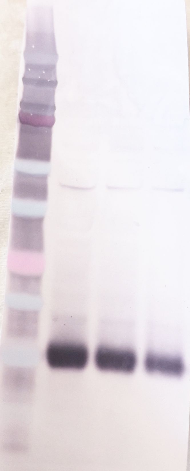
Detection of Human Cystatin C by Western Blot
Cystatin E/M and C secretions from various cell lines: Common laboratory non-melanoma and established laboratory melanoma cell lines (top), and primary and metastatic melanoma cell lines established from patients, as well as skin control (bottom). (A) Equal amounts of serum free media were collected from 5 × 10⁵ cells 48 h after changing from ordinary growth media and secreted proteins were concentrated by TCA-precipitation and subjected to SDS-PAGE and immunoblotting. The filters were stained with a cystatin E/M-specific (upper panels) or a cystatin C-specific (lower panel) antibody, respectively. (B) Inhibitory activity against legumain was measured in the conditioned media as residual legumain activity. A partially purified legumain fraction from rat kidney was mixed with conditioned media and the ability to cleave the substrate Z-Ala-Ala-Asn-NHMec was measured by fluorometry. Control bar (100%) represents activity in the rat legumain fraction without addition of conditioned media. Image collected and cropped by CiteAb from the following open publication (https://pubmed.ncbi.nlm.nih.gov/20074384), licensed under a CC-BY license. Not internally tested by R&D Systems.Applications for Human Cystatin C Antibody
Application
Recommended Usage
Immunohistochemistry
5-15 µg/mL
Sample: Immersion fixed paraffin-embedded sections of human breast
Sample: Immersion fixed paraffin-embedded sections of human breast
Immunoprecipitation
25 µg/mL
Sample: Conditioned cell culture medium spiked with Recombinant Human Cystatin C (Catalog # 1196-PI), see our available Western blot detection antibodies
Sample: Conditioned cell culture medium spiked with Recombinant Human Cystatin C (Catalog # 1196-PI), see our available Western blot detection antibodies
Simple Western
10 µg/mL
Sample: Human hepatocellular carcinoma cell line
Sample: Human hepatocellular carcinoma cell line
Western Blot
0.25-2 µg/mL
Sample: HepG2 human hepatocellular carcinoma cell line, human liver tissue, human serum, human placenta tissue and PC‑3 human prostate cancer cell line
Sample: HepG2 human hepatocellular carcinoma cell line, human liver tissue, human serum, human placenta tissue and PC‑3 human prostate cancer cell line
Reviewed Applications
Read 2 reviews rated 4.5 using AF1196 in the following applications:
Formulation, Preparation, and Storage
Purification
Antigen Affinity-purified
Reconstitution
Reconstitute at 0.2 mg/mL in sterile PBS. For liquid material, refer to CoA for concentration.
Loading...
Formulation
Lyophilized from a 0.2 μm filtered solution in PBS with Trehalose. See Certificate of Analysis for details.
*Small pack size (-SP) is supplied either lyophilized or as a 0.2 µm filtered solution in PBS.
*Small pack size (-SP) is supplied either lyophilized or as a 0.2 µm filtered solution in PBS.
Shipping
Lyophilized product is shipped at ambient temperature. Liquid small pack size (-SP) is shipped with polar packs. Upon receipt, store immediately at the temperature recommended below.
Stability & Storage
Use a manual defrost freezer and avoid repeated freeze-thaw cycles.
- 12 months from date of receipt, -20 to -70 °C as supplied.
- 1 month, 2 to 8 °C under sterile conditions after reconstitution.
- 6 months, -20 to -70 °C under sterile conditions after reconstitution.
Calculators
Background: Cystatin C
References
- Reed, C.H. (2000) British J. Biomed. Sci. 57:323.
- Janowski, R. et al. (2001) Nat. Struct. Biol. 8:316.
- Abrahamson, M. (1994) Methods Enzymol. 244:685.
- Abrahamson, M. et al. (1992) Hum. Genet. 89:377.
- Laterza, O.F. et al. (2002) Clin. Chem. 48:699.
Alternate Names
ARMD11, CST3, Gamma-trace, Neuroendocrine basic polypeptide, Post-gamma-globulin
Gene Symbol
CST3
UniProt
Additional Cystatin C Products
Product Documents for Human Cystatin C Antibody
Certificate of Analysis
To download a Certificate of Analysis, please enter a lot or batch number in the search box below.
Note: Certificate of Analysis not available for kit components.
Product Specific Notices for Human Cystatin C Antibody
For research use only
Related Research Areas
Citations for Human Cystatin C Antibody
 Powered by Bioz
Customer Reviews for Human Cystatin C Antibody (2)
4.5 out of 5
2 Customer Ratings
Have you used Human Cystatin C Antibody?
Submit a review and receive an Amazon gift card!
$25/€18/£15/$25CAN/¥2500 Yen for a review with an image
$10/€7/£6/$10CAN/¥1110 Yen for a review without an image
Submit a review
Customer Images
Showing
1
-
2 of
2 reviews
Showing All
Filter By:
-
Application: Western BlotSample Tested: HepG2 human hepatocellular carcinoma cell lineSpecies: HumanVerified Customer | Posted 05/17/2022Good signal at 40 ug Incubated ON at 4 C Dilution of 1:500

-
Application: ELISASample Tested: Recombinant proteinSpecies: HumanVerified Customer | Posted 12/17/2020
There are no reviews that match your criteria.
Protocols
Find general support by application which include: protocols, troubleshooting, illustrated assays, videos and webinars.
- Antigen Retrieval Protocol (PIER)
- Antigen Retrieval for Frozen Sections Protocol
- Appropriate Fixation of IHC/ICC Samples
- Cellular Response to Hypoxia Protocols
- Chromogenic IHC Staining of Formalin-Fixed Paraffin-Embedded (FFPE) Tissue Protocol
- Chromogenic Immunohistochemistry Staining of Frozen Tissue
- ClariTSA™ Fluorophore Kits
- Detection & Visualization of Antibody Binding
- Fluorescent IHC Staining of Frozen Tissue Protocol
- Graphic Protocol for Heat-induced Epitope Retrieval
- Graphic Protocol for the Preparation and Fluorescent IHC Staining of Frozen Tissue Sections
- Graphic Protocol for the Preparation and Fluorescent IHC Staining of Paraffin-embedded Tissue Sections
- Graphic Protocol for the Preparation of Gelatin-coated Slides for Histological Tissue Sections
- IHC Sample Preparation (Frozen sections vs Paraffin)
- Immunofluorescent IHC Staining of Formalin-Fixed Paraffin-Embedded (FFPE) Tissue Protocol
- Immunohistochemistry (IHC) and Immunocytochemistry (ICC) Protocols
- Immunohistochemistry Frozen Troubleshooting
- Immunohistochemistry Paraffin Troubleshooting
- Immunoprecipitation Protocol
- Preparing Samples for IHC/ICC Experiments
- Preventing Non-Specific Staining (Non-Specific Binding)
- Primary Antibody Selection & Optimization
- Protocol for Heat-Induced Epitope Retrieval (HIER)
- Protocol for Making a 4% Formaldehyde Solution in PBS
- Protocol for VisUCyte™ HRP Polymer Detection Reagent
- Protocol for the Preparation & Fixation of Cells on Coverslips
- Protocol for the Preparation and Chromogenic IHC Staining of Frozen Tissue Sections
- Protocol for the Preparation and Chromogenic IHC Staining of Frozen Tissue Sections - Graphic
- Protocol for the Preparation and Chromogenic IHC Staining of Paraffin-embedded Tissue Sections
- Protocol for the Preparation and Chromogenic IHC Staining of Paraffin-embedded Tissue Sections - Graphic
- Protocol for the Preparation and Fluorescent IHC Staining of Frozen Tissue Sections
- Protocol for the Preparation and Fluorescent IHC Staining of Paraffin-embedded Tissue Sections
- Protocol for the Preparation of Gelatin-coated Slides for Histological Tissue Sections
- R&D Systems Quality Control Western Blot Protocol
- TUNEL and Active Caspase-3 Detection by IHC/ICC Protocol
- The Importance of IHC/ICC Controls
- Troubleshooting Guide: Immunohistochemistry
- Troubleshooting Guide: Western Blot Figures
- Western Blot Conditions
- Western Blot Protocol
- Western Blot Protocol for Cell Lysates
- Western Blot Troubleshooting
- Western Blot Troubleshooting Guide
- View all Protocols, Troubleshooting, Illustrated assays and Webinars
Loading...
Associated Pathways
