DDR2, also known as TYR010 and TKT, is a widely expressed 130 kDa type I transmembrane glycoprotein belonging to the discoidin-like domain containing subfamily of receptor tyrosine kinases (1). Mature human DDR2 consists of a 378 amino acid (aa) extracellular domain (ECD) that includes the discoidin-like domain, a 22 aa transmembrane segment, and a 434 aa cytoplasmic domain that includes the kinase domain (2). Within the ECD, human DDR2 shares 53% aa sequence identity with DDR1 and 97% aa sequence identity with mouse DDR2. The discoidin-like domain mediates DDR2 interactions with collagens I, III, and X (3‑5). Collagens II and V are less efficacious ligands (3). DDR2 selectively recognizes the triple helical structure of collagen compared to monomeric or denatured collagen (3, 5, 6). Within collagen II, the D2 period is required for DDR2 binding, and the D1 period is additionally required to trigger DDR2 autophosphorylation (6). The ECD of DDR2 exists as a non-covalent dimer in solution, and dimerization of the receptor greatly enhances collagen binding (4, 7). DDR2 interaction with collagen I inhibits collagen fibrillogenesis and alters collagen fiber morphology (7). Ligand binding induces DDR2 autophosphorylation in the cytoplasmic domain (3, 5, 8), which promotes associations with Shc and Src (9). In addition to the above mechanism, DDR2 exhibits a distinct interaction with collagen X. A region other than the discoidin-like domain of DDR2 recognizes the non-helical NC1 domain of collagen X, and this interaction does not lead to receptor autophosphorylation (5). Activation of DDR2 by collagen induces upregulation of MMP-1, -2, and -13 as well as DDR2 itself (3, 8, 10). DDR2 is implicated in collagenous matrix destruction and cell invasiveness (8, 10). DDR2 is also upregulated in several pathological conditions, including hepatic fibrosis following injury, rheumatoid and osteoarthritis, and smooth muscle cell hyperplasia (8, 10‑12).

Key Product Details
Species Reactivity
Validated:
Human
Cited:
Human, Mouse
Applications
Validated:
Immunohistochemistry, Western Blot
Cited:
Immunohistochemistry, Immunohistochemistry-Paraffin, Western Blot, Immunocytochemistry, Immunoprecipitation, Bioassay, ELISA Detection
Label
Unconjugated
Antibody Source
Monoclonal Mouse IgG2B Clone # 290814
Loading...
Product Specifications
Immunogen
Mouse myeloma cell line NS0-derived recombinant human DDR2
Gln24-Arg399
Accession # Q16832
Gln24-Arg399
Accession # Q16832
Specificity
Detects human DDR2 in direct ELISAs and Western blots. In direct ELISAs and Western blots, no cross-reactivity with recombinant human DDR1 is observed.
Clonality
Monoclonal
Host
Mouse
Isotype
IgG2B
Scientific Data Images for Human DDR2 Antibody (290814)
DDR2 in Human Lung.
DDR2 was detected in immersion fixed paraffin-embedded sections of human lung using 25 µg/mL Mouse Anti-Human DDR2 Monoclonal Antibody (Catalog # MAB2538) overnight at 4 °C. Tissue was stained with the Anti-Mouse HRP-DAB Cell & Tissue Staining Kit (brown; Catalog # CTS002) and counterstained with hematoxylin (blue). Specific labeling was localized to to cells in connective tissue. View our protocol for Chromogenic IHC Staining of Paraffin-embedded Tissue Sections.Applications for Human DDR2 Antibody (290814)
Application
Recommended Usage
Immunohistochemistry
8-25 µg/mL
Sample: Immersion fixed paraffin-embedded sections of human lung
Sample: Immersion fixed paraffin-embedded sections of human lung
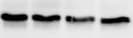
Western Blot
1 µg/mL
Sample: Recombinant Human DDR2 (Catalog # 2538-DR)
Sample: Recombinant Human DDR2 (Catalog # 2538-DR)
Reviewed Applications
Read 1 review rated 5 using MAB2538 in the following applications:
Formulation, Preparation, and Storage
Purification
Protein A or G purified from hybridoma culture supernatant
Reconstitution
Reconstitute at 0.5 mg/mL in sterile PBS. For liquid material, refer to CoA for concentration.
Loading...
Formulation
Lyophilized from a 0.2 μm filtered solution in PBS with Trehalose. *Small pack size (SP) is supplied either lyophilized or as a 0.2 µm filtered solution in PBS.
Shipping
Lyophilized product is shipped at ambient temperature. Liquid small pack size (-SP) is shipped with polar packs. Upon receipt, store immediately at the temperature recommended below.
Stability & Storage
Use a manual defrost freezer and avoid repeated freeze-thaw cycles.
- 12 months from date of receipt, -20 to -70 °C as supplied.
- 1 month, 2 to 8 °C under sterile conditions after reconstitution.
- 6 months, -20 to -70 °C under sterile conditions after reconstitution.
Calculators
Background: DDR2
References
- Vogel, W.F. et al. (2006) Cell. Signal. 18:1108.
- Karn, T. et al. (1993) Oncogene 8:3433.
- Vogel, W. et al. (1997) Mol. Cell 1:13.
- Leitinger, B. (2003) J. Biol. Chem. 278:16761.
- Leitinger, B. and A.P.L Kwan (2006) Matrix Biol. 25:355.
- Leitinger, B. et al. (2004) J. Mol. Biol. 344:993.
- Mihai, C. et al. (2006) J. Mol. Biol. 361:864.
- Olaso, E. et al. (2001) J. Clin. Invest. 108:1369.
- Ikeda, K. et al. (2002) J. Biol. Chem. 277:19206.
- Xu, L. et al. (2005) J. Biol. Chem. 280:548.
- Wang, J. et al. (2002) J. Autoimmun. 19:161.
- Ferri, N. et al. (2004) Am. J. Pathol. 164:1575.
Long Name
Discoidin Domain Receptor 2
Alternate Names
TKT, Trk3, Tyro-10
Gene Symbol
DDR2
UniProt
Additional DDR2 Products
Product Documents for Human DDR2 Antibody (290814)
Certificate of Analysis
To download a Certificate of Analysis, please enter a lot or batch number in the search box below.
Note: Certificate of Analysis not available for kit components.
Product Specific Notices for Human DDR2 Antibody (290814)
For research use only
Related Research Areas
Citations for Human DDR2 Antibody (290814)
 Powered by Bioz
Customer Reviews for Human DDR2 Antibody (290814) (1)
5 out of 5
1 Customer Rating
Have you used Human DDR2 Antibody (290814)?
Submit a review and receive an Amazon gift card!
$25/€18/£15/$25CAN/¥2500 Yen for a review with an image
$10/€7/£6/$10CAN/¥1110 Yen for a review without an image
Submit a review
Customer Images
Showing
1
-
1 of
1 review
Showing All
Filter By:
-
Application: Western BlotSample Tested: joint tissueSpecies: HumanVerified Customer | Posted 10/25/2021

There are no reviews that match your criteria.
Protocols
Find general support by application which include: protocols, troubleshooting, illustrated assays, videos and webinars.
- Antigen Retrieval Protocol (PIER)
- Antigen Retrieval for Frozen Sections Protocol
- Appropriate Fixation of IHC/ICC Samples
- Cellular Response to Hypoxia Protocols
- Chromogenic IHC Staining of Formalin-Fixed Paraffin-Embedded (FFPE) Tissue Protocol
- Chromogenic Immunohistochemistry Staining of Frozen Tissue
- ClariTSA™ Fluorophore Kits
- Detection & Visualization of Antibody Binding
- Fluorescent IHC Staining of Frozen Tissue Protocol
- Graphic Protocol for Heat-induced Epitope Retrieval
- Graphic Protocol for the Preparation and Fluorescent IHC Staining of Frozen Tissue Sections
- Graphic Protocol for the Preparation and Fluorescent IHC Staining of Paraffin-embedded Tissue Sections
- Graphic Protocol for the Preparation of Gelatin-coated Slides for Histological Tissue Sections
- IHC Sample Preparation (Frozen sections vs Paraffin)
- Immunofluorescent IHC Staining of Formalin-Fixed Paraffin-Embedded (FFPE) Tissue Protocol
- Immunohistochemistry (IHC) and Immunocytochemistry (ICC) Protocols
- Immunohistochemistry Frozen Troubleshooting
- Immunohistochemistry Paraffin Troubleshooting
- Preparing Samples for IHC/ICC Experiments
- Preventing Non-Specific Staining (Non-Specific Binding)
- Primary Antibody Selection & Optimization
- Protocol for Heat-Induced Epitope Retrieval (HIER)
- Protocol for Making a 4% Formaldehyde Solution in PBS
- Protocol for VisUCyte™ HRP Polymer Detection Reagent
- Protocol for the Preparation & Fixation of Cells on Coverslips
- Protocol for the Preparation and Chromogenic IHC Staining of Frozen Tissue Sections
- Protocol for the Preparation and Chromogenic IHC Staining of Frozen Tissue Sections - Graphic
- Protocol for the Preparation and Chromogenic IHC Staining of Paraffin-embedded Tissue Sections
- Protocol for the Preparation and Chromogenic IHC Staining of Paraffin-embedded Tissue Sections - Graphic
- Protocol for the Preparation and Fluorescent IHC Staining of Frozen Tissue Sections
- Protocol for the Preparation and Fluorescent IHC Staining of Paraffin-embedded Tissue Sections
- Protocol for the Preparation of Gelatin-coated Slides for Histological Tissue Sections
- R&D Systems Quality Control Western Blot Protocol
- TUNEL and Active Caspase-3 Detection by IHC/ICC Protocol
- The Importance of IHC/ICC Controls
- Troubleshooting Guide: Immunohistochemistry
- Troubleshooting Guide: Western Blot Figures
- Western Blot Conditions
- Western Blot Protocol
- Western Blot Protocol for Cell Lysates
- Western Blot Troubleshooting
- Western Blot Troubleshooting Guide
- View all Protocols, Troubleshooting, Illustrated assays and Webinars
Loading...
Associated Pathways
