DNAX accessory molecule-1 (DNAM-1), also known as CD226, is a 65 kDa type I transmembrane glycoprotein in the immunoglobulin superfamily (1). Mature human DNAM-1 contains a 236 amino acid (aa) extracellular domain (ECD) with two Ig-like C2-set domains and a 61 aa cytoplasmic region that contains motifs for binding PDZ domains and band 4.1 family proteins (1, 2). Within the ECD, human DNAM-1 shares 50% and 52% aa sequence identity with mouse and rat DNAM-1, respectively. DNAM-1 is expressed on multiple lymphoid and myeloid cell types and interacts with CD155/PVR and Nectin-2/CD112 (3, 4). Ligation of DNAM-1 promotes the activation of NK cells, CD8+ T cells, and mast cells (2‑6), dendritic cell maturation, megakaryocyte and activated platelet adhesion to vascular endothelial cells, and monocyte extravasation; it inhibits the formation of osteoclasts (7‑10). Platelet-endothelium interactions mediated by DNAM-1 enable the metastasis of tumor cells to the lung (11). In activated, but not in resting NK, T, and mast cells, the cis association of DNAM-1 with CD18 contributes to the tyrosine and serine phosphorylation of DNAM-1 during activation (6, 9, 12‑14).
Human DNAM-1/CD226 Antibody (102511)
R&D Systems | Catalog # MAB666

Key Product Details
Species Reactivity
Validated:
Human
Cited:
Human, Mouse
Applications
Validated:
Western Blot, Neutralization, Flow Cytometry, CyTOF-ready
Cited:
Neutralization, Flow Cytometry, Bioassay
Label
Unconjugated
Antibody Source
Monoclonal Mouse IgG1 Clone # 102511
Loading...
Product Specifications
Immunogen
Mouse myeloma cell line NS0-derived recombinant human DNAM-1/CD226
Glu19-Asn247 (predicted)
Accession # Q15762
Glu19-Asn247 (predicted)
Accession # Q15762
Specificity
Detects human DNAM-1/CD226 in direct ELISAs and Western blots.
Clonality
Monoclonal
Host
Mouse
Isotype
IgG1
Endotoxin Level
<0.10 EU per 1 μg of the antibody by the LAL method.
Scientific Data Images for Human DNAM-1/CD226 Antibody (102511)
Detection of DNAM-1/CD226 in Human PBMCs by Flow Cytometry.
Human peripheral blood mononuclear cells (PBMCs) gated on CD3-cells were stained with Mouse Anti-Human NCAM-1/CD56 APC-conjugated Monoclonal Antibody (Catalog # FAB2408A) and either (A) Mouse Anti-Human DNAM-1/CD226 Monoclonal Antibody (Catalog # MAB666) or (B) Mouse IgG1Isotype Control (Catalog # MAB002) followed by Phycoerythrin-conjugated Anti-Mouse IgG Secondary Antibody (Catalog # F0102B).
Neutralization of DNAM-1/CD226 Fc Chimera by Human DNAM-1/CD226 Antibody
In a functional ELISA, Human DNAM-1/CD226 Antibody (Catalog # MAB666) blocks the binding of Recombinant Human DNAM-1/CD226 Fc Chimera (666-DN) to Biotinylated Recombinant Human CD155/PVR Fc Chimera Avi-tag (AVI9174). The Neutralization Dose (ND50) for this effect is typically 0.300-6.00 µg/mL.Applications for Human DNAM-1/CD226 Antibody (102511)
Application
Recommended Usage
CyTOF-ready
Ready to be labeled using established conjugation methods. No BSA or other carrier proteins that could interfere with conjugation.
Flow Cytometry
0.25 µg/106 cells
Sample: Human peripheral blood mononuclear cells (PBMCs) gated on CD3- cells
Sample: Human peripheral blood mononuclear cells (PBMCs) gated on CD3- cells
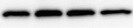
Western Blot
1 µg/mL
Sample: Recombinant Human DNAM-1/CD226 Fc Chimera (Catalog # 666-DN) under non-reducing conditions only
Sample: Recombinant Human DNAM-1/CD226 Fc Chimera (Catalog # 666-DN) under non-reducing conditions only
Neutralization
In a functional ELISA, Human
DNAM-1/CD226 Antibody (Catalog # MAB666) blocks the binding of Recombinant
Human DNAM-1/CD226 Fc Chimera (Catalog # 666-DN) to Biotinylated Recombinant
Human CD155/PVR Fc Chimera Avi-tag (Catalog # AVI9174). The Neutralization Dose
(ND50) for this effect is typically 0.300-6.00 µg/mL.
Reviewed Applications
Read 1 review rated 5 using MAB666 in the following applications:
Flow Cytometry Panel Builder
Bio-Techne Knows Flow Cytometry
Save time and reduce costly mistakes by quickly finding compatible reagents using the Panel Builder Tool.
Advanced Features
- Spectra Viewer - Custom analysis of spectra from multiple fluorochromes
- Spillover Popups - Visualize the spectra of individual fluorochromes
- Antigen Density Selector - Match fluorochrome brightness with antigen density

Formulation, Preparation, and Storage
Purification
Protein A or G purified from ascites
Reconstitution
Reconstitute at 0.5 mg/mL in sterile PBS. For liquid material, refer to CoA for concentration.
Loading...
Formulation
Lyophilized from a 0.2 μm filtered solution in PBS with Trehalose. See Certificate of Analysis for details.
*Small pack size (-SP) is supplied either lyophilized or as a 0.2 µm filtered solution in PBS.
*Small pack size (-SP) is supplied either lyophilized or as a 0.2 µm filtered solution in PBS.
Shipping
Lyophilized product is shipped at ambient temperature. Liquid small pack size (-SP) is shipped with polar packs. Upon receipt, store immediately at the temperature recommended below.
Stability & Storage
Use a manual defrost freezer and avoid repeated freeze-thaw cycles.
- 12 months from date of receipt, -20 to -70 °C as supplied.
- 1 month, 2 to 8 °C under sterile conditions after reconstitution.
- 6 months, -20 to -70 °C under sterile conditions after reconstitution.
Calculators
Background: DNAM-1/CD226
References
- Fuchs, A. and M. Colonna (2006) Semin. Cancer Biol. 16:359.
- Shibuya, A. et al. (1996) Immunity 4:573.
- Bottino, C. et al. (2003) J. Exp. Med. 198:557.
- Tahara-Hanaoka, S. et al. (2004) Int. Immunol. 16:533.
- Dardalhon, V. et al. (2005) J. Immunol. 175:1558.
- Bachelet, I. et al. (2006) J. Biol. Chem. 281:27190.
- Reymond, N. et al. (2004) J. Exp. Med. 199:1331.
- Kakehi, S. et al. (2007) Mol. Cell. Biochem. 301:209.
- Kojima, H. et al. (2003) J. Biol. Chem. 278:36748.
- Tahara-Hanaoka, S. et al. (2006) Blood 107:1491.
- Morimoto, K. et al. (2007) Oncogene July 16 epub.
- Shibuya, K. et al. (1999) Immunity 11:615.
- Shibuya, K. et al. (2003) J. Exp. Med. 198:1829.
- Shibuya, A. et al. (1998) J. Immunol. 166:1671.
Long Name
DNAX Accessory Molecule 1
Alternate Names
CD226, DNAM1, PTA1, TLiSA1
Gene Symbol
CD226
UniProt
Additional DNAM-1/CD226 Products
Product Documents for Human DNAM-1/CD226 Antibody (102511)
Certificate of Analysis
To download a Certificate of Analysis, please enter a lot or batch number in the search box below.
Note: Certificate of Analysis not available for kit components.
Product Specific Notices for Human DNAM-1/CD226 Antibody (102511)
For research use only
Citations for Human DNAM-1/CD226 Antibody (102511)
 Powered by Bioz
Customer Reviews for Human DNAM-1/CD226 Antibody (102511) (1)
5 out of 5
1 Customer Rating
Have you used Human DNAM-1/CD226 Antibody (102511)?
Submit a review and receive an Amazon gift card!
$25/€18/£15/$25CAN/¥2500 Yen for a review with an image
$10/€7/£6/$10CAN/¥1110 Yen for a review without an image
Submit a review
Customer Images
Showing
1
-
1 of
1 review
Showing All
Filter By:
-
Application: Western BlotSample Tested: Mesenchymal stromal cellsSpecies: HumanVerified Customer | Posted 10/24/2021Mesenchymal stromal cellsprimary antibody dilution was 1:1000, secondary 1:4000

There are no reviews that match your criteria.
Protocols
Find general support by application which include: protocols, troubleshooting, illustrated assays, videos and webinars.
- 7-Amino Actinomycin D (7-AAD) Cell Viability Flow Cytometry Protocol
- Cellular Response to Hypoxia Protocols
- Extracellular Membrane Flow Cytometry Protocol
- Flow Cytometry Protocol for Cell Surface Markers
- Flow Cytometry Protocol for Staining Membrane Associated Proteins
- Flow Cytometry Staining Protocols
- Flow Cytometry Troubleshooting Guide
- Intracellular Flow Cytometry Protocol Using Alcohol (Methanol)
- Intracellular Flow Cytometry Protocol Using Detergents
- Intracellular Nuclear Staining Flow Cytometry Protocol Using Detergents
- Intracellular Staining Flow Cytometry Protocol Using Alcohol Permeabilization
- Intracellular Staining Flow Cytometry Protocol Using Detergents to Permeabilize Cells
- Propidium Iodide Cell Viability Flow Cytometry Protocol
- Protocol for Liperfluo
- Protocol for the Characterization of Human Th22 Cells
- Protocol for the Characterization of Human Th9 Cells
- Protocol: Annexin V and PI Staining by Flow Cytometry
- Protocol: Annexin V and PI Staining for Apoptosis by Flow Cytometry
- R&D Systems Quality Control Western Blot Protocol
- Troubleshooting Guide: Fluorokine Flow Cytometry Kits
- Troubleshooting Guide: Western Blot Figures
- Western Blot Conditions
- Western Blot Protocol
- Western Blot Protocol for Cell Lysates
- Western Blot Troubleshooting
- Western Blot Troubleshooting Guide
- View all Protocols, Troubleshooting, Illustrated assays and Webinars
Loading...
Associated Pathways
