EphA2, also known as Eck, Myk2, and Sek2, is a member of the Eph receptor tyrosine kinase family which binds Ephrins A1, 2, 3, 4, and 5 (1, 2, 3, 4). A and B class Eph proteins have a common structural organization. The human EphA2 cDNA encodes a 976 amino acid (aa) precursor including a 24 aa signal sequence, a 510 aa extracellular domain (ECD), a 24 aa transmembrane segment, and a 418 aa cytoplasmic domain. The ECD contains an N-terminal globular domain, a cysteine-rich domain, and two fibronectin type III domains (5). The cytoplasmic domain contains a juxtamembrane motif with two tyrosine residues, which are the major autophosphorylation sites, a kinase domain, and a sterile alpha motif (SAM) (5). The ECD of human EphA2 shares 90‑94% aa sequence identity with mouse, bovine, and canine EphA2, and approximately 45% aa sequence identity with human EphA1, 3, 4, 5, 7, and 8. EphA2 becomes autophosphorylated following ligand binding (6, 7) and then interacts with SH2 domain-containing PI3-kinase to activate MAPK pathways (8, 9). Reverse signaling is also propagated through the Ephrin ligand. Transcription of EphA2 is dependent on the expression of E-Cadherin (10), and can be induced by p53 family transcription factors (11). EphA2 is upregulated in breast, prostate, and colon cancer vascular endothelium. Its ligand, EphrinA1, is expressed by the local tumor cells (12, 13). In some cases, EphA2 and EphrinA1 are expressed on the same blood vessels (14). EphA2 signaling cooperates with VEGF receptor signaling in promoting endothelial cell migration (13). The gene encoding human EphA2 maps to a region on chromosome 1 which is frequently deleted in neuroectodermal tumors (15).

Key Product Details
Validated by
Knockout/Knockdown
Species Reactivity
Validated:
Human
Cited:
Human, Recombinant Protein, Yeast
Applications
Validated:
Knockout Validated, Flow Cytometry, Immunocytochemistry, CyTOF-ready
Cited:
Immunohistochemistry, Western Blot, Flow Cytometry, ELISA Capture
Label
Unconjugated
Antibody Source
Monoclonal Mouse IgG2A Clone # 371805
Loading...
Product Specifications
Immunogen
Mouse myeloma cell line NS0-derived recombinant human EphA2
Gln25-Asn534
Accession # P29317
Gln25-Asn534
Accession # P29317
Specificity
Detects human EphA2 in direct ELISAs and Western blots. In direct ELISAs, no cross-reactivity with recombinant mouse EphA4, A5, A6, A7, A8, or recombinant rat EphB1 is observed.
Clonality
Monoclonal
Host
Mouse
Isotype
IgG2A
Scientific Data Images for Human EphA2 Antibody (371805)
Detection of EphA2 in A431 Human Cell Line by Flow Cytometry.
A431 human epithelial carcinoma cell line was stained with Mouse Anti-Human EphA2 Monoclonal Antibody (Catalog # MAB3035, filled histogram) or isotype control antibody (Catalog # MAB003, open histogram), followed by Phycoerythrin-conjugated Anti-Mouse IgG Secondary Antibody (Catalog # F0102B). View our protocol for Staining Membrane-associated Proteins.
EphA2 in A431 Human Cell Line.
EphA2 was detected in immersion fixed A431 human epithelial carcinoma cell line using Mouse Anti-Human EphA2 Monoclonal Antibody (Catalog # MAB3035) at 10 µg/mL for 3 hours at room temperature. Cells were stained using the NorthernLights™ 557-conjugated Anti-Mouse IgG Secondary Antibody (red; Catalog # NL007) and counterstained with DAPI(blue). Specific staining was localized to the cell surface. View our protocol for Fluorescent ICC Staining of Cells on Coverslips.
EphA2 Specificity is Shown by Immunocytochemistry in Knockout Cell Line.
EphA2 was detected in immersion fixed A431 human epithelial carcinoma cell line but is not detected in EphA2 knockout (KO) A431 Human Cell Line cell line using Mouse Anti-Human EphA2 Monoclonal Antibody (Catalog # MAB3035) at 5 µg/mL for 3 hours at room temperature. Cells were stained using the NorthernLights™ 493-conjugated Anti-Mouse IgG Secondary Antibody (green; Catalog # NL009) and counterstained with DAPI (blue). Specific staining was localized to cell membranes. View our protocol for Fluorescent ICC Staining of Cells on Coverslips.
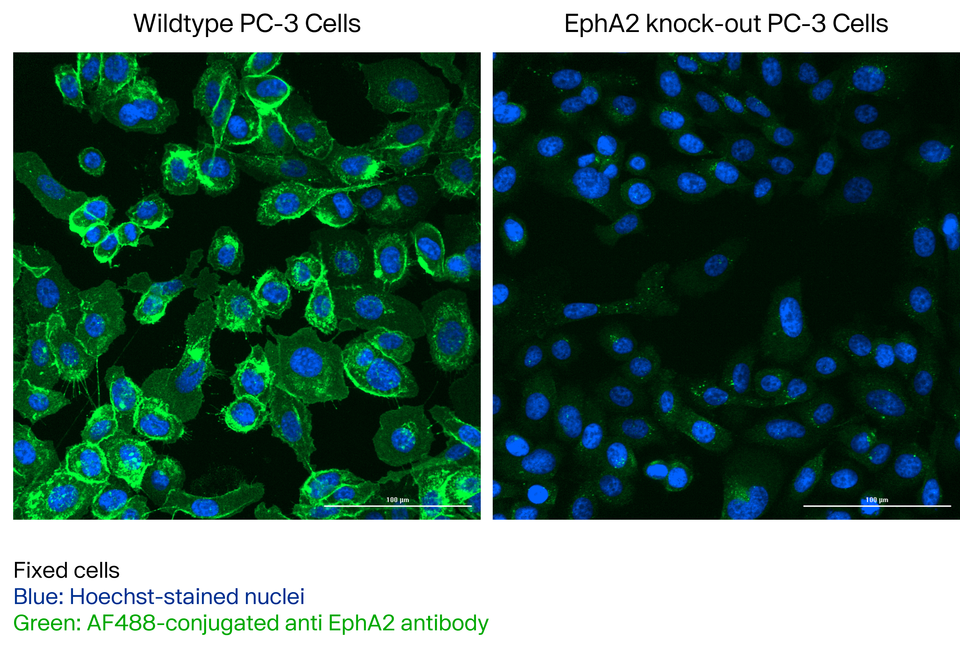
EphA2 in PC-3 Human Prostate Cancer Cell Line.
Fixed PC-3 cell stained with MAB3035 and detected with a secondary anti-mouse AlexaFluor488-conjugated antibody. Left: wildtype PC-3 cells (EphA2 expressing); right: PC-3 cells EphA2 Knock-out. Image from a verified customer review.
Detection of EphA2 by Flow Cytometry
Identification of non-competing anti-EphA2 mAbs.(A) Histograms for EphA2-positive cell lines HEC-1-A and PC-3 first stained with unlabeled EphA2 mAb 1C1 or 3035, an IgG control, or buffer only, followed by staining with either labeled 1C1 (1C1-A488) or 3035 (3035-A488). A488 fluorescence is detected by conducting flow cytometry on 10,000 live, single cells. (B) Percent staining by 1C1-A488 or 3035-A488 relative to the HEC-1-A or PC-3 samples first incubated with buffer only. Image collected and cropped by CiteAb from the following open publication (https://pubmed.ncbi.nlm.nih.gov/25894652), licensed under a CC-BY license. Not internally tested by R&D Systems.Applications for Human EphA2 Antibody (371805)
Application
Recommended Usage
CyTOF-ready
Ready to be labeled using established conjugation methods. No BSA or other carrier proteins that could interfere with conjugation.
Flow Cytometry
0.25 µg/106 cells
Sample: A431 human epithelial carcinoma cell line
Sample: A431 human epithelial carcinoma cell line
Immunocytochemistry
8-25 µg/mL
Sample: Immersion fixed A431 human epithelial carcinoma cell line
Sample: Immersion fixed A431 human epithelial carcinoma cell line
Knockout Validated
EphA2 was detected in immersion fixed A431 human cell line but is not detected in EphA2 knockout (KO) A431 human cell line.
Reviewed Applications
Read 4 reviews rated 4.5 using MAB3035 in the following applications:
Flow Cytometry Panel Builder
Bio-Techne Knows Flow Cytometry
Save time and reduce costly mistakes by quickly finding compatible reagents using the Panel Builder Tool.
Advanced Features
- Spectra Viewer - Custom analysis of spectra from multiple fluorochromes
- Spillover Popups - Visualize the spectra of individual fluorochromes
- Antigen Density Selector - Match fluorochrome brightness with antigen density

Formulation, Preparation, and Storage
Purification
Protein A or G purified from hybridoma culture supernatant
Reconstitution
Reconstitute at 0.5 mg/mL in sterile PBS. For liquid material, refer to CoA for concentration.
Loading...
Formulation
Lyophilized from a 0.2 μm filtered solution in PBS with Trehalose. *Small pack size (SP) is supplied either lyophilized or as a 0.2 µm filtered solution in PBS.
Shipping
Lyophilized product is shipped at ambient temperature. Liquid small pack size (-SP) is shipped with polar packs. Upon receipt, store immediately at the temperature recommended below.
Stability & Storage
Use a manual defrost freezer and avoid repeated freeze-thaw cycles.
- 12 months from date of receipt, -20 to -70 °C as supplied.
- 1 month, 2 to 8 °C under sterile conditions after reconstitution.
- 6 months, -20 to -70 °C under sterile conditions after reconstitution.
Calculators
Background: EphA2
References
- Poliakov, A. et al. (2004) Dev. Cell 7:465.
- Surawska, H. et al. (2004) Cytokine Growth Factor Rev. 15:419.
- Pasquale, E.B. (2005) Nat. Rev. Mol. Cell Biol. 6:462.
- Davy, A. and P. Soriano (2005) Dev. Dyn. 232:1.
- Bohme, B et al. (1993) Oncogene 8:2857.
- Pandey, A. et al. (1995) Science 268:567.
- Bartley, T.D. et al. (1994) Nature 368:558.
- Pandey, A. et al. (1994) J. Biol. Chem. 269:30154.
- Miao, H. et al. (2001) Nat. Cell Biol. 3:527.
- Orsulic, S. and R. Kemler (2000) J. Cell Sci. 113:1793.
- Dohn, M. et al. (2001) Oncogene 20:6503.
- Zelinski, D.P. et al. (2001) Cancer Res. 61:2301.
- Brantley, D.M. et al. (2002) Oncogene 21:7011.
- Ogawa, K. et al. (2000) Oncogene 19:6043.
- Sulman, E.P. et al. (1997) Genomics 40:371.
Alternate Names
Eck, Myk2, Sek2
Gene Symbol
EPHA2
UniProt
Additional EphA2 Products
Product Documents for Human EphA2 Antibody (371805)
Certificate of Analysis
To download a Certificate of Analysis, please enter a lot or batch number in the search box below.
Note: Certificate of Analysis not available for kit components.
Product Specific Notices for Human EphA2 Antibody (371805)
For research use only
Related Research Areas
Citations for Human EphA2 Antibody (371805)
 Powered by Bioz
Customer Reviews for Human EphA2 Antibody (371805) (4)
4.5 out of 5
4 Customer Ratings
Have you used Human EphA2 Antibody (371805)?
Submit a review and receive an Amazon gift card!
$25/€18/£15/$25CAN/¥2500 Yen for a review with an image
$10/€7/£6/$10CAN/¥1110 Yen for a review without an image
Submit a review
Customer Images

Showing
1
-
4 of
4 reviews
Showing All
Filter By:
-
Application: ImmunocytochemistrySample Tested: PC-3 human prostate cancer cell lineSpecies: HumanVerified Customer | Posted 08/19/2025Fixed PC-3 cell stained with MAB3035 and detected with a secondary anti-mouse AlexaFluor488-conjugated antibody. Left: wildtype PC-3 cells (EphA2 expressing); right: PC-3 cells EphA2 Knock-out.Cells were fixed with 4% PFA for 20 minutes at room temperature and then permeabilised with Triton X-100 0.2%. The cells were stained with MAB3035 diluted 1:200 in PBS (containing 3% BSA) for 1 hour at room temperature. The antibody was then detected using a fluorescent anti-mouse secondary antibody.

-
Application: ELISASample Tested: Serum and PlasmaSpecies: HumanVerified Customer | Posted 07/05/2022
-
Application: Western BlotSample Tested: See PMID 21245385Species: HumanVerified Customer | Posted 02/19/2015
-
Application: Western BlotSample Tested: See PMID 24036371Species: OtherVerified Customer | Posted 02/19/2015
There are no reviews that match your criteria.
Protocols
Find general support by application which include: protocols, troubleshooting, illustrated assays, videos and webinars.
- 7-Amino Actinomycin D (7-AAD) Cell Viability Flow Cytometry Protocol
- Appropriate Fixation of IHC/ICC Samples
- Cellular Response to Hypoxia Protocols
- ClariTSA™ Fluorophore Kits
- Detection & Visualization of Antibody Binding
- Extracellular Membrane Flow Cytometry Protocol
- Flow Cytometry Protocol for Cell Surface Markers
- Flow Cytometry Protocol for Staining Membrane Associated Proteins
- Flow Cytometry Staining Protocols
- Flow Cytometry Troubleshooting Guide
- ICC Cell Smear Protocol for Suspension Cells
- ICC Immunocytochemistry Protocol Videos
- ICC for Adherent Cells
- Immunocytochemistry (ICC) Protocol
- Immunocytochemistry Troubleshooting
- Immunofluorescence of Organoids Embedded in Cultrex Basement Membrane Extract
- Immunohistochemistry (IHC) and Immunocytochemistry (ICC) Protocols
- Intracellular Flow Cytometry Protocol Using Alcohol (Methanol)
- Intracellular Flow Cytometry Protocol Using Detergents
- Intracellular Nuclear Staining Flow Cytometry Protocol Using Detergents
- Intracellular Staining Flow Cytometry Protocol Using Alcohol Permeabilization
- Intracellular Staining Flow Cytometry Protocol Using Detergents to Permeabilize Cells
- Preparing Samples for IHC/ICC Experiments
- Preventing Non-Specific Staining (Non-Specific Binding)
- Primary Antibody Selection & Optimization
- Propidium Iodide Cell Viability Flow Cytometry Protocol
- Protocol for Liperfluo
- Protocol for VisUCyte™ HRP Polymer Detection Reagent
- Protocol for the Characterization of Human Th22 Cells
- Protocol for the Characterization of Human Th9 Cells
- Protocol for the Fluorescent ICC Staining of Cell Smears - Graphic
- Protocol for the Fluorescent ICC Staining of Cultured Cells on Coverslips - Graphic
- Protocol for the Preparation and Fluorescent ICC Staining of Cells on Coverslips
- Protocol for the Preparation and Fluorescent ICC Staining of Non-adherent Cells
- Protocol for the Preparation and Fluorescent ICC Staining of Stem Cells on Coverslips
- Protocol for the Preparation of a Cell Smear for Non-adherent Cell ICC - Graphic
- Protocol: Annexin V and PI Staining by Flow Cytometry
- Protocol: Annexin V and PI Staining for Apoptosis by Flow Cytometry
- TUNEL and Active Caspase-3 Detection by IHC/ICC Protocol
- The Importance of IHC/ICC Controls
- Troubleshooting Guide: Fluorokine Flow Cytometry Kits
- View all Protocols, Troubleshooting, Illustrated assays and Webinars
Loading...