Growth Differentiation Factor 15 (GDF-15), also called Macrophage inhibitory cytokine-1 (MIC-1), placental transforming growth factor-beta, prostate-derived factor, and placental bone morphogenetic protein, is a divergent member of the transforming growth factor beta (TGF-beta ) superfamily. GDF-15 is highly expressed in placenta and is expressed at lower levels in kidney, pancreas, prostate and colon. It is also widely expressed in brain. Similar to other TGF-beta family proteins, GDF-15 is synthesized as a large precursor protein that is cleaved at the dibasic cleavage site (RXXR) to release the carboxy-terminal domain. The carboxy-terminal domain of GDF-15 contains the characteristic seven conserved cysteine residues necessary for the formation of the cysteine knot and the single interchain disulfide bond. Furthermore, the carboxy-terminal domain contains two additional cysteine residues that form a fourth intrachain disulfide bond. Biologically active GDF-15 is a disulfide-linked homodimer of the carboxy-terminal 112 amino acid residues. Mature human GDF-15 shares 66.1% and 68.7% amino acid sequence similarity with rat and mouse GDF-15, respectively, which are remarkably low homologies between species in TGF-beta superfamily. GDF-15 has been shown to have various functions, including inhibition of production of tumor necrosis factor alpha (TNF-alpha ) from lipopolysaccharide-stimulated macrophages, induction of cartilage formation, early-stage endochonadal bone formation, and promotion of neuronal survival.

Key Product Details
Species Reactivity
Validated:
Human
Cited:
Human
Applications
Validated:
Western Blot, Simple Western
Cited:
Immunohistochemistry, Immunohistochemistry-Paraffin, Western Blot, Neutralization, Immunocytochemistry, ELISA Development
Label
Unconjugated
Antibody Source
Polyclonal Goat IgG
Loading...
Product Specifications
Immunogen
Chinese hamster ovary cell line CHO-derived recombinant human GDF-15
Ala197-Ile308
Accession # Q99988
Ala197-Ile308
Accession # Q99988
Specificity
Detects human GDF-15 in direct ELISAs and Western blots. In direct ELISAs and Western blots, approximately 65% cross-reactivity with recombinant mouse (rm) GDF-15 is observed, and less than 1% cross-reactivity with rmGDF‑1, rmGDF-3, and rmGDF-11 is observed.
Clonality
Polyclonal
Host
Goat
Isotype
IgG
Scientific Data Images for Human GDF-15 Antibody
Detection of Human GDF‑15 by Western Blot.
Western blot shows lysate of HT1080 human fibrosarcoma cell line. PVDF membrane was probed with 0.5 µg/mL of Goat Anti-Human GDF-15 Antigen Affinity-purified Polyclonal Antibody (Catalog # AF957) followed by HRP-conjugated Anti-Goat IgG Secondary Antibody (Catalog # HAF017). A specific band was detected for GDF-15 at approximately 35 kDa (as indicated). This experiment was conducted under reducing conditions and using Immunoblot Buffer Group 1.
Detection of Human GDF‑15 by Simple WesternTM.
Simple Western lane view shows lysate of HT1080 human fibrosarcoma cell line, loaded at 0.2 mg/mL. A specific band was detected for GDF-15 at approximately 47 kDa (as indicated) using 5 µg/mL of Goat Anti-Human GDF-15 Antigen Affinity-purified Polyclonal Antibody (Catalog # AF957) followed by 1:50 dilution of HRP-conjugated Anti-Goat IgG Secondary Antibody (Catalog # HAF109). This experiment was conducted under reducing conditions and using the 12-230 kDa separation system.
Detection of Human GDF-15 by Western Blot
Upregulation of GDF-15 by hypoxia in endothelial cells. Human pulmonary microvascular endothelial cells (HPMEC) were subjected to hypoxia for various time periods (2 h to 24 h). The mRNA and protein levels of GDF-15 (secreted form) were determined either by quantitative RT-PCR (panel A), immunoradiometric sandwich assay - IRMA (panel B) or Western Blot analysis (panel C). Hypoxia increased GDF-15 expression in a time dependent manner, which was initially detected after 2 hours on mRNA level and after 4 hours on protein level. Data from n = 4 each group are shown as mean ± SD. *p < 0.05 compared to control. Image collected and cropped by CiteAb from the following open publication (https://pubmed.ncbi.nlm.nih.gov/21548946), licensed under a CC-BY license. Not internally tested by R&D Systems.
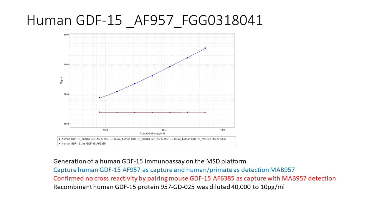
Human GDF-15 ELISA Standard Curve
Recombinant Human GDF‑15 (Catalog # 957-GD) was serially diluted and captured by Mouse Anti-Human/Primate GDF‑15 Monoclonal Antibody (Catalog # MAB957) coated on a Clear Polystyrene Microplate (Catalog # DY990). Goat Anti-Human GDF‑15 Antigen Affinity-purified Polyclonal Antibody (Catalog # AF957) was biotinylated and incubated with the protein captured on the plate. Detection of the standard curve was achieved by incubating Streptavidin-HRP (Catalog # DY998)Applications for Human GDF-15 Antibody
Application
Recommended Usage
Simple Western
5 µg/mL
Sample: HT1080 human fibrosarcoma cell line
Sample: HT1080 human fibrosarcoma cell line
Western Blot
0.5 µg/mL
Sample: HT1080 human fibrosarcoma cell line
Sample: HT1080 human fibrosarcoma cell line
Reviewed Applications
Read 2 reviews rated 5 using AF957 in the following applications:
Formulation, Preparation, and Storage
Purification
Antigen Affinity-purified
Reconstitution
Reconstitute at 0.2 mg/mL in sterile PBS. For liquid material, refer to CoA for concentration.
Loading...
Formulation
Lyophilized from a 0.2 μm filtered solution in PBS with Trehalose. *Small pack size (SP) is supplied either lyophilized or as a 0.2 µm filtered solution in PBS.
Shipping
Lyophilized product is shipped at ambient temperature. Liquid small pack size (-SP) is shipped with polar packs. Upon receipt, store immediately at the temperature recommended below.
Stability & Storage
Use a manual defrost freezer and avoid repeated freeze-thaw cycles.
- 12 months from date of receipt, -20 to -70 °C as supplied.
- 1 month, 2 to 8 °C under sterile conditions after reconstitution.
- 6 months, -20 to -70 °C under sterile conditions after reconstitution.
Calculators
Background: GDF-15
References
- Bootcov, M.R. et al. (1997) Proc. Natl. Acad. Sci. USA 94:11514.
- Böttner, M. et al. (1999) Gene 237:105.
- Fairlie, W.D. et al. (1998) J. Leukoc. Biol 65:2.
- Fairlie, W.D. et al. (2001) J B.C 20:16911.
- Bauskin, A.R. et al. (2000) EMBO J. 19:2212.
- Strelau, J. et al. (2000) J. Neurosci. 20:8597.
- Schober, A. et al. (2001) J. Comp. Neurol. 439:32.
Long Name
Growth Differentiation Factor 15
Alternate Names
GDF15, MIC-1, NAG-1, PDF, PLAB, PTGF-beta
Gene Symbol
GDF15
UniProt
Additional GDF-15 Products
Product Documents for Human GDF-15 Antibody
Certificate of Analysis
To download a Certificate of Analysis, please enter a lot or batch number in the search box below.
Note: Certificate of Analysis not available for kit components.
Product Specific Notices for Human GDF-15 Antibody
For research use only
Related Research Areas
Citations for Human GDF-15 Antibody
 Powered by Bioz
Customer Reviews for Human GDF-15 Antibody (2)
5 out of 5
2 Customer Ratings
Have you used Human GDF-15 Antibody?
Submit a review and receive an Amazon gift card!
$25/€18/£15/$25CAN/¥2500 Yen for a review with an image
$10/€7/£6/$10CAN/¥1110 Yen for a review without an image
Submit a review
Customer Images
Showing
1
-
2 of
2 reviews
Showing All
Filter By:
-
Application: ELISASample Tested: Recombinant proteinSpecies: HumanVerified Customer | Posted 07/08/2020

-
Application: ELISASample Tested: EDTA PlasmaSpecies: HumanVerified Customer | Posted 12/20/2017
There are no reviews that match your criteria.
Protocols
Find general support by application which include: protocols, troubleshooting, illustrated assays, videos and webinars.
- Cellular Response to Hypoxia Protocols
- R&D Systems Quality Control Western Blot Protocol
- Troubleshooting Guide: Western Blot Figures
- Western Blot Conditions
- Western Blot Protocol
- Western Blot Protocol for Cell Lysates
- Western Blot Troubleshooting
- Western Blot Troubleshooting Guide
- View all Protocols, Troubleshooting, Illustrated assays and Webinars
Loading...