Growth Differentiation Factor 15 (GDF-15), also called Macrophage inhibitory cytokine-1 (MIC-1), placental transforming growth factor-beta, prostate-derived factor, and placental bone morphogenetic protein, is a divergent member of the transforming growth factor beta (TGF-beta ) superfamily. GDF-15 is highly expressed in placenta and is expressed at lower levels in kidney, pancreas, prostate and colon. It is also widely expressed in brain. Similarly to other TGF-beta family proteins, GDF-15 is synthesized as a large precursor protein that is cleaved at the dibasic cleavage site (RXXR) to release the carboxy-terminal domain. The carboxy-terminal domain of GDF-15 contains the characteristic seven conserved cysteine residues necessary for the formation of the cysteine knot and the single interchain disulfide bond. Furthermore, the carboxy-terminal domain contains two additional cysteine residues that form a fourth intrachain disulfide bond. Biologically active GDF-15 is a disulfide-linked homodimer of the carboxy-terminal 112 amino acid residues. Mature human GDF-15 shares 66.1% and 68.7% amino acid sequence similarity with rat and mouse GDF-15, respectively, which are remarkably low homologies between species in TGF-beta superfamily. GDF-15 has been shown to have various functions, including inhibition of production of tumor necrosis factor alpha (TNF-alpha ) from lipopolysaccharide-stimulated macrophages, induction of cartilage formation, early-stage endochonadal bone formation, and promotion of neuronal survival.

Key Product Details
Species Reactivity
Validated:
Human, Primate
Cited:
Human, Mouse
Applications
Validated:
Western Blot, ELISA Capture (Matched Antibody Pair)
Cited:
Western Blot, Neutralization, ELISA Capture, ELISA Development, ELISA Development (Capture), In vivo assay, Immunoassay Development, Luminex Development
Label
Unconjugated
Antibody Source
Monoclonal Mouse IgG2B Clone # 147627
Loading...
Product Specifications
Immunogen
Chinese hamster ovary cell line CHO-derived recombinant human GDF-15
Ala197-Ile308
Accession # Q99988
Ala197-Ile308
Accession # Q99988
Specificity
Detects human GDF-15 in ELISAs and Western blots. In ELISAs, no cross-reactivity with recombinant human GDF‑11, recombinant mouse (rm) GDF‑5, rmGDF‑6, rmGDF‑7, or rmGDF‑8 is observed.
Clonality
Monoclonal
Host
Mouse
Isotype
IgG2B
Scientific Data Images for Human/Primate GDF‑15 Antibody
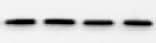
Detection of Human GDF-15 by Western Blot
AML cell lines highly express GDF15. a RT-qPCR analysis of different cytokines associated with the regulation of adipogenesis in AML cell lines (K562, THP-1 and HL-60). GAPDH was used as a housekeeping gene. *P < 0.05, ***P < 0.001. b and c, RT-qPCR (b) and Western blotting (c) analysis of GDF15 in different cell lines (Kasumi, HL-60, THP-1, K562 and HEL). The densitometry values of protein expression changes were indicated. beta -actin was used as an internal control for RT-qPCR and Western blotting analysis. d ELISA detection of GDF15 expression in the supernatant of THP-1 cells with different cell densities Image collected and cropped by CiteAb from the following open publication (https://pubmed.ncbi.nlm.nih.gov/29566722), licensed under a CC-BY license. Not internally tested by R&D Systems.
Detection of Human GDF-15 by Immunohistochemistry
The relationship of GDF15 expression and small marrow adipocytes in AML patients. a RT-qPCR analysis of GDF15 mRNA expression in BM from AML patients (n = 15) and the controls (n = 12). The results shown are from three independent experiments. *P < 0.05. b Western blotting analysis of GDF15 protein levels in BM from AML patients and the controls. The densitometry values of protein expression changes were indicated. beta -actin protein was used as an internal control for Western blotting analysis. c and d Representative confocal images showed the expression of GDF15 and leukemic cell markers CD34 (c) or CD117 (d) in BM sections of AML patients. DAPI was used to stain the nuclei. White triangles showed the leukemic cells with GDF15+. White arrows showed the non-leukemic cells with GDF15. Scale bar represents 40 μm. e and f Scatter plot showed the positive correlation of small adipocyte volume (e) or small adipocyte number (f) with the level of GDF15 in BM of AML (n = 20, R = 0.6679, P = 0.0013, or n = 20, R = 0.7205, P = 0.003, Spearman correlation test) Image collected and cropped by CiteAb from the following open publication (https://pubmed.ncbi.nlm.nih.gov/29566722), licensed under a CC-BY license. Not internally tested by R&D Systems.Applications for Human/Primate GDF‑15 Antibody
Application
Recommended Usage
Western Blot
1 µg/mL
Sample: Recombinant Human GDF‑15 (Catalog # 957-GD)
under non-reducing conditions only
Sample: Recombinant Human GDF‑15 (Catalog # 957-GD)
under non-reducing conditions only
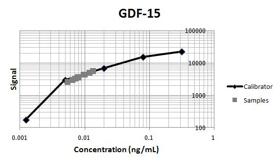
Human/Primate GDF-15 Sandwich Immunoassay
Please Note: Optimal dilutions of this antibody should be experimentally determined.
Reviewed Applications
Read 7 reviews rated 4.6 using MAB957 in the following applications:
Formulation, Preparation, and Storage
Purification
Protein A or G purified from hybridoma culture supernatant
Reconstitution
Reconstitute at 0.5 mg/mL in sterile PBS. For liquid material, refer to CoA for concentration.
Loading...
Formulation
Lyophilized from a 0.2 μm filtered solution in PBS with Trehalose. *Small pack size (SP) is supplied either lyophilized or as a 0.2 µm filtered solution in PBS.
Shipping
Lyophilized product is shipped at ambient temperature. Liquid small pack size (-SP) is shipped with polar packs. Upon receipt, store immediately at the temperature recommended below.
Stability & Storage
Use a manual defrost freezer and avoid repeated freeze-thaw cycles.
- 12 months from date of receipt, -20 to -70 °C as supplied.
- 1 month, 2 to 8 °C under sterile conditions after reconstitution.
- 6 months, -20 to -70 °C under sterile conditions after reconstitution.
Calculators
Background: GDF-15
References
- Bootcov, M.R. et al. (1997) Proc. Natl. Acad. Sci. USA 94:11514.
- Böttner, M. et al. (1999) Gene 237:105.
- Fairlie, W.D. et al. (1998) J. Leukoc. Biol 65:2.
- Fairlie, W.D. et al. (2001) J B.C 20:16911.
- Bauskin, A.R. et al. (2000) EMBO J. 19:2212.
- Strelau, J. et al. (2000) J. Neurosci. 20:8597.
- Schober, A. et al. (2001) J. Comp. Neurol. 439:32.
Long Name
Growth Differentiation Factor 15
Alternate Names
GDF15, MIC-1, NAG-1, PDF, PLAB, PTGF-beta
Gene Symbol
GDF15
UniProt
Additional GDF-15 Products
Product Documents for Human/Primate GDF‑15 Antibody
Certificate of Analysis
To download a Certificate of Analysis, please enter a lot or batch number in the search box below.
Note: Certificate of Analysis not available for kit components.
Product Specific Notices for Human/Primate GDF‑15 Antibody
For research use only
Related Research Areas
Citations for Human/Primate GDF‑15 Antibody
 Powered by Bioz
Customer Reviews for Human/Primate GDF‑15 Antibody (7)
4.6 out of 5
7 Customer Ratings
Have you used Human/Primate GDF‑15 Antibody?
Submit a review and receive an Amazon gift card!
$25/€18/£15/$25CAN/¥2500 Yen for a review with an image
$10/€7/£6/$10CAN/¥1110 Yen for a review without an image
Submit a review
Customer Images

Showing
1
-
5 of
7 reviews
Showing All
Filter By:
-
Application: Western BlotSample Tested: EDTA PlasmaSpecies: HumanVerified Customer | Posted 12/26/2021

-
Application: Western BlotSample Tested: SerumSpecies: HumanVerified Customer | Posted 08/31/2021
-
Application: ELISASample Tested: Recombinant proteinSpecies: HumanVerified Customer | Posted 07/08/2020
-
Application: ELISASample Tested: Serum and PlasmaSpecies: HumanVerified Customer | Posted 11/08/2019This antibody was used as capture in an ELISA with BAF940 as the detection. Human Serum and Plasma samples were measurable.
-
Application: ELISASample Tested: Serum and PlasmaSpecies: HumanVerified Customer | Posted 06/11/2019Together with cat# BAF940 this antibody was used in an immunoassay to measure GDF-15 in human serum samples.
-
Application: ELISASample Tested: EDTA PlasmaSpecies: HumanVerified Customer | Posted 12/20/2017
-
Application: Block/NeutralizeSample Tested: Extravillous trophoblasts conditioned mediumSpecies: MouseVerified Customer | Posted 01/22/2017Human uterine endothelial cells were cultured in Extravillous trophoblasts conditioned medium. Antibody was used to neutralize Extravillous trophoblast secreted GDF-15.
There are no reviews that match your criteria.
Protocols
Find general support by application which include: protocols, troubleshooting, illustrated assays, videos and webinars.
- Cellular Response to Hypoxia Protocols
- R&D Systems Quality Control Western Blot Protocol
- Troubleshooting Guide: Western Blot Figures
- Western Blot Conditions
- Western Blot Protocol
- Western Blot Protocol for Cell Lysates
- Western Blot Troubleshooting
- Western Blot Troubleshooting Guide
- View all Protocols, Troubleshooting, Illustrated assays and Webinars
Loading...