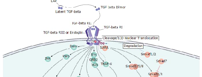
Hepatocyte Nuclear Factor 4 alpha (HNF4 alpha; NR2A1) is a member of the orphan nuclear receptor family. HNF4 alpha is expressed in the liver, kidney, intestine and pancreas. Mutation of HNF4 alpha in humans has been associated with maturity-onset diabetes of the young type 1 (MODY1). HNF4 binds to DNA as an exclusive homodimer. The HNF4 alpha gene is alternatively spliced and may generate up to nine different isoforms, HNF4 alpha 1 through HNF4 alpha 9. The expression of HNF-4 gamma (NR2A2) is observed in the pancreas, kidney, small intestine, and testes with no expression detected in the liver. The level of HNF-4 gamma is less 10% than that of HNF-4 alpha.
Human HNF-4 gamma/NR2A2 Antibody (N3224)
R&D Systems | Catalog # PP-N3224-00

Scientific images may be resized, cropped, or adjusted for brightness or contrast for presentation purposes. These updates are limited to presentation and do not affect the underlying scientific interpretation of the data. Where available, additional source data may be provided upon request.
Loading...
Key Product Details
Species Reactivity
Human
Applications
Immunohistochemistry, Western Blot
Label
Unconjugated
Antibody Source
Monoclonal Mouse IgG2A Clone # N3224
Loading...
Product Specifications
Immunogen
Human HNF-4 gamma/NR2A2
Clonality
Monoclonal
Host
Mouse
Isotype
IgG2A
Scientific Data Images for Human HNF-4 gamma/NR2A2 Antibody (N3224)
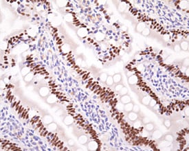
HNF‑4 gamma /NR2A2 in Rat Small Intestine.
HNF‑4 gamma /NR2A2 was detected in perfusion fixed paraffin-embedded sections of rat small intestine using HNF‑4 gamma /NR2A2 Monoclonal Antibody (Catalog # PP‑N3224‑00) overnight at 4 °C. Tissue was stained (brown) and counterstained with hematoxylin (blue). View our protocol for Chromogenic IHC Staining of Paraffin-embedded Tissue Sections.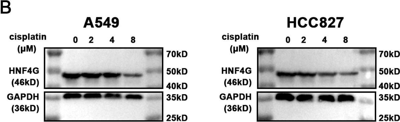
Detection of HNF-4 gamma/NR2A2 by Western Blot
HNF4G promotes resistance to cisplatin in lung adenocarcinoma cells.(A) qRT-PCR and (B) western blot results of HNF4G expression after cisplatin treatment in A549 and HCC827 cells. (C) qRT-PCR results of HNF4G expression in HNF4G overexpression and knockdown cells. (D) Cisplatin sensitivity curves of HNF4G overexpression and knockdown cells. * p < 0.05, ** p < 0.01, *** p < 0.001. Image collected and cropped by CiteAb from the following open publication (https://pubmed.ncbi.nlm.nih.gov/36923501), licensed under a CC-BY license. Not internally tested by R&D Systems.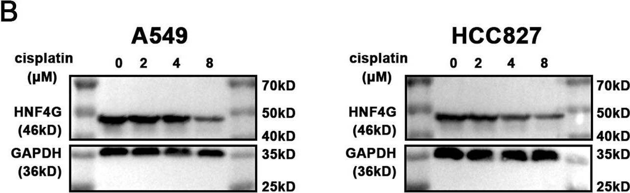
Detection of HNF-4 gamma/NR2A2 by Western Blot
HNF4G promotes resistance to cisplatin in lung adenocarcinoma cells.(A) qRT-PCR and (B) western blot results of HNF4G expression after cisplatin treatment in A549 and HCC827 cells. (C) qRT-PCR results of HNF4G expression in HNF4G overexpression and knockdown cells. (D) Cisplatin sensitivity curves of HNF4G overexpression and knockdown cells. * p < 0.05, ** p < 0.01, *** p < 0.001. Image collected and cropped by CiteAb from the following open publication (https://pubmed.ncbi.nlm.nih.gov/36923501), licensed under a CC-BY license. Not internally tested by R&D Systems.Applications for Human HNF-4 gamma/NR2A2 Antibody (N3224)
Application
Recommended Usage
Immunohistochemistry
5-15 µg/mL
Sample: Perfusion fixed paraffin-embedded sections of rat small intestine
Sample: Perfusion fixed paraffin-embedded sections of rat small intestine
Western Blot
Optimal dilutions of this antibody should be experimentally determined.
Reviewed Applications
Read 1 review rated 5 using PP-N3224-00 in the following applications:
Formulation, Preparation, and Storage
Formulation
A liquid formulation in physiologic saline with 0.1% NaN<sub>3</sub>.
Shipping
The product is shipped with dry ice or equivalent. Upon receipt, store it immediately at the temperature recommended below.
Stability & Storage
Store the unopened product at -70 °C. Use a manual defrost freezer and avoid repeated freeze-thaw cycles. Do not use past expiration date.
Background: HNF-4 gamma/NR2A2
Long Name
Hepatocyte Nuclear Factor 4 gamma
Alternate Names
HNF4 gamma, HNF4G, NR2A2
Entrez Gene IDs
3174 (Human)
Gene Symbol
HNF4G
Additional HNF-4 gamma/NR2A2 Products
Product Documents for Human HNF-4 gamma/NR2A2 Antibody (N3224)
Certificate of Analysis
To download a Certificate of Analysis, please enter a lot or batch number in the search box below.
Note: Certificate of Analysis not available for kit components.
Product Specific Notices for Human HNF-4 gamma/NR2A2 Antibody (N3224)
For research use only
Related Research Areas
Citations for Human HNF-4 gamma/NR2A2 Antibody (N3224)
 Powered by Bioz
Customer Reviews for Human HNF-4 gamma/NR2A2 Antibody (N3224) (1)
5 out of 5
1 Customer Rating
Have you used Human HNF-4 gamma/NR2A2 Antibody (N3224)?
Submit a review and receive an Amazon gift card!
$25/€18/£15/$25CAN/¥2500 for a review with an image
$10/€7/£6/$10CAN/¥1110 for a review without an image
Submit a review
Customer Images
Showing
1
-
1 of
1 review
Showing All
Filter By:
-
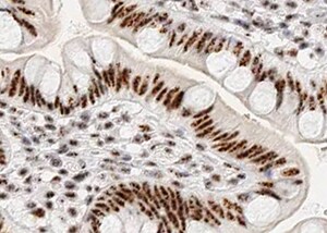
Application: ImmunohistochemistrySample Tested: Small intestine tissueSpecies: HumanVerified Customer | Posted 10/23/2022

There are no reviews that match your criteria.
Protocols
Find general support by application which include: protocols, troubleshooting, illustrated assays, videos and webinars.
- Antigen Retrieval Protocol (PIER)
- Antigen Retrieval for Frozen Sections Protocol
- Appropriate Fixation of IHC/ICC Samples
- Cellular Response to Hypoxia Protocols
- Chromogenic IHC Staining of Formalin-Fixed Paraffin-Embedded (FFPE) Tissue Protocol
- Chromogenic Immunohistochemistry Staining of Frozen Tissue
- ClariTSA™ Fluorophore Kits
- Detection & Visualization of Antibody Binding
- Fluorescent IHC Staining of Frozen Tissue Protocol
- Graphic Protocol for Heat-induced Epitope Retrieval
- Graphic Protocol for the Preparation and Fluorescent IHC Staining of Frozen Tissue Sections
- Graphic Protocol for the Preparation and Fluorescent IHC Staining of Paraffin-embedded Tissue Sections
- Graphic Protocol for the Preparation of Gelatin-coated Slides for Histological Tissue Sections
- IHC Sample Preparation (Frozen sections vs Paraffin)
- Immunofluorescent IHC Staining of Formalin-Fixed Paraffin-Embedded (FFPE) Tissue Protocol
- Immunohistochemistry (IHC) and Immunocytochemistry (ICC) Protocols
- Immunohistochemistry Frozen Troubleshooting
- Immunohistochemistry Paraffin Troubleshooting
- Preparing Samples for IHC/ICC Experiments
- Preventing Non-Specific Staining (Non-Specific Binding)
- Primary Antibody Selection & Optimization
- Protocol for Heat-Induced Epitope Retrieval (HIER)
- Protocol for Making a 4% Formaldehyde Solution in PBS
- Protocol for VisUCyte™ HRP Polymer Detection Reagent
- Protocol for the Preparation & Fixation of Cells on Coverslips
- Protocol for the Preparation and Chromogenic IHC Staining of Frozen Tissue Sections
- Protocol for the Preparation and Chromogenic IHC Staining of Frozen Tissue Sections - Graphic
- Protocol for the Preparation and Chromogenic IHC Staining of Paraffin-embedded Tissue Sections
- Protocol for the Preparation and Chromogenic IHC Staining of Paraffin-embedded Tissue Sections - Graphic
- Protocol for the Preparation and Fluorescent IHC Staining of Frozen Tissue Sections
- Protocol for the Preparation and Fluorescent IHC Staining of Paraffin-embedded Tissue Sections
- Protocol for the Preparation of Gelatin-coated Slides for Histological Tissue Sections
- R&D Systems Quality Control Western Blot Protocol
- TUNEL and Active Caspase-3 Detection by IHC/ICC Protocol
- The Importance of IHC/ICC Controls
- Troubleshooting Guide: Immunohistochemistry
- Troubleshooting Guide: Western Blot Figures
- Western Blot Conditions
- Western Blot Protocol
- Western Blot Protocol for Cell Lysates
- Western Blot Troubleshooting
- Western Blot Troubleshooting Guide
- View all Protocols, Troubleshooting, Illustrated assays and Webinars
Loading...
Associated Pathways