Ikaros (also LyF1) is a 60 kDa member of the C2H2-type zinc-finger protein family. It is found in both T and B cells and serves as a context‑dependent activator or repressor of genes. Human Ikaros is 519 amino acids (aa) in length. It possesses two zinc‑finger domains, one at the N‑terminus that contains four zinc-finger motifs (aa 117‑224) and one at the C‑terminus that contains two zinc‑finger motifs (aa 462‑514). The C‑terminal motifs mediate homo- or heterodimerization, while the N‑terminal motifs bind DNA. Multiple splice forms of 37 kDa to 47 kDa exist (Ikaros 2‑8) with reduced numbers of N‑terminal zinc-finger motifs. At least three are needed for DNA binding, and a heterodimer with a two‑motif monomer is suggested to be transcriptionally inert. Over aa 427‑519 (which are not spliced), human Ikaros shows 88% aa identity to mouse Ikaros.

Key Product Details
Validated by
Biological Validation
Species Reactivity
Validated:
Human
Cited:
Human
Applications
Validated:
Western Blot, Intracellular Staining by Flow Cytometry, Immunocytochemistry, Simple Western, Chromatin Immunoprecipitation (ChIP), CyTOF-ready
Cited:
Western Blot
Label
Unconjugated
Antibody Source
Polyclonal Goat IgG
Loading...
Product Specifications
Immunogen
E. coli-derived recombinant human lkaros
Ser427-Ser519
Accession # Q13422
Ser427-Ser519
Accession # Q13422
Specificity
Detects human lkaros in direct ELISAs and Western blots. In direct ELISAs and Western blots, less than 1% cross‑reactivity with recombinat human (rh) ZIC-1, rhZNF-24, and rhZNF-206 is observed.
Clonality
Polyclonal
Host
Goat
Isotype
IgG
Scientific Data Images for Human Ikaros Antibody
Detection of Human Ikaros by Western Blot.
Western blot shows lysates of Nalm-6 human Pre-B acute lymphocytic leukemia cell line. PVDF membrane was probed with 1 µg/mL of Goat Anti-Human Ikaros Antigen Affinity-purified Polyclonal Antibody (Catalog # AF4984) followed by HRP-conjugated Anti-Goat IgG Secondary Antibody (Catalog # HAF019). Bands were detected for Ikaros (1-8 spice forms) at approximately 37 to 63 kDa (as indicated). This experiment was conducted under reducing conditions and using Immunoblot Buffer Group 8.
Detection of Ikaros-regulated Genes by Chromatin Immunoprecipitation.
Jurkat human acute T cell leukemia cell line treated with 50 ng/mL PMA and 200 ng/mL calcium ionomycin for 30 minutes was fixed using formaldehyde, resuspended in lysis buffer, and sonicated to shear chromatin. Ikaros/DNA complexes were immunoprecipitated using 5 µg Goat Anti-Human Ikaros Antigen Affinity-purified Polyclonal Antibody (Catalog # AF4984) or control antibody (Catalog # AB-108-C) for 15 minutes in an ultrasonic bath, followed by Biotinylated Anti-Goat IgG Secondary Antibody (Catalog # BAF109). Immunocomplexes were captured using 50 µL of MagCellect Streptavidin Ferrofluid (Catalog # MAG999) and DNA was purified using chelating resin solution. TheVPACpromoter was detected by standard PCR.
Detection of Ikaros in Jurkat Human Cell Line by Flow Cytometry.
Jurkat human acute T cell leukemia cell line was stained with Goat Anti-Human Ikaros Antigen Affinity-purified Polyclonal Antibody (Catalog # AF4984, filled histogram) or control antibody (Catalog # AB-108-C, open histogram), followed by Phycoerythrin-conjugated Anti-Goat IgG Secondary Antibody (Catalog # F0107). To facilitate intracellular staining, cells were fixed with paraformaldehyde and permeabilized with methanol.
Detection of Human Ikaros by Simple WesternTM.
Simple Western lane view shows lysates of Nalm-6 human Pre-B acute lymphocytic leukemia cell line, loaded at 0.2 mg/mL. Specific bands were detected for Ikaros at approximately 63-77 kDa (as indicated) using 10 µg/mL of Goat Anti-Human Ikaros Antigen Affinity-purified Polyclonal Antibody (Catalog # AF4984) followed by 1:50 dilution of HRP-conjugated Anti-Goat IgG Secondary Antibody (Catalog # HAF109). This experiment was conducted under reducing conditions and using the 12-230 kDa separation system.Applications for Human Ikaros Antibody
Application
Recommended Usage
Chromatin Immunoprecipitation (ChIP)
5 µg/5 x 106 cells
Sample: PMA and calcium ionomycin treated Jurkat human acute T cell leukemia cell line chromatin, VPAC promoter detected by standard PCR
Sample: PMA and calcium ionomycin treated Jurkat human acute T cell leukemia cell line chromatin, VPAC promoter detected by standard PCR
CyTOF-ready
Ready to be labeled using established conjugation methods. No BSA or other carrier proteins that could interfere with conjugation.
Immunocytochemistry
5-15 µg/mL
Sample: Immersion fixed Jurkat human acute T cell leukemia cell line
Sample: Immersion fixed Jurkat human acute T cell leukemia cell line
Intracellular Staining by Flow Cytometry
2.5 µg/106 cells
Sample: Jurkat human acute T cell leukemia cell line fixed with paraformaldehyde and permeabilized with methanol
Sample: Jurkat human acute T cell leukemia cell line fixed with paraformaldehyde and permeabilized with methanol
Simple Western
10 µg/mL
Sample: Nalm‑6 human Pre-B acute lymphocytic leukemia cell line
Sample: Nalm‑6 human Pre-B acute lymphocytic leukemia cell line
Western Blot
1 µg/mL
Sample: Nalm‑6 human Pre‑B acute lymphocytic leukemia cell line
Sample: Nalm‑6 human Pre‑B acute lymphocytic leukemia cell line
Reviewed Applications
Read 1 review rated 4 using AF4984 in the following applications:
Flow Cytometry Panel Builder
Bio-Techne Knows Flow Cytometry
Save time and reduce costly mistakes by quickly finding compatible reagents using the Panel Builder Tool.
Advanced Features
- Spectra Viewer - Custom analysis of spectra from multiple fluorochromes
- Spillover Popups - Visualize the spectra of individual fluorochromes
- Antigen Density Selector - Match fluorochrome brightness with antigen density

Formulation, Preparation, and Storage
Purification
Antigen Affinity-purified
Reconstitution
Reconstitute at 0.2 mg/mL in sterile PBS. For liquid material, refer to CoA for concentration.
Loading...
Formulation
Lyophilized from a 0.2 μm filtered solution in PBS with Trehalose. *Small pack size (SP) is supplied either lyophilized or as a 0.2 µm filtered solution in PBS.
Shipping
Lyophilized product is shipped at ambient temperature. Liquid small pack size (-SP) is shipped with polar packs. Upon receipt, store immediately at the temperature recommended below.
Stability & Storage
Use a manual defrost freezer and avoid repeated freeze-thaw cycles.
- 12 months from date of receipt, -20 to -70 °C as supplied.
- 1 month, 2 to 8 °C under sterile conditions after reconstitution.
- 6 months, -20 to -70 °C under sterile conditions after reconstitution.
Calculators
Background: Ikaros
Long Name
DNA-binding protein Ikaros
Alternate Names
IK1, IKZF1, LyF-1, LYF1, PRO0758, ZNFN1A1
Gene Symbol
IKZF1
UniProt
Additional Ikaros Products
Product Documents for Human Ikaros Antibody
Certificate of Analysis
To download a Certificate of Analysis, please enter a lot or batch number in the search box below.
Note: Certificate of Analysis not available for kit components.
Product Specific Notices for Human Ikaros Antibody
For research use only
Related Research Areas
Citations for Human Ikaros Antibody
 Powered by Bioz
Customer Reviews for Human Ikaros Antibody (1)
4 out of 5
1 Customer Rating
Have you used Human Ikaros Antibody?
Submit a review and receive an Amazon gift card!
$25/€18/£15/$25CAN/¥2500 Yen for a review with an image
$10/€7/£6/$10CAN/¥1110 Yen for a review without an image
Submit a review
Customer Images
Showing
1
-
1 of
1 review
Showing All
Filter By:
-
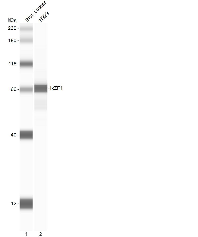
Application: Analysis of H929 whole cell lysatesSample Tested: Cell LysatesSpecies: HumanVerified Customer | Posted 01/20/2023H929 whole cell lysates, 0.4mg/ml protein concentration, Anti Ikaros 1:50, Goat HRP 1:200

There are no reviews that match your criteria.
Protocols
Find general support by application which include: protocols, troubleshooting, illustrated assays, videos and webinars.
- 7-Amino Actinomycin D (7-AAD) Cell Viability Flow Cytometry Protocol
- Appropriate Fixation of IHC/ICC Samples
- Cellular Response to Hypoxia Protocols
- ChIP Protocol Video
- Chromatin Immunoprecipitation (ChIP) Protocol
- Chromatin Immunoprecipitation Protocol
- ClariTSA™ Fluorophore Kits
- Detection & Visualization of Antibody Binding
- Extracellular Membrane Flow Cytometry Protocol
- Flow Cytometry Protocol for Cell Surface Markers
- Flow Cytometry Protocol for Staining Membrane Associated Proteins
- Flow Cytometry Staining Protocols
- Flow Cytometry Troubleshooting Guide
- ICC Cell Smear Protocol for Suspension Cells
- ICC Immunocytochemistry Protocol Videos
- ICC for Adherent Cells
- Immunocytochemistry (ICC) Protocol
- Immunocytochemistry Troubleshooting
- Immunofluorescence of Organoids Embedded in Cultrex Basement Membrane Extract
- Immunohistochemistry (IHC) and Immunocytochemistry (ICC) Protocols
- Intracellular Flow Cytometry Protocol Using Alcohol (Methanol)
- Intracellular Flow Cytometry Protocol Using Detergents
- Intracellular Nuclear Staining Flow Cytometry Protocol Using Detergents
- Intracellular Staining Flow Cytometry Protocol Using Alcohol Permeabilization
- Intracellular Staining Flow Cytometry Protocol Using Detergents to Permeabilize Cells
- Preparing Samples for IHC/ICC Experiments
- Preventing Non-Specific Staining (Non-Specific Binding)
- Primary Antibody Selection & Optimization
- Propidium Iodide Cell Viability Flow Cytometry Protocol
- Protocol for Liperfluo
- Protocol for VisUCyte™ HRP Polymer Detection Reagent
- Protocol for the Characterization of Human Th22 Cells
- Protocol for the Characterization of Human Th9 Cells
- Protocol for the Fluorescent ICC Staining of Cell Smears - Graphic
- Protocol for the Fluorescent ICC Staining of Cultured Cells on Coverslips - Graphic
- Protocol for the Preparation and Fluorescent ICC Staining of Cells on Coverslips
- Protocol for the Preparation and Fluorescent ICC Staining of Non-adherent Cells
- Protocol for the Preparation and Fluorescent ICC Staining of Stem Cells on Coverslips
- Protocol for the Preparation of a Cell Smear for Non-adherent Cell ICC - Graphic
- Protocol: Annexin V and PI Staining by Flow Cytometry
- Protocol: Annexin V and PI Staining for Apoptosis by Flow Cytometry
- R&D Systems Quality Control Western Blot Protocol
- TUNEL and Active Caspase-3 Detection by IHC/ICC Protocol
- The Importance of IHC/ICC Controls
- Troubleshooting Guide: Fluorokine Flow Cytometry Kits
- Troubleshooting Guide: Western Blot Figures
- Western Blot Conditions
- Western Blot Protocol
- Western Blot Protocol for Cell Lysates
- Western Blot Troubleshooting
- Western Blot Troubleshooting Guide
- View all Protocols, Troubleshooting, Illustrated assays and Webinars
Loading...
Associated Pathways

