IL-27 is a heterodimeric group 2 receptor ligand molecule that belongs to the IL-6/IL-12 family of long type I cytokines (1). It is composed of EBI3 (EBV-induced gene 3), also known as IL27B, a 34 kDa glycoprotein that is related to the p40 subunit of IL-12 and IL-23, and p28, also known as IL27A, the 28 kDa glycoprotein that is related to the p35 chain of IL-12 (2-4). The human EBI3 gene encodes a 229 amino acid (aa) precursor that contains a 20 aa signal peptide and 209 aa mature protein (5). The mature region contains two potential N-linked glycosylation sites, two fibronectin type III domains, and two pairs of conserved cysteine residues with a WSXWS-like motif that places the molecule in the hematopoietin receptor family (5). Although p40, the EBI3 counterpart in IL-12, is known to form homodimers, there is no evidence to date that EBI3 also homodimerizes. Human EBI3 is 61% aa identical to mouse EBI3. The human p28 gene encodes a 243 aa precursor that contains a 28 aa signal sequence and 215 aa mature region (6). The mature region is characterized by the presence of four alpha -helices, placing it in the IL-6 family of helical cytokines. Human p28 is 74% aa identical to mouse p28. IL-27 is expressed by monocytes, endothelial cells and dendritic cells (7). IL-27 binds to and signals through a heterodimeric receptor complex composed of WSX-1 (TCCR) and gp130. Evidence suggests IL-27 interacts only with WSX-1 (6, 8, 9). IL-27 has both anti- and proinflammatory properties. As an anti‑inflammatory, IL-27 seems to induce a general negative feedback program that limits T and NK-T cell activity (3, 7). At the onset of infection, IL-27 induces an IL‑12 receptor on naïve CD4+ T cells, making them susceptible to subsequent IL-12 activity (and possible Th1 development) (10).

Key Product Details
Species Reactivity
Validated:
Human
Cited:
Human
Applications
Validated:
Western Blot, ELISA Capture (Matched Antibody Pair), Neutralization
Cited:
Immunohistochemistry, Western Blot, Neutralization, Bioassay, ELISA Development, ELISA Development (Capture)
Label
Unconjugated
Antibody Source
Polyclonal Goat IgG
Loading...
Product Specifications
Immunogen
Mouse myeloma cell line NS0-derived recombinant human IL‑27 (R&D Systems, Catalog # 2526-IL)
Arg21-Lys229 (IL-27 EBI-3 subunit), Phe29-Pro243 (IL-27 p28 subunit)
Accession # Q14213 (EBI-3 subunit), AAM34498 (p28 subunit)
Arg21-Lys229 (IL-27 EBI-3 subunit), Phe29-Pro243 (IL-27 p28 subunit)
Accession # Q14213 (EBI-3 subunit), AAM34498 (p28 subunit)
Specificity
Detects human IL-27 in ELISAs and Western blots.
Clonality
Polyclonal
Host
Goat
Isotype
IgG
Endotoxin Level
<0.10 EU per 1 μg of the antibody by the LAL method.
Scientific Data Images for Human IL-27 Antibody
Detection of Human IL‑27 by Western Blot.
Western blot shows lysates of MT-2 human T cell line and CHO Chinese hamster ovary cell line either mock transfected or transfected with human IL-27 p28/IL-27A. PVDF membrane was probed with 1 µg/mL of Goat Anti-Human IL-27 Antigen Affinity-purified Polyclonal Antibody (Catalog # AF2526) followed by HRP-conjugated Anti-Goat IgG Secondary Antibody (HAF109). Specific bands were detected for the IL-27 EBI3/IL-27B at approximately 32 kDa and IL-27 p28/IL-27A at approximately 28 kDa (as indicated). This experiment was conducted under reducing conditions and using Immunoblot Buffer Group 1.
IL‑27 Inhibition of EMCV-induced Cytopathy and Neutralization by Human IL‑27 Antibody.
Recombinant Human IL-27 (Catalog # 2526-IL) reduces the Encephalo-myocarditis Virus (EMCV)-induced cytopathy in the HepG2 human hepatocellular carcinoma cell line in a dose-dependent manner (orange line). Inhibition of EMCV activity elicited by Recombinant Human IL-27 (25 ng/mL) is neutralized (green line) by increasing concen-trations of Goat Anti-Human IL-27 Antigen Affinity-purified Polyclonal Antibody (Catalog # AF2526). The ND50 is typically 0.200-2.40 µg/mL.
Detection of Human IL-27 by Flow Cytometry
IL-27 secreted by TFH cells support plasmablasts and plasma cell formation. (A–C) Mechanism of TFH mediated B cell response was determined by autologous TFH-B cell co-culture (CHB: n=5, HC-vacc: n=5). To imitate HBV-specific interactions between TFH and B cells, FACS-sorted TFH cells were first primed with HBsAg for 3 h, washed and then incubated with autologous CD19+CD27+ memory and CD19+CD27-IgD+ naïve B cells with and without of IL-21 and IL-27 neutralizing antibodies for 5 days. Generation of plasmablasts and plasma cells was analyzed by flow cytometry (D) expression of Blimp-1 (CHB: n=5, HC-vacc: n=5). Plasmablasts were gated as CD27+CD38+ cells and plasma cells were defined based on CD27+CD38+CD138. (E) Incubation of memory B and naïve B cells with rIL-27 for 5 days showed increased plasmablasts and plasma cell formation (CHB: n=5). Statistical analysis was performed using either Kruskal-Wallis test (ANOVA) with Dunn’s post hoc test for multiple comparisons or paired t test. Bars indicates mean and error bars designate standard deviation. * indicates p < 0.05, **p < 0.01 and ***p < 0.001. Image collected and cropped by CiteAb from the following open publication (https://pubmed.ncbi.nlm.nih.gov/33584666), licensed under a CC-BY license. Not internally tested by R&D Systems.
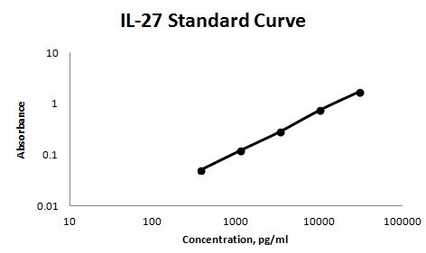
Human IL-27 ELISA Standard Curve
Recombinant Human IL‑27 (Catalog # 2526-IL) was serially diluted and captured by Goat Anti-Human IL‑27 Antigen Affinity-purified Polyclonal Antibody (Catalog # AF2526) coated on a Clear Polystyrene Microplate (Catalog # DY990). Goat Anti-Human IL‑27 Antigen Affinity-purified Polyclonal Antibody (Catalog # AF2526) was biotinylated and incubated with the protein captured on the plate. Detection of the standard curve was achieved by incubating Streptavidin-HRP (Catalog # DY998)
Human IL-27 ELISA Standard Curve
Recombinant Human IL‑27 (Catalog # 2526-IL) was serially diluted and captured by Goat Anti-Human IL‑27 Antigen Affinity-purified Polyclonal Antibody (Catalog # AF2526) coated on a Clear Polystyrene Microplate (Catalog # DY990). Goat Anti-Human IL‑27 Antigen Affinity-purified Polyclonal Antibody (Catalog # AF2526) was biotinylated and incubated with the protein captured on the plate. Detection of the standard curve was achieved by incubating Streptavidin-HRP (Catalog # DY998)Applications for Human IL-27 Antibody
Application
Recommended Usage
Western Blot
1 µg/mL
Sample: MT‑2 human T cell line and CHO Chinese hamster ovary cell line transfected with human IL-27 p28
Sample: MT‑2 human T cell line and CHO Chinese hamster ovary cell line transfected with human IL-27 p28
Neutralization
Measured by its ability to neutralize IL‑27 inhibition of EMCV-induced cytopathy in the HepG2 human hepatocellular carcinoma cell line.
Bender, H. et al. (2009) Hepatology 50:585. The Neutralization Dose (ND50) is typically 0.200‑2.40 µg/mL in the presence of 25 ng/mL Recombinant Human IL‑27.
Human IL-27 Sandwich Immunoassay
Please Note: Optimal dilutions of this antibody should be experimentally determined.
Reviewed Applications
Read 1 review rated 4 using AF2526 in the following applications:
Formulation, Preparation, and Storage
Purification
Antigen Affinity-purified
Reconstitution
Reconstitute at 0.2 mg/mL in sterile PBS. For liquid material, refer to CoA for concentration.
Loading...
Formulation
Lyophilized from a 0.2 μm filtered solution in PBS with Trehalose. See Certificate of Analysis for details.
*Small pack size (-SP) is supplied either lyophilized or as a 0.2 µm filtered solution in PBS.
*Small pack size (-SP) is supplied either lyophilized or as a 0.2 µm filtered solution in PBS.
Shipping
Lyophilized product is shipped at ambient temperature. Liquid small pack size (-SP) is shipped with polar packs. Upon receipt, store immediately at the temperature recommended below.
Stability & Storage
Use a manual defrost freezer and avoid repeated freeze-thaw cycles.
- 12 months from date of receipt, -20 to -70 °C as supplied.
- 1 month, 2 to 8 °C under sterile conditions after reconstitution.
- 6 months, -20 to -70 °C under sterile conditions after reconstitution.
Calculators
Background: IL-27
References
- Boulay, J-L. et al. (2003) Immunity 19:159.
- Trinchieri, G. et al. (2003) Immunity 19:641.
- Murakami, M. et al. (2004) Growth Factors 22:75.
- Cordoba-Rodriguez, R. and D.M. Frucht (2003) Exp. Opin. Biol. Ther. 3:715.
- Devergne, O. et al. (1996) J. Virology 70:1143.
- Pflanz, S. et al. (2002) Immunity 16:779.
- Villarino, A.V. et al. (2004) J. Immunol. 173:715.
- Pflanz, S. et al. (2004) J Immunol 172:2225.
- Scheller, J. et al. (2005) Biochem. Biophys. Res. Commun. 326:724.
- Holscher, C. (2004) Med. Microbiol. Immunol. (Berl).193:1.
Long Name
Interleukin 27
Alternate Names
IL27
Gene Symbol
IL27
Additional IL-27 Products
Product Documents for Human IL-27 Antibody
Certificate of Analysis
To download a Certificate of Analysis, please enter a lot or batch number in the search box below.
Note: Certificate of Analysis not available for kit components.
Product Specific Notices for Human IL-27 Antibody
For research use only
Related Research Areas
Citations for Human IL-27 Antibody
 Powered by Bioz
Customer Reviews for Human IL-27 Antibody (1)
4 out of 5
1 Customer Rating
Have you used Human IL-27 Antibody?
Submit a review and receive an Amazon gift card!
$25/€18/£15/$25CAN/¥2500 Yen for a review with an image
$10/€7/£6/$10CAN/¥1110 Yen for a review without an image
Submit a review
Customer Images
Showing
1
-
1 of
1 review
Showing All
Filter By:
-
Application: ELISASample Tested: Serum and PlasmaSpecies: HumanVerified Customer | Posted 11/27/2017AF2526 was used as the solid phase antibody, BAF2526 was used as the detection antibody. 2526-IL was used as the immunoassay standard. Sensitivity of the assay was around 100 pg/ml.

There are no reviews that match your criteria.
Protocols
Find general support by application which include: protocols, troubleshooting, illustrated assays, videos and webinars.
- Cellular Response to Hypoxia Protocols
- R&D Systems Quality Control Western Blot Protocol
- Troubleshooting Guide: Western Blot Figures
- Western Blot Conditions
- Western Blot Protocol
- Western Blot Protocol for Cell Lysates
- Western Blot Troubleshooting
- Western Blot Troubleshooting Guide
- View all Protocols, Troubleshooting, Illustrated assays and Webinars
Loading...
Associated Pathways
