
Key Product Details
Species Reactivity
Validated:
Human
Cited:
Human, Mouse, Primate - Macaca mulatta (Rhesus Macaque)
Applications
Validated:
Immunohistochemistry, Neutralization
Cited:
Immunohistochemistry, Immunohistochemistry-Paraffin, Western Blot, Neutralization, Immunocytochemistry
Label
Unconjugated
Antibody Source
Polyclonal Goat IgG
Loading...
Product Specifications
Immunogen
E. coli-derived recombinant human LIF
Ser23-Phe202
Accession # P15018
Ser23-Phe202
Accession # P15018
Specificity
Detects human LIF in direct ELISAs.
Clonality
Polyclonal
Host
Goat
Isotype
IgG
Endotoxin Level
<0.10 EU per 1 μg of the antibody by the LAL method.
Scientific Data Images for Human LIF Antibody
LIF in Human Lung.
LIF was detected in immersion fixed paraffin-embedded sections of human lung array using Goat Anti-Human LIF Antigen Affinity-purified Polyclonal Antibody (Catalog # AF-250-NA) at 25 µg/mL overnight at 4 °C. Tissue was stained using the Anti-Goat HRP-DAB Cell & Tissue Staining Kit (brown; CTS008) and counterstained with hematoxylin (blue). Lower panel shows a lack of labeling if primary antibodies are omitted and tissue is stained only with secondary antibody followed by incubation with detection reagents. View our protocol for Chromogenic IHC Staining of Paraffin-embedded Tissue Sections.
LIF in Human Alzheimer's Brain.
LIF was detected in immersion fixed paraffin-embedded sections of human Alzheimer's brain using Goat Anti-Human LIF Antigen Affinity-purified Polyclonal Antibody (Catalog # AF-250-NA) at 10 µg/mL overnight at 4 °C. Before incubation with the primary antibody, tissue was subjected to heat-induced epitope retrieval using Antigen Retrieval Reagent-Basic CTS013). Tissue was stained using the Anti-Goat HRP-DAB Cell & Tissue Staining Kit (brown CTS008) and counterstained with hematoxylin (blue). Specific staining was localized to neuronal cytoplasm. View our protocol for Chromogenic IHC Staining of Paraffin-embedded Tissue Sections.
Cell Proliferation Induced by LIF and Neutralization by Human LIF Antibody.
Recombinant Human LIF (7734-LF) stimulates proliferation in the TF-1 human erythroleukemic cell line in a dose-depend-ent manner (orange line), as measured by Resazurin (AR002). Proliferation elicited by 3 ng/mL Recombinant Human LIF is neutralized (green line) by increasing concentrations of Goat Anti-Human LIF Antigen Affinity-purified Polyclonal Antibody (Catalog # AF-250-NA). The ND50 is typically 0.04-0.08 µg/mL.
Detection of Human LIF by Western Blot
LIF promotes Glut1 PM translocation through the activation of AKT signaling.A LIF activated the AKT signaling, which can be blocked by MK2206, an AKT inhibitor (upper panel), or Wortmannin (Wort), a PI3K inhibitor (lower panel), in MCF7, MDA-MB 231 and T47D cells. The levels of p-AKTSer473 (p-AKT), which reflect the activity of AKT were measured by Western-blot assays. B Blocking AKT signaling by MK2206 (left) or Wortmannin (right) largely abolished the promoting effect of LIF on Glut1 PM translocation in breast cancer cells. C, D Blocking AKT signaling by expression of dominant-negative AKT (AKT-DN) largely abolished the promoting effect of LIF on Glut1 PM translocation in breast cancer cells. Cells were transfected with vectors expressing AKT-DN. The levels of p-AKT and total AKT (C), as well as Glut1 PM translocation (D), were determined by Western-blot assays. Uncropped Western-blot images are shown in Supplementary Fig 3. Image collected and cropped by CiteAb from the following open publication (https://pubmed.ncbi.nlm.nih.gov/35440095), licensed under a CC-BY license. Not internally tested by R&D Systems.
Detection of Human LIF by Western Blot
LIF promotes Glut1 PM translocation through the activation of AKT signaling.A LIF activated the AKT signaling, which can be blocked by MK2206, an AKT inhibitor (upper panel), or Wortmannin (Wort), a PI3K inhibitor (lower panel), in MCF7, MDA-MB 231 and T47D cells. The levels of p-AKTSer473 (p-AKT), which reflect the activity of AKT were measured by Western-blot assays. B Blocking AKT signaling by MK2206 (left) or Wortmannin (right) largely abolished the promoting effect of LIF on Glut1 PM translocation in breast cancer cells. C, D Blocking AKT signaling by expression of dominant-negative AKT (AKT-DN) largely abolished the promoting effect of LIF on Glut1 PM translocation in breast cancer cells. Cells were transfected with vectors expressing AKT-DN. The levels of p-AKT and total AKT (C), as well as Glut1 PM translocation (D), were determined by Western-blot assays. Uncropped Western-blot images are shown in Supplementary Fig 3. Image collected and cropped by CiteAb from the following open publication (https://pubmed.ncbi.nlm.nih.gov/35440095), licensed under a CC-BY license. Not internally tested by R&D Systems.Applications for Human LIF Antibody
Application
Recommended Usage
Immunohistochemistry
5-15 µg/mL
Sample: Immersion fixed paraffin-embedded sections of human lung and human Alzheimer's brain subjected to heat-induced epitope retrieval using Antigen Retrieval Reagent-Basic (Catalog # CTS013).
Sample: Immersion fixed paraffin-embedded sections of human lung and human Alzheimer's brain subjected to heat-induced epitope retrieval using Antigen Retrieval Reagent-Basic (Catalog # CTS013).
Neutralization
Measured by its ability to neutralize LIF-induced proliferation in the TF‑1 human erythroleukemic cell line. Kitamura, T. et al. (1989) J. Cell Physiol. 140:323. The Neutralization Dose (ND50) is typically 0.04-0.08 µg/mL in the presence of 3 ng/mL Recombinant Human LIF.
Reviewed Applications
Read 1 review rated 4 using AF-250-NA in the following applications:
Formulation, Preparation, and Storage
Purification
Antigen Affinity-purified
Reconstitution
Reconstitute at 0.2 mg/mL in sterile PBS. For liquid material, refer to CoA for concentration.
Loading...
Formulation
Lyophilized from a 0.2 μm filtered solution in PBS with Trehalose. *Small pack size (SP) is supplied either lyophilized or as a 0.2 µm filtered solution in PBS.
Shipping
Lyophilized product is shipped at ambient temperature. Liquid small pack size (-SP) is shipped with polar packs. Upon receipt, store immediately at the temperature recommended below.
Stability & Storage
Use a manual defrost freezer and avoid repeated freeze-thaw cycles.
- 12 months from date of receipt, -20 to -70 °C as supplied.
- 1 month, 2 to 8 °C under sterile conditions after reconstitution.
- 6 months, -20 to -70 °C under sterile conditions after reconstitution.
Calculators
Background: LIF
Long Name
Leukemia Inhibitory Factor
Alternate Names
D Factor, Emfilermin, HILDA, MLPLI
Gene Symbol
LIF
UniProt
Additional LIF Products
Product Documents for Human LIF Antibody
Certificate of Analysis
To download a Certificate of Analysis, please enter a lot or batch number in the search box below.
Note: Certificate of Analysis not available for kit components.
Product Specific Notices for Human LIF Antibody
For research use only
Related Research Areas
Citations for Human LIF Antibody
 Powered by Bioz
Customer Reviews for Human LIF Antibody (1)
4 out of 5
1 Customer Rating
Have you used Human LIF Antibody?
Submit a review and receive an Amazon gift card!
$25/€18/£15/$25CAN/¥2500 Yen for a review with an image
$10/€7/£6/$10CAN/¥1110 Yen for a review without an image
Submit a review
Customer Images
Showing
1
-
1 of
1 review
Showing All
Filter By:
-
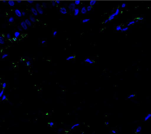
Application: Immunocytochemistry/ImmunofluorescenceSample Tested: Skin tissueSpecies: HumanVerified Customer | Posted 06/19/2021

There are no reviews that match your criteria.
Protocols
Find general support by application which include: protocols, troubleshooting, illustrated assays, videos and webinars.
- Antigen Retrieval Protocol (PIER)
- Antigen Retrieval for Frozen Sections Protocol
- Appropriate Fixation of IHC/ICC Samples
- Cellular Response to Hypoxia Protocols
- Chromogenic IHC Staining of Formalin-Fixed Paraffin-Embedded (FFPE) Tissue Protocol
- Chromogenic Immunohistochemistry Staining of Frozen Tissue
- ClariTSA™ Fluorophore Kits
- Detection & Visualization of Antibody Binding
- Fluorescent IHC Staining of Frozen Tissue Protocol
- Graphic Protocol for Heat-induced Epitope Retrieval
- Graphic Protocol for the Preparation and Fluorescent IHC Staining of Frozen Tissue Sections
- Graphic Protocol for the Preparation and Fluorescent IHC Staining of Paraffin-embedded Tissue Sections
- Graphic Protocol for the Preparation of Gelatin-coated Slides for Histological Tissue Sections
- IHC Sample Preparation (Frozen sections vs Paraffin)
- Immunofluorescent IHC Staining of Formalin-Fixed Paraffin-Embedded (FFPE) Tissue Protocol
- Immunohistochemistry (IHC) and Immunocytochemistry (ICC) Protocols
- Immunohistochemistry Frozen Troubleshooting
- Immunohistochemistry Paraffin Troubleshooting
- Preparing Samples for IHC/ICC Experiments
- Preventing Non-Specific Staining (Non-Specific Binding)
- Primary Antibody Selection & Optimization
- Protocol for Heat-Induced Epitope Retrieval (HIER)
- Protocol for Making a 4% Formaldehyde Solution in PBS
- Protocol for VisUCyte™ HRP Polymer Detection Reagent
- Protocol for the Preparation & Fixation of Cells on Coverslips
- Protocol for the Preparation and Chromogenic IHC Staining of Frozen Tissue Sections
- Protocol for the Preparation and Chromogenic IHC Staining of Frozen Tissue Sections - Graphic
- Protocol for the Preparation and Chromogenic IHC Staining of Paraffin-embedded Tissue Sections
- Protocol for the Preparation and Chromogenic IHC Staining of Paraffin-embedded Tissue Sections - Graphic
- Protocol for the Preparation and Fluorescent IHC Staining of Frozen Tissue Sections
- Protocol for the Preparation and Fluorescent IHC Staining of Paraffin-embedded Tissue Sections
- Protocol for the Preparation of Gelatin-coated Slides for Histological Tissue Sections
- TUNEL and Active Caspase-3 Detection by IHC/ICC Protocol
- The Importance of IHC/ICC Controls
- Troubleshooting Guide: Immunohistochemistry
- View all Protocols, Troubleshooting, Illustrated assays and Webinars
Loading...
Associated Pathways
