The immunoglobulin-like transcript (ILT) comprise a family of activating and inhibitory type immunoreceptors whose genes are located in the same locus that encodes killer cell Ig-like receptors (KIR) (1‑3). ILT4, also known as LIR-2 and LILRB2, is a type I transmembrane protein expressed primarily on monocytes and dendritic cells (DC) (4). Human ILT4 is produced as a 598 amino acid (aa) precursor including a 21 aa signal sequence, a 440 aa extracellular domain (ECD), a 21 aa transmembrane segment, and a 116 aa cytoplasmic domain. The ECD contains four Ig-like domains, and the cytoplasmic domain contains three immunoreceptor tyrosine-based inhibitory motifs (ITIM) (5). The ECD of human ILT4 shares 76% aa identity with chimpanzee ILT4 and 74%, 81%, 33%, 52%, 77%, 61%, and 64 % aa identity with human ILT1, 2, 3, 5, 6, 7, and 8, respectively. ILT4 binds to classical MHC I proteins as well as the non-classical HLA-G1 and HLA-F molecules (5‑9). It competes with CD8 alpha for MHC I binding but does not compete with KIR2DL1 (7). Ligation of ILT4 induces Tyr phosphorylation within its cytoplasmic ITIMs, a requirement for association with SHP-1 (4, 6). Activation of ILT4 inhibits signaling through Fc gamma RI (4) and Fc epsilon RI (6) and causes DC to become tolerogenic by downregulation of costimulatory molecules (10, 11). ILT4 mediates tolerogenic DC-induced CD4+ T cell energy in vitro and in vivo (10‑12).
Human LILRB2/CD85d/ILT4 Antibody (287219)
R&D Systems | Catalog # MAB2078

Key Product Details
Species Reactivity
Validated:
Human
Cited:
Human, Mouse
Applications
Validated:
Western Blot, Neutralization, Flow Cytometry, CyTOF-ready
Cited:
Immunohistochemistry, Neutralization, Flow Cytometry, Immunocytochemistry
Label
Unconjugated
Antibody Source
Monoclonal Mouse IgG2A Clone # 287219
Loading...
Product Specifications
Immunogen
Mouse myeloma cell line NS0-derived recombinant human LILRB2/CD85d/ILT4
Gly24-His458
Accession # ACT64556
Gly24-His458
Accession # ACT64556
Specificity
Detects human LILRB2/CD85d/ILT4 in direct ELISAs and Western blots. In direct ELISAs and Western blots, no cross-reactivity with recombinant human (rh) ILT1, rhILT2, rhILT3, rhILT6, rhILT7, rhILT11, rhLIR6 or rhLIR8 is observed.
Clonality
Monoclonal
Host
Mouse
Isotype
IgG2A
Endotoxin Level
<0.10 EU per 1 μg of the antibody by the LAL method.
Scientific Data Images for Human LILRB2/CD85d/ILT4 Antibody (287219)
Detection of LILRB2/CD85d/ILT4 in Human Blood Monocytes by Flow Cytometry.
Human peripheral blood monocytes were stained with Mouse Anti-Human CD14 PE-conjugated Monoclonal Antibody (Catalog # FAB3832P) and either (A) Mouse Anti-Human LILRB2/CD85d/ILT4 Monoclonal Antibody (Catalog # MAB2078) or (B) Mouse IgG2AIsotype Control (Catalog # MAB003) followed by Allophycocyanin-conjugated Anti-Mouse IgG Secondary Antibody (Catalog # F0101B).
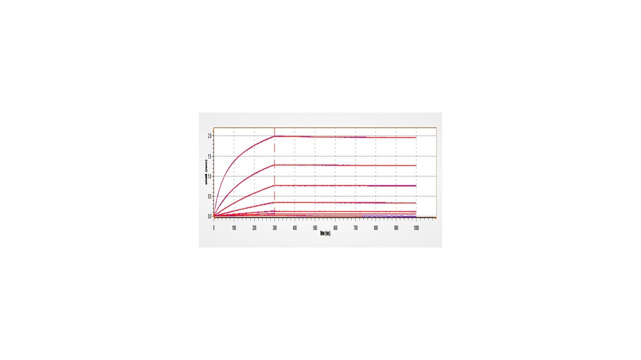
Cell Adhesion Mediated by LILRB2/CD85d/ILT4 and Neutralization by Human LILRB2/CD85d/ILT4 Antibody.
Recombinant Human LILRB2/CD85d/ILT4 Fc Chimera (Catalog # 2078-T4), immobilized onto a microplate, supports the adhesion of the HSB2 human peripheral blood acute lymphoblastic leukemia cell line in a dose-dependent manner (orange line). Adhesion elicited by Recombinant Human LILRB2/CD85d/ILT4 Fc Chimera (35 µg/mL) is neutralized (green line) by increasing concentrations of Mouse Anti-Human LILRB2/CD85d/ILT4 Monoclonal Antibody (Catalog # MAB2078). The ND50 is typically 0.2-0.8 µg/mL.Applications for Human LILRB2/CD85d/ILT4 Antibody (287219)
Application
Recommended Usage
CyTOF-ready
Ready to be labeled using established conjugation methods. No BSA or other carrier proteins that could interfere with conjugation.
Flow Cytometry
0.25 µg/106 cells
Sample: Human peripheral blood monocytes
Sample: Human peripheral blood monocytes
Western Blot
1 µg/mL
Sample: Recombinant Human LILRB2/CD85d/ILT4 Fc Chimera (Catalog # 2078-T4)
under non-reducing conditions only
Sample: Recombinant Human LILRB2/CD85d/ILT4 Fc Chimera (Catalog # 2078-T4)
under non-reducing conditions only
Neutralization
Measured by its ability to neutralize LILRB2/CD85d/ILT4-mediated adhesion of the HSB2 human peripheral blood acute lymphoblastic leukemia cell line. The Neutralization Dose (ND50) is typically 0.2-0.8 µg/mL in the presence of 35 µg/mL Recombinant Human LILRB2/CD85d/ILT4 Fc Chimera.
Reviewed Applications
Read 2 reviews rated 4.5 using MAB2078 in the following applications:
Flow Cytometry Panel Builder
Bio-Techne Knows Flow Cytometry
Save time and reduce costly mistakes by quickly finding compatible reagents using the Panel Builder Tool.
Advanced Features
- Spectra Viewer - Custom analysis of spectra from multiple fluorochromes
- Spillover Popups - Visualize the spectra of individual fluorochromes
- Antigen Density Selector - Match fluorochrome brightness with antigen density

Formulation, Preparation, and Storage
Purification
Protein A or G purified from hybridoma culture supernatant
Reconstitution
Reconstitute at 0.5 mg/mL in sterile PBS. For liquid material, refer to CoA for concentration.
Loading...
Formulation
Lyophilized from a 0.2 μm filtered solution in PBS with Trehalose. *Small pack size (SP) is supplied either lyophilized or as a 0.2 µm filtered solution in PBS.
Shipping
Lyophilized product is shipped at ambient temperature. Liquid small pack size (-SP) is shipped with polar packs. Upon receipt, store immediately at the temperature recommended below.
Stability & Storage
Use a manual defrost freezer and avoid repeated freeze-thaw cycles.
- 12 months from date of receipt, -20 to -70 °C as supplied.
- 1 month, 2 to 8 °C under sterile conditions after reconstitution.
- 6 months, -20 to -70 °C under sterile conditions after reconstitution.
Calculators
Background: LILRB2/CD85d/ILT4
References
- Suciu-Foca, N. et al. (2005) Int. Immunopharmacol. 5:7.
- Hofmeister, V. and E.H. Weiss (2003) Semin. Canc. Biol. 13:317.
- Hunt, J.S. et al. (2005) FASEB J. 19:681.
- Finger, N.A. et al. (1998) Eur. J. Immunol. 28:3423.
- Borges, L. et al. (1997) J. Immunol. 159:5192.
- Colonna, M. et al. (1998) J. Immunol. 160:3096.
- Shiroishi, M. et al. (2003) Proc. Natl. Acad. Sci. 100:8856.
- Lepin, E.J.M. et al. (2000) Eur. J. Immunol. 30:3552.
- Allen, R.L. et al. (2001) J. Immunol. 167:5543.
- Chang, C.C. et al. (2002) Nat. Immunol. 3:237.
- Ristich, V. et al. (2005) Eur. J. Immunol. 35:1133.
- Manavalan, J.S. et al. (2003) Transpl. Immunol. 11:245.
Long Name
Leukocyte Immunoglobulin-like Receptor, Subfamily B (with TM and ITIM Domains), Member 5
Alternate Names
CD85d, ILT4, LIR2, MIR10
Entrez Gene IDs
10288 (Human)
Gene Symbol
LILRB2
UniProt
Additional LILRB2/CD85d/ILT4 Products
Product Documents for Human LILRB2/CD85d/ILT4 Antibody (287219)
Certificate of Analysis
To download a Certificate of Analysis, please enter a lot or batch number in the search box below.
Note: Certificate of Analysis not available for kit components.
Product Specific Notices for Human LILRB2/CD85d/ILT4 Antibody (287219)
For research use only
Citations for Human LILRB2/CD85d/ILT4 Antibody (287219)
 Powered by Bioz
Customer Reviews for Human LILRB2/CD85d/ILT4 Antibody (287219) (2)
4.5 out of 5
2 Customer Ratings
Have you used Human LILRB2/CD85d/ILT4 Antibody (287219)?
Submit a review and receive an Amazon gift card!
$25/€18/£15/$25CAN/¥2500 Yen for a review with an image
$10/€7/£6/$10CAN/¥1110 Yen for a review without an image
Submit a review
Customer Images

Showing
1
-
2 of
2 reviews
Showing All
Filter By:
-
Application: Flow CytometrySample Tested: M2c macrophagesSpecies: HumanVerified Customer | Posted 06/20/2026Detection of ILT4 in M2c macrophages. The cells were incubated with 2 μl of the human ILT4 antibody (Red) or isotype control (blue) for 30 minutes on ice, followed by a secondary a-mouse IgG APCThe antibody was used to detect the expression of ILT4 by flow cytometry in monocyte-derived human macrophages polarized to M2c phenotype

-
Application: affinity bindingSample Tested: 293T human embryonic kidney cell lineSpecies: HumanVerified Customer | Posted 08/25/2022
There are no reviews that match your criteria.
Protocols
Find general support by application which include: protocols, troubleshooting, illustrated assays, videos and webinars.
- 7-Amino Actinomycin D (7-AAD) Cell Viability Flow Cytometry Protocol
- Cellular Response to Hypoxia Protocols
- Extracellular Membrane Flow Cytometry Protocol
- Flow Cytometry Protocol for Cell Surface Markers
- Flow Cytometry Protocol for Staining Membrane Associated Proteins
- Flow Cytometry Staining Protocols
- Flow Cytometry Troubleshooting Guide
- Intracellular Flow Cytometry Protocol Using Alcohol (Methanol)
- Intracellular Flow Cytometry Protocol Using Detergents
- Intracellular Nuclear Staining Flow Cytometry Protocol Using Detergents
- Intracellular Staining Flow Cytometry Protocol Using Alcohol Permeabilization
- Intracellular Staining Flow Cytometry Protocol Using Detergents to Permeabilize Cells
- Propidium Iodide Cell Viability Flow Cytometry Protocol
- Protocol for Liperfluo
- Protocol for the Characterization of Human Th22 Cells
- Protocol for the Characterization of Human Th9 Cells
- Protocol: Annexin V and PI Staining by Flow Cytometry
- Protocol: Annexin V and PI Staining for Apoptosis by Flow Cytometry
- R&D Systems Quality Control Western Blot Protocol
- Troubleshooting Guide: Fluorokine Flow Cytometry Kits
- Troubleshooting Guide: Western Blot Figures
- Western Blot Conditions
- Western Blot Protocol
- Western Blot Protocol for Cell Lysates
- Western Blot Troubleshooting
- Western Blot Troubleshooting Guide
- View all Protocols, Troubleshooting, Illustrated assays and Webinars
Loading...