
Key Product Details
Species Reactivity
Validated:
Human
Cited:
Human, Mouse, Primate - Macaca mulatta (Rhesus Macaque), Primate - Pan troglodytes (Chimpanzee), Transgenic Mouse
Applications
Validated:
Western Blot, Immunocytochemistry, Simple Western
Cited:
Immunohistochemistry, Immunohistochemistry-Paraffin, Western Blot, Immunocytochemistry, Simple Western
Label
Unconjugated
Antibody Source
Polyclonal Goat IgG
Loading...
Product Specifications
Immunogen
E. coli-derived recombinant human LIN-28A
Met1-Asn209
Accession # Q9H9Z2
Met1-Asn209
Accession # Q9H9Z2
Specificity
Detects human LIN-28A in direct ELISAs and Western blots.
Clonality
Polyclonal
Host
Goat
Isotype
IgG
Scientific Data Images for Human LIN-28A Antibody
Detection of Human LIN‑28A by Western Blot.
Western blot shows lysates of JAR human choriocarcinoma cell line and NTera-2 human testicular embryonic carcinoma cell line. PVDF membrane was probed with 0.1 µg/mL of Goat Anti-Human LIN-28A Antigen Affinity-purified Polyclonal Antibody (Catalog # AF3757) followed by HRP-conjugated Anti-Goat IgG Secondary Antibody (Catalog # HAF109). A specific band was detected for LIN-28A at approximately 30 kDa (as indicated). This experiment was conducted under reducing conditions and using Immunoblot Buffer Group 1.
LIN‑28A in BG01V Human Stem Cells.
LIN-28A was detected in immersion fixed BG01V human embryonic stem cells using 10 µg/mL Goat Anti-Human LIN-28A Antigen Affinity-purified Polyclonal Antibody (Catalog # AF3757) for 3 hours at room temperature. Cells were stained with the NorthernLights™ 557-conjugated Anti-Goat IgG Secondary Antibody (red; Catalog # NL001) and counter-stained with DAPI (blue). View our protocol for Fluorescent ICC Staining of Cells on Coverslips.
LIN‑28A in D3 Mouse Stem Cells.
LIN-28A was detected in immersion fixed D3 mouse embryonic stem cell line using Goat Anti-Human LIN-28A Antigen Affinity-purified Polyclonal Antibody (Catalog # AF3757) at 10 µg/mL for 3 hours at room temperature. Cells were stained using the Northern-Lights™ 557-conjugated Anti-Goat IgG Secondary Antibody (red; Catalog # NL001) and counterstained with DAPI (blue). Specific staining was localized to cytoplasm. View our protocol for Fluorescent ICC Staining of Cells on Coverslips.
Detection of Human LIN-28A by Simple WesternTM.
Simple Western lane view shows lysates of NTera‑2 human testicular embryonic carcinoma cell line and JAR human choriocarcinoma cell line, loaded at 0.2 mg/mL. A specific band was detected for LIN-28A at approximately 45 kDa (as indicated) using 10 µg/mL of Goat Anti-Human LIN-28A Antigen Affinity-purified Polyclonal Antibody (Catalog # AF3757). This experiment was conducted under reducing conditions and using the 12-230 kDa separation system.
Detection of LIN-28A by Western Blot
Expression of LIN28 in mouse testis. Western blot analysis was performed on 20 μg of protein extracts for each sample. beta -actin served as a control. Molecular weight standards were marked in kDa. (A) Western blot analysis of LIN28 in adult mouse tissues. (B) Absence of LIN28 in germ cell-deficient XXY* testes. Testes were collected from adult and post-natal day 10-old mice. V6.5 mouse embryonic stem (ES) cells served as a positive control. LIN28 was absent in fibroblast feeder cells. (C) Developmental expression of LIN28 in postnatal testes. Testes were collected from mice of postnatal day 1 through adulthood. Image collected and cropped by CiteAb from the following open publication (https://pubmed.ncbi.nlm.nih.gov/19563657), licensed under a CC-BY license. Not internally tested by R&D Systems.
Detection of LIN-28A by Western Blot
Expression and siRNA knockdown of LIN28 in cultured spermatogonia highly enriched for spermatogonial stem cells (SSCs). (A) Immunostaining of SSCs with anti-LIN28 and anti-PLZF or anti-GFRA1 antibodies. Scale bar, 50 μm. (B) Quantitative PCR measurement of Lin28 mRNA levels (n = 3, mean ± SE) in SSCs after siRNA treatment for 30 hours. (C) Decreased LIN28 protein abundance (43% compared to the control) in SSCs after 30 hours of siRNA treatment. The control SSCs were not treated with Lin28 siRNA. Feeder cells served as a negative control. beta -actin served as a loading control. (D) The number of SSCs (n = 3, mean ± SE) with and without Lin28 siRNA treatment. (E) Quantitative measurement of mature let-7g miRNA levels (n = 3, mean ± SE) in SSCs after siRNA treatment for 30 hours. Image collected and cropped by CiteAb from the following open publication (https://pubmed.ncbi.nlm.nih.gov/19563657), licensed under a CC-BY license. Not internally tested by R&D Systems.Applications for Human LIN-28A Antibody
Application
Recommended Usage
Immunocytochemistry
5-15 µg/mL
Sample: Immersion fixed BG01V human embryonic stem cells and D3 mouse embryonic stem cells
Sample: Immersion fixed BG01V human embryonic stem cells and D3 mouse embryonic stem cells
Simple Western
10 µg/mL
Sample: NTera‑2 human testicular embryonic carcinoma cell line and JAR human choriocarcinoma cell line
Sample: NTera‑2 human testicular embryonic carcinoma cell line and JAR human choriocarcinoma cell line
Western Blot
0.1 µg/mL
Sample: JAR human choriocarcinoma cell line and NTera‑2 human testicular embryonic carcinoma cell line
Sample: JAR human choriocarcinoma cell line and NTera‑2 human testicular embryonic carcinoma cell line
Reviewed Applications
Read 1 review rated 4 using AF3757 in the following applications:
Formulation, Preparation, and Storage
Purification
Antigen Affinity-purified
Reconstitution
Reconstitute at 0.2 mg/mL in sterile PBS. For liquid material, refer to CoA for concentration.
Loading...
Formulation
Lyophilized from a 0.2 μm filtered solution in PBS with Trehalose. *Small pack size (SP) is supplied either lyophilized or as a 0.2 µm filtered solution in PBS.
Shipping
Lyophilized product is shipped at ambient temperature. Liquid small pack size (-SP) is shipped with polar packs. Upon receipt, store immediately at the temperature recommended below.
Stability & Storage
Use a manual defrost freezer and avoid repeated freeze-thaw cycles.
- 12 months from date of receipt, -20 to -70 °C as supplied.
- 1 month, 2 to 8 °C under sterile conditions after reconstitution.
- 6 months, -20 to -70 °C under sterile conditions after reconstitution.
Calculators
Background: LIN-28A
Long Name
RNA-binding Protein LIN-28
Alternate Names
CSDD1, LIN28, LIN28A, Tex17, ZCCHC1
Gene Symbol
LIN28A
UniProt
Additional LIN-28A Products
Product Documents for Human LIN-28A Antibody
Certificate of Analysis
To download a Certificate of Analysis, please enter a lot or batch number in the search box below.
Note: Certificate of Analysis not available for kit components.
Product Specific Notices for Human LIN-28A Antibody
For research use only
Related Research Areas
Citations for Human LIN-28A Antibody
 Powered by Bioz
Customer Reviews for Human LIN-28A Antibody (1)
4 out of 5
1 Customer Rating
Have you used Human LIN-28A Antibody?
Submit a review and receive an Amazon gift card!
$25/€18/£15/$25CAN/¥2500 Yen for a review with an image
$10/€7/£6/$10CAN/¥1110 Yen for a review without an image
Submit a review
Customer Images
Showing
1
-
1 of
1 review
Showing All
Filter By:
-
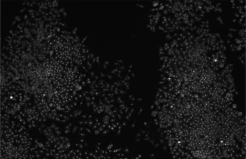
Application: ImmunocytochemistrySample Tested: R1 wild type Mouse embryonic Stem CellsSpecies: MouseVerified Customer | Posted 08/04/2016lin-28a in R1 wild type mouse embryonic stem cells

There are no reviews that match your criteria.
Protocols
Find general support by application which include: protocols, troubleshooting, illustrated assays, videos and webinars.
- Appropriate Fixation of IHC/ICC Samples
- Cellular Response to Hypoxia Protocols
- ClariTSA™ Fluorophore Kits
- Detection & Visualization of Antibody Binding
- ICC Cell Smear Protocol for Suspension Cells
- ICC Immunocytochemistry Protocol Videos
- ICC for Adherent Cells
- Immunocytochemistry (ICC) Protocol
- Immunocytochemistry Troubleshooting
- Immunofluorescence of Organoids Embedded in Cultrex Basement Membrane Extract
- Immunohistochemistry (IHC) and Immunocytochemistry (ICC) Protocols
- Preparing Samples for IHC/ICC Experiments
- Preventing Non-Specific Staining (Non-Specific Binding)
- Primary Antibody Selection & Optimization
- Protocol for VisUCyte™ HRP Polymer Detection Reagent
- Protocol for the Fluorescent ICC Staining of Cell Smears - Graphic
- Protocol for the Fluorescent ICC Staining of Cultured Cells on Coverslips - Graphic
- Protocol for the Preparation and Fluorescent ICC Staining of Cells on Coverslips
- Protocol for the Preparation and Fluorescent ICC Staining of Non-adherent Cells
- Protocol for the Preparation and Fluorescent ICC Staining of Stem Cells on Coverslips
- Protocol for the Preparation of a Cell Smear for Non-adherent Cell ICC - Graphic
- R&D Systems Quality Control Western Blot Protocol
- TUNEL and Active Caspase-3 Detection by IHC/ICC Protocol
- The Importance of IHC/ICC Controls
- Troubleshooting Guide: Western Blot Figures
- Western Blot Conditions
- Western Blot Protocol
- Western Blot Protocol for Cell Lysates
- Western Blot Troubleshooting
- Western Blot Troubleshooting Guide
- View all Protocols, Troubleshooting, Illustrated assays and Webinars
Loading...
Associated Pathways
