MCM7/minichromosome maintenance complex component 7, is a highly conserved mini-chromosome maintenance protein, with ATP binding, DNA helicase activity and single-stranded DNA binding. MCM7 forms a hexameric protein complex with MCM2, 4 and 6 proteins that has DNA helicase activity essential for the initiation of gene replication. MCM7 also binds to RAD17 to mediate ATR recruitment to damaged DNA. MCM7 expression is repressed in quiescent cells but induced in late G1 to S phase by growth factor stimulation. It is up-regulated in many cancers including colorectal, T cell lymphomas, glioblastoma, and has prognostic value for hepatocellular carcinoma and gastric adenocarcinoma.
Human MCM7 Antibody (2068B)
R&D Systems | Catalog # MAB9217
Recombinant Monoclonal Antibody.

Key Product Details
Species Reactivity
Human
Applications
Immunohistochemistry, Western Blot, Intracellular Staining by Flow Cytometry, Immunocytochemistry
Label
Unconjugated
Antibody Source
Recombinant Monoclonal Rabbit IgG Clone # 2068B
Loading...
Product Specifications
Immunogen
E. coli-derived recombinant human MCM7
Gly188-Ala328
Accession # P33993
Gly188-Ala328
Accession # P33993
Specificity
Detects human MCM7 in direct ELISAs and Western blots.
Clonality
Monoclonal
Host
Rabbit
Isotype
IgG
Scientific Data Images for Human MCM7 Antibody (2068B)
Detection of Human MCM7 by Western Blot.
Western blot shows lysates of HeLa human cervical epithelial carcinoma cell line, MCF-7 human breast cancer cell line, Daudi human Burkitt's lymphoma cell line, and Jurkat human acute T cell leukemia cell line. PVDF membrane was probed with 0.2 µg/mL of Rabbit Anti-Human MCM7 Monoclonal Antibody (Catalog # MAB9217) followed by HRP-conjugated Anti-Rabbit IgG Secondary Antibody (Catalog # HAF008). A specific band was detected for MCM7 at approximately 80 kDa (as indicated). This experiment was conducted under reducing conditions and using Immunoblot Buffer Group 1.
MCM7 in HeLa Human Cell Line.
MCM7 was detected in immersion fixed HeLa human cervical epithelial carcinoma cell line using Rabbit Anti-Human MCM7 Monoclonal Antibody (Catalog # MAB9217) at 1 µg/mL for 3 hours at room temperature. Cells were stained using the NorthernLights™ 557-conjugated Anti-Rabbit IgG Secondary Antibody (red; Catalog # NL004) and counterstained with DAPI (blue). Specific staining was localized to nuclei. View our protocol for Fluorescent ICC Staining of Cells on Coverslips.
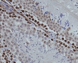
MCM7 in Human Mesothelioma Tissue.
MCM7 was detected in immersion fixed paraffin-embedded sections of human mesothelioma tissue using Rabbit Anti-Human MCM7 Monoclonal Antibody (Catalog # MAB9217) at 3 µg/mL for 1 hour at room temperature followed by incubation with the Anti-Rabbit IgG VisUCyte™ HRP Polymer Antibody (Catalog # VC003). Tissue was stained using DAB (brown) and counterstained with hematoxylin (blue). Specific staining was localized to nuclei. View our protocol for IHC Staining with VisUCyte HRP Polymer Detection Reagents.
Detection of MCM7 in HeLa Human Cell Line by Flow Cytometry.
HeLa human cervical epithelial carcinoma cell line was stained with Rabbit Anti-Human MCM7 Monoclonal Antibody (Catalog # MAB9217, filled histogram) or isotype control antibody (Catalog # AB-105-C, open histogram), followed by Allophycocyanin-conjugated Anti-Rabbit IgG Secondary Antibody (Catalog # F0111). To facilitate intracellular staining, cells were fixed and permeabilized with FlowX FoxP3 Fixation & Permeabilization Buffer Kit (Catalog # FC012). View our protocol for Staining Intracellular Molecules.Applications for Human MCM7 Antibody (2068B)
Application
Recommended Usage
Immunocytochemistry
1-25 µg/mL
Sample: Immersion fixed HeLa human cervical epithelial carcinoma cell line
Sample: Immersion fixed HeLa human cervical epithelial carcinoma cell line
Immunohistochemistry
3-25 µg/mL
Sample: Immersion fixed paraffin-embedded sections of human mesothelioma tissue
Sample: Immersion fixed paraffin-embedded sections of human mesothelioma tissue
Intracellular Staining by Flow Cytometry
0.25 µg/106 cells
Sample: HeLa human cervical epithelial carcinoma cell line fixed and permeabilized with FlowX FoxP3 Fixation & Permeabilization Buffer Kit (Catalog # FC012)
Sample: HeLa human cervical epithelial carcinoma cell line fixed and permeabilized with FlowX FoxP3 Fixation & Permeabilization Buffer Kit (Catalog # FC012)
Western Blot
0.2 µg/mL
Sample: HeLa human cervical epithelial carcinoma cell line, MCF‑7 human breast cancer cell line, Daudi human Burkitt's lymphoma cell line, and Jurkat human acute T cell leukemia cell line
Sample: HeLa human cervical epithelial carcinoma cell line, MCF‑7 human breast cancer cell line, Daudi human Burkitt's lymphoma cell line, and Jurkat human acute T cell leukemia cell line
Reviewed Applications
Read 1 review rated 5 using MAB9217 in the following applications:
Flow Cytometry Panel Builder
Bio-Techne Knows Flow Cytometry
Save time and reduce costly mistakes by quickly finding compatible reagents using the Panel Builder Tool.
Advanced Features
- Spectra Viewer - Custom analysis of spectra from multiple fluorochromes
- Spillover Popups - Visualize the spectra of individual fluorochromes
- Antigen Density Selector - Match fluorochrome brightness with antigen density

Formulation, Preparation, and Storage
Purification
Protein A or G purified from cell culture supernatant
Reconstitution
For liquid material, refer to CoA for concentration.
Formulation
Lyophilized from a 0.2 μm filtered solution in PBS with Trehalose. *Small pack size (SP) is supplied either lyophilized or as a 0.2 µm filtered solution in PBS.
Shipping
Lyophilized product is shipped at ambient temperature. Liquid small pack size (-SP) is shipped with polar packs. Upon receipt, store immediately at the temperature recommended below.
Stability & Storage
Use a manual defrost freezer and avoid repeated freeze-thaw cycles.
- 12 months from date of receipt, -20 to -70 °C as supplied.
- 1 month, 2 to 8 °C under sterile conditions after reconstitution.
- 6 months, -20 to -70 °C under sterile conditions after reconstitution.
Calculators
Background: MCM7
Long Name
Minichromosome Maintenance Protein 7
Alternate Names
CDC47, MCM2, P1CDC47, P85MCM, PNAS146, PPP1R104, Protein Phosphatase 1
Gene Symbol
MCM7
UniProt
Additional MCM7 Products
Product Documents for Human MCM7 Antibody (2068B)
Certificate of Analysis
To download a Certificate of Analysis, please enter a lot or batch number in the search box below.
Note: Certificate of Analysis not available for kit components.
Product Specific Notices for Human MCM7 Antibody (2068B)
For research use only
Related Research Areas
Customer Reviews for Human MCM7 Antibody (2068B) (1)
5 out of 5
1 Customer Rating
Have you used Human MCM7 Antibody (2068B)?
Submit a review and receive an Amazon gift card!
$25/€18/£15/$25CAN/¥2500 Yen for a review with an image
$10/€7/£6/$10CAN/¥1110 Yen for a review without an image
Submit a review
Customer Images
Showing
1
-
1 of
1 review
Showing All
Filter By:
-
Application: ImmunohistochemistrySample Tested: Testicular tissueSpecies: HumanVerified Customer | Posted 11/26/2021

There are no reviews that match your criteria.
Protocols
Find general support by application which include: protocols, troubleshooting, illustrated assays, videos and webinars.
- 7-Amino Actinomycin D (7-AAD) Cell Viability Flow Cytometry Protocol
- Antigen Retrieval Protocol (PIER)
- Antigen Retrieval for Frozen Sections Protocol
- Appropriate Fixation of IHC/ICC Samples
- Cellular Response to Hypoxia Protocols
- Chromogenic IHC Staining of Formalin-Fixed Paraffin-Embedded (FFPE) Tissue Protocol
- Chromogenic Immunohistochemistry Staining of Frozen Tissue
- ClariTSA™ Fluorophore Kits
- Detection & Visualization of Antibody Binding
- Extracellular Membrane Flow Cytometry Protocol
- Flow Cytometry Protocol for Cell Surface Markers
- Flow Cytometry Protocol for Staining Membrane Associated Proteins
- Flow Cytometry Staining Protocols
- Flow Cytometry Troubleshooting Guide
- Fluorescent IHC Staining of Frozen Tissue Protocol
- Graphic Protocol for Heat-induced Epitope Retrieval
- Graphic Protocol for the Preparation and Fluorescent IHC Staining of Frozen Tissue Sections
- Graphic Protocol for the Preparation and Fluorescent IHC Staining of Paraffin-embedded Tissue Sections
- Graphic Protocol for the Preparation of Gelatin-coated Slides for Histological Tissue Sections
- ICC Cell Smear Protocol for Suspension Cells
- ICC Immunocytochemistry Protocol Videos
- ICC for Adherent Cells
- IHC Sample Preparation (Frozen sections vs Paraffin)
- Immunocytochemistry (ICC) Protocol
- Immunocytochemistry Troubleshooting
- Immunofluorescence of Organoids Embedded in Cultrex Basement Membrane Extract
- Immunofluorescent IHC Staining of Formalin-Fixed Paraffin-Embedded (FFPE) Tissue Protocol
- Immunohistochemistry (IHC) and Immunocytochemistry (ICC) Protocols
- Immunohistochemistry Frozen Troubleshooting
- Immunohistochemistry Paraffin Troubleshooting
- Intracellular Flow Cytometry Protocol Using Alcohol (Methanol)
- Intracellular Flow Cytometry Protocol Using Detergents
- Intracellular Nuclear Staining Flow Cytometry Protocol Using Detergents
- Intracellular Staining Flow Cytometry Protocol Using Alcohol Permeabilization
- Intracellular Staining Flow Cytometry Protocol Using Detergents to Permeabilize Cells
- Preparing Samples for IHC/ICC Experiments
- Preventing Non-Specific Staining (Non-Specific Binding)
- Primary Antibody Selection & Optimization
- Propidium Iodide Cell Viability Flow Cytometry Protocol
- Protocol for Heat-Induced Epitope Retrieval (HIER)
- Protocol for Liperfluo
- Protocol for Making a 4% Formaldehyde Solution in PBS
- Protocol for VisUCyte™ HRP Polymer Detection Reagent
- Protocol for the Characterization of Human Th22 Cells
- Protocol for the Characterization of Human Th9 Cells
- Protocol for the Fluorescent ICC Staining of Cell Smears - Graphic
- Protocol for the Fluorescent ICC Staining of Cultured Cells on Coverslips - Graphic
- Protocol for the Preparation & Fixation of Cells on Coverslips
- Protocol for the Preparation and Chromogenic IHC Staining of Frozen Tissue Sections
- Protocol for the Preparation and Chromogenic IHC Staining of Frozen Tissue Sections - Graphic
- Protocol for the Preparation and Chromogenic IHC Staining of Paraffin-embedded Tissue Sections
- Protocol for the Preparation and Chromogenic IHC Staining of Paraffin-embedded Tissue Sections - Graphic
- Protocol for the Preparation and Fluorescent ICC Staining of Cells on Coverslips
- Protocol for the Preparation and Fluorescent ICC Staining of Non-adherent Cells
- Protocol for the Preparation and Fluorescent ICC Staining of Stem Cells on Coverslips
- Protocol for the Preparation and Fluorescent IHC Staining of Frozen Tissue Sections
- Protocol for the Preparation and Fluorescent IHC Staining of Paraffin-embedded Tissue Sections
- Protocol for the Preparation of Gelatin-coated Slides for Histological Tissue Sections
- Protocol for the Preparation of a Cell Smear for Non-adherent Cell ICC - Graphic
- Protocol: Annexin V and PI Staining by Flow Cytometry
- Protocol: Annexin V and PI Staining for Apoptosis by Flow Cytometry
- R&D Systems Quality Control Western Blot Protocol
- TUNEL and Active Caspase-3 Detection by IHC/ICC Protocol
- The Importance of IHC/ICC Controls
- Troubleshooting Guide: Fluorokine Flow Cytometry Kits
- Troubleshooting Guide: Immunohistochemistry
- Troubleshooting Guide: Western Blot Figures
- Western Blot Conditions
- Western Blot Protocol
- Western Blot Protocol for Cell Lysates
- Western Blot Troubleshooting
- Western Blot Troubleshooting Guide
- View all Protocols, Troubleshooting, Illustrated assays and Webinars
Loading...