Epithelial (E)‑Cadherin (ECAD), also known as Cadherin-1, cell-CAM120/80 in the human, uvomorulin in the mouse, Arc-1 in the dog, and L-CAM in the chicken, is a member of the Cadherin family of cell adhesion molecules (gene name CDH1). Cadherins are calcium-dependent transmembrane proteins which bind to one another in a homophilic manner. On their cytoplasmic side, they associate with the three catenins, alpha, beta, and gamma (plakoglobin). This association links the cadherin protein to the cytoskeleton. Without association with the catenins, the cadherins are non-adhesive. Cadherins play a role in development, specifically in tissue formation. They may also help to maintain tissue architecture in the adult. E-Cadherin may also play a role in tumor development, as loss of E-Cadherin has been associated with tumor invasiveness. E-Cadherin is a classical cadherin molecule. Classical cadherins consist of a large extracellular domain which contains DXD and DXNDN repeats responsible for mediating calcium‑dependent adhesion, a single-pass transmembrane domain, and a short carboxy-terminal cytoplasmic domain responsible for interacting with the catenins. E‑Cadherin contains five extracellular calcium-binding domains of approximately 110 amino acids each (amino acids 155-697).

Key Product Details
Species Reactivity
Human, Mouse
Applications
Multiplex Immunofluorescence, Immunohistochemistry, Western Blot, COMET
Label
Unconjugated
Antibody Source
Monoclonal Mouse IgG1 Clone # 1006132
Loading...
Product Specifications
Immunogen
Synthetic peptide containing human E‑Cadherin
Accession # P12830
Accession # P12830
Specificity
Detects human E-Cadherin, P-Cadherin, N-Cadherin and mouse E-Cadherin in Western blots. Due to the high homology of the immunogen used, this antibody is designed to be pan specific to Cadherins.
Clonality
Monoclonal
Host
Mouse
Isotype
IgG1
Scientific Data Images for Cadherin Pan Specific Antibody (1006132)
Detection of Cadherin in Human Liver via Multiplex Immunofluorescence staining on COMET™
Cadherin was detected in immersion fixed paraffin-embedded sections of human liver using Mouse Anti-Human/Mouse Cadherin Pan Specific Monoclonal Antibody (MAB18385) at 15 μg/mL at 37° Celsius for 8 minutes. Before incubation with the primary antibody, tissue underwent an all-in-one dewaxing and antigen retrieval preprocessing using PreTreatment Module (PT Module) and Dewax and HIER Buffer H (pH 9). Tissue was stained using the Alexa Fluor™ 647 Goat anti-Mouse IgG Secondary Antibody at 1:200 at 37° Celsius for 8 minutes. (Yellow; Lunaphore Catalog # DR647MS) and counterstained with DAPI (blue; Lunaphore Catalog # DR100). Specific staining was localized to the membrane. Protocol available in COMET™ Panel Builder.
Detection of Cadherin in Human Colon via Multiplex Immunofluorescence staining on COMET™
Cadherin was detected in immersion fixed paraffin-embedded sections of human colon using Mouse Anti-Human/Mouse Cadherin Pan Specific Monoclonal Antibody (MAB18385) at 15 μg/mL at 37° Celsius for 8 minutes. Before incubation with the primary antibody, tissue underwent an all-in-one dewaxing and antigen retrieval preprocessing using PreTreatment Module (PT Module) and Dewax and HIER Buffer H (pH 9). Tissue was stained using the Alexa Fluor™ 647 Goat anti-Mouse IgG Secondary Antibody at 1:200 at 37° Celsius for 8 minutes. (Yellow; Lunaphore Catalog # DR647MS) and counterstained with DAPI (blue; Lunaphore Catalog # DR100). Specific staining was localized to the membrane. Protocol available in COMET™ Panel Builder.
Detection of Cadherin in Human Colon Cancer via Multiplex Immunofluorescence staining on COMET™
Cadherin was detected in immersion fixed paraffin-embedded sections of human colon cancer using Mouse Anti-Human/Mouse Cadherin Pan Specific Monoclonal Antibody (MAB18385) at 15 μg/mL at 37 ° Celsius for 8 minutes. Before incubation with the primary antibody, tissue underwent an all-in-one dewaxing and antigen retrieval preprocessing using PreTreatment Module (PT Module) and Dewax and HIER Buffer H (pH 9). Tissue was stained using the Alexa Fluor™ 647 Goat anti-Mouse IgG Secondary Antibody at 1:200 at 37° Celsius for 8 minutes. (Yellow; Lunaphore Catalog # DR647MS) and counterstained with DAPI (blue; Lunaphore Catalog # DR100). Specific staining was localized to the membrane. Protocol available in COMET™ Panel Builder.
Detection of Cadherin in Mouse Stomach via seqIF™ staining on COMET™
Cadherin Antibody was detected in immersion fixed paraffin-embedded sections of Mouse Stomach using Mouse Anti-Human/Mouse E-Cadherin, Monoclonal Antibody (Catalog #MAB18385) at 15ug/mL at 37 ° Celsius for 4 minutes. Before incubation with the primary antibody, tissue underwent an all-in-one dewaxing and antigen retrieval preprocessing using PreTreatment Module (PT Module) and Dewax and HIER Buffer H (pH 9; Epredia Catalog # TA-999-DHBH). Tissue was stained using the Alexa Fluor™ 647 Goat anti-Mouse IgG Secondary Antibody at 1:200 at 37 ° Celsius for 2 minutes. (Yellow; Lunaphore Catalog # DR647MS) and counterstained with DAPI (blue; Lunaphore Catalog # DR100). Specific staining was localized to the membrane. Protocol available in COMET™ Panel Builder.
Detection of Human and Mouse Cadherin by Western Blot.
Western blot shows lysates of A431 human epithelial carcinoma cell line, A549 human lung carcinoma cell line, HepG2 human hepatocellular carcinoma cell line, MCF-7 human breast cancer cell line, and C2C12 mouse myoblast cell line. PVDF membrane was probed with 1 µg/mL of Mouse Anti-Human/Mouse Cadherin Pan Specific Monoclonal Antibody (Catalog # MAB18385) followed by HRP-conjugated Anti-Mouse IgG Secondary Antibody (HAF018). Specific bands were detected for Cadherin at approximately 135 kDa (as indicated). This experiment was conducted under reducing conditions and using Immunoblot Buffer Group 1.
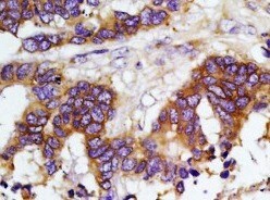
Cadherin in Human Cystadenocarcinoma.
Cadherin was detected in immersion fixed paraffin-embedded sections of human papillary serous cystadenocarcinoma of the colon using Mouse Anti-Human/Mouse Cadherin Pan Specific Monoclonal Antibody (Catalog # MAB18385) at 5 µg/mL for 1 hour at room temperature followed by incubation with the Anti-Mouse IgG VisUCyte™ HRP Polymer Antibody (VC001). Before incubation with the primary antibody, tissue was subjected to heat-induced epitope retrieval using Antigen Retrieval Reagent-Basic (CTS013). Tissue was stained using DAB (brown) and counterstained with hematoxylin (blue). Specific staining was localized to plasma membrane. View our protocol for IHC Staining with VisUCyte HRP Polymer Detection Reagents.
Detection of Human Cadherins by Western Blot.
Western blot shows lysates of HEK293T human embryonic kidney cell line mock transfected or transfected with human E-Cadherin and NS0 mouse myeloma cell line transfected with Human P-Cadherin, N-Cadherin, or mock transfected. PVDF membrane was probed with 1 µg/mL of Mouse Anti-Human/Mouse Cadherin Pan Specific Monoclonal Antibody (Catalog # MAB18385) followed by HRP-conjugated Anti-Mouse IgG Secondary Antibody (HAF018). A specific band was detected for Cadherin at approximately 135 kDa (as indicated). This experiment was conducted under reducing conditions and using Immunoblot Buffer Group 1.Applications for Cadherin Pan Specific Antibody (1006132)
Application
Recommended Usage
COMET
Optimal dilutions of this antibody should be experimentally determined.
Immunohistochemistry
5-25 µg/mL
Sample: Immersion fixed human papillary serous cystadenocarcinoma of the colon
Sample: Immersion fixed human papillary serous cystadenocarcinoma of the colon
Multiplex Immunofluorescence
15 µg/mL
Sample: Immersion fixed paraffin embedded sections of human liver, human colon, human colon cancer, and mouse stomach
Sample: Immersion fixed paraffin embedded sections of human liver, human colon, human colon cancer, and mouse stomach
Western Blot
1 µg/mL
Sample: A431 human epithelial carcinoma cell line, A549 human lung carcinoma cell line, HepG2 human hepatocellular carcinoma cell line, MCF‑7 human breast cancer cell line, C2C12 mouse myoblast cell line, HEK293T human embryonic kidney cell line, HEK293T human embryonic kidney cell line transfected with human E-Cadherin, and NS0 mouse myeloma cell line transfected with Human P-Cadherin or N-Cadherin
Sample: A431 human epithelial carcinoma cell line, A549 human lung carcinoma cell line, HepG2 human hepatocellular carcinoma cell line, MCF‑7 human breast cancer cell line, C2C12 mouse myoblast cell line, HEK293T human embryonic kidney cell line, HEK293T human embryonic kidney cell line transfected with human E-Cadherin, and NS0 mouse myeloma cell line transfected with Human P-Cadherin or N-Cadherin
Reviewed Applications
Read 1 review rated 5 using MAB18385 in the following applications:
Formulation, Preparation, and Storage
Purification
Protein A or G purified from hybridoma culture supernatant
Reconstitution
Reconstitute at 0.5 mg/mL in sterile PBS. For liquid material, refer to CoA for concentration.
Loading...
Formulation
Lyophilized from a 0.2 μm filtered solution in PBS with Trehalose. See Certificate of Analysis for details.
*Small pack size (-SP) is supplied either lyophilized or as a 0.2 µm filtered solution in PBS.
*Small pack size (-SP) is supplied either lyophilized or as a 0.2 µm filtered solution in PBS.
Shipping
Lyophilized product is shipped at ambient temperature. Liquid small pack size (-SP) is shipped with polar packs. Upon receipt, store immediately at the temperature recommended below.
Stability & Storage
Use a manual defrost freezer and avoid repeated freeze-thaw cycles.
- 12 months from date of receipt, -20 to -70 °C as supplied.
- 1 month, 2 to 8 °C under sterile conditions after reconstitution.
- 6 months, -20 to -70 °C under sterile conditions after reconstitution.
Calculators
Background: Cadherin
References
- Bussemakers, M.J.G. et al. (1993) Mol. Biol. Reports 17:123.
- Overduin, M. et al. (1995) Science 267:386.
- Takeichi, M. (1991) Science 251:1451.
Long Name
Cadherin Pan Specific
Alternate Names
Arc-1, cadherin 1, E-cadherin (epithelial), cadherin 1, type 1, E-cadherin (epithelial), cadherin-1, calcium-dependent adhesion protein, epithelial, CAM 120/80, CD324, CD324 antigen, CDHE, cell-CAM 120/80, ECAD, E-cadherin, Epithelial cadherin, LCAM, UVOE-Cadherin, uvomorulin
Entrez Gene IDs
999 (Human)
Gene Symbol
CDH1
UniProt
Additional Cadherin Products
Product Documents for Cadherin Pan Specific Antibody (1006132)
Certificate of Analysis
To download a Certificate of Analysis, please enter a lot or batch number in the search box below.
Note: Certificate of Analysis not available for kit components.
Product Specific Notices for Cadherin Pan Specific Antibody (1006132)
For research use only
Related Research Areas
Customer Reviews for Cadherin Pan Specific Antibody (1006132) (1)
5 out of 5
1 Customer Rating
Have you used Cadherin Pan Specific Antibody (1006132)?
Submit a review and receive an Amazon gift card!
$25/€18/£15/$25CAN/¥2500 Yen for a review with an image
$10/€7/£6/$10CAN/¥1110 Yen for a review without an image
Submit a review
Customer Images
Showing
1
-
1 of
1 review
Showing All
Filter By:
-
Application: ImmunohistochemistrySample Tested: Colon tissueSpecies: MouseVerified Customer | Posted 12/24/2021

There are no reviews that match your criteria.
Protocols
Find general support by application which include: protocols, troubleshooting, illustrated assays, videos and webinars.
- Antigen Retrieval Protocol (PIER)
- Antigen Retrieval for Frozen Sections Protocol
- Appropriate Fixation of IHC/ICC Samples
- Cellular Response to Hypoxia Protocols
- Chromogenic IHC Staining of Formalin-Fixed Paraffin-Embedded (FFPE) Tissue Protocol
- Chromogenic Immunohistochemistry Staining of Frozen Tissue
- ClariTSA™ Fluorophore Kits
- Detection & Visualization of Antibody Binding
- Fluorescent IHC Staining of Frozen Tissue Protocol
- Graphic Protocol for Heat-induced Epitope Retrieval
- Graphic Protocol for the Preparation and Fluorescent IHC Staining of Frozen Tissue Sections
- Graphic Protocol for the Preparation and Fluorescent IHC Staining of Paraffin-embedded Tissue Sections
- Graphic Protocol for the Preparation of Gelatin-coated Slides for Histological Tissue Sections
- IHC Sample Preparation (Frozen sections vs Paraffin)
- Immunofluorescent IHC Staining of Formalin-Fixed Paraffin-Embedded (FFPE) Tissue Protocol
- Immunohistochemistry (IHC) and Immunocytochemistry (ICC) Protocols
- Immunohistochemistry Frozen Troubleshooting
- Immunohistochemistry Paraffin Troubleshooting
- Preparing Samples for IHC/ICC Experiments
- Preventing Non-Specific Staining (Non-Specific Binding)
- Primary Antibody Selection & Optimization
- Protocol for Heat-Induced Epitope Retrieval (HIER)
- Protocol for Making a 4% Formaldehyde Solution in PBS
- Protocol for VisUCyte™ HRP Polymer Detection Reagent
- Protocol for the Preparation & Fixation of Cells on Coverslips
- Protocol for the Preparation and Chromogenic IHC Staining of Frozen Tissue Sections
- Protocol for the Preparation and Chromogenic IHC Staining of Frozen Tissue Sections - Graphic
- Protocol for the Preparation and Chromogenic IHC Staining of Paraffin-embedded Tissue Sections
- Protocol for the Preparation and Chromogenic IHC Staining of Paraffin-embedded Tissue Sections - Graphic
- Protocol for the Preparation and Fluorescent IHC Staining of Frozen Tissue Sections
- Protocol for the Preparation and Fluorescent IHC Staining of Paraffin-embedded Tissue Sections
- Protocol for the Preparation of Gelatin-coated Slides for Histological Tissue Sections
- R&D Systems Quality Control Western Blot Protocol
- TUNEL and Active Caspase-3 Detection by IHC/ICC Protocol
- The Importance of IHC/ICC Controls
- Troubleshooting Guide: Immunohistochemistry
- Troubleshooting Guide: Western Blot Figures
- Western Blot Conditions
- Western Blot Protocol
- Western Blot Protocol for Cell Lysates
- Western Blot Troubleshooting
- Western Blot Troubleshooting Guide
- View all Protocols, Troubleshooting, Illustrated assays and Webinars
Loading...