Cyclooxygenase-2 (COX-2) also known as prostaglandin G/H synthase 2 (PGHS2) is a 70 kDa microsomal enzyme that belongs to the prostaglandin G/H synthase family. It is inducibly-expressed by a number of cell types, including fibroblasts, vascular smooth muscle cells, endothelium, and monocytes. Functionally, COX-2 is a homodimer that catalyzes two steps in the conversion of arachadonic acid to prostaglandin H2. Mature human COX-2 is 587 amino acids (aa) in length and contains one EGF-like domain (aa 18‑55), a potential membrane interacting region (aa 277‑292) and a globular catalytic domain (aa 293‑604). At least one splice form exists that shows an 11 aa substitution for the C-terminal 451 amino acids. Mature human COX-2 shows 87% aa identity to mouse COX-2.

Key Product Details
Validated by
Biological Validation
Species Reactivity
Validated:
Human, Mouse
Cited:
Human, Mouse, Xenograft
Applications
Validated:
Western Blot, Intracellular Staining by Flow Cytometry, Immunocytochemistry, CyTOF-ready
Cited:
Immunohistochemistry, Immunohistochemistry-Paraffin, Immunohistochemistry-Frozen, Western Blot, Simple Western
Label
Unconjugated
Antibody Source
Polyclonal Goat IgG
Loading...
Product Specifications
Immunogen
E. coli-derived recombinant human COX-2
Ala18-Leu604
Accession # P35354
Ala18-Leu604
Accession # P35354
Specificity
Detects human and mouse COX-2 in Western blots. In Western blots, less than 1% cross-reactivity with recombinant human COX-1 is observed.
Clonality
Polyclonal
Host
Goat
Isotype
IgG
Scientific Data Images for COX-2 Antibody
Detection of Human and Mouse COX‑2 by Western Blot.
Western blot shows lysates of human peripheral blood mononuclear cell (PBMC) and RAW 264.7 mouse monocyte/macrophage cell line untreated (-) or treated (+) with 1 ug/mL LPS for 24 hours and U937 human histiocytic lymphoma cell line untreated or treated with 100 nM PMA and 1 ug/mL LPS for 48 hours and 24 hours, respectively. PVDF membrane was probed with 1 µg/mL of Human/Mouse COX-2 Polyclonal Antibody (Catalog # AF4198), followed by HRP-conjugated Anti-Goat IgG Secondary Antibody (Catalog # HAF109). A specific band was detected for COX-2 at approximately 75 kDa (as indicated). This experiment was conducted under reducing conditions and using Immunoblot Buffer Group 2.
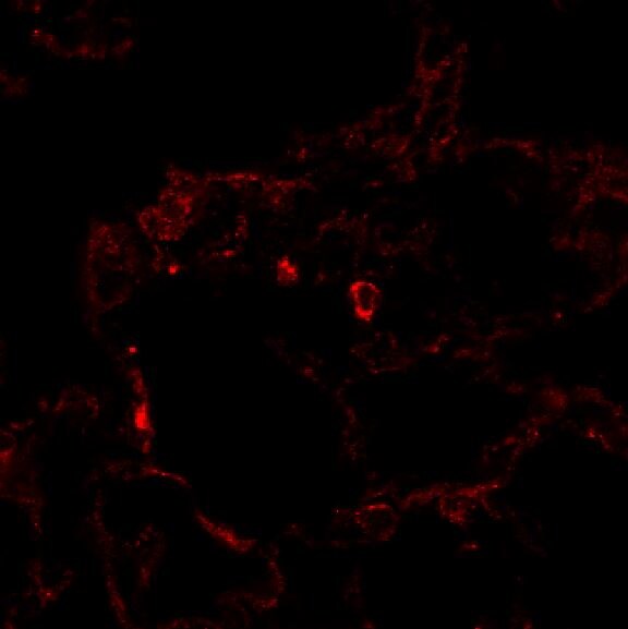
COX‑2 in HUVEC Human Cells.
COX-2 was detected in immersion fixed HUVEC human umbilical vein endothelial cells using 10 µg/mL Human/Mouse COX-2 Antigen Affinity-purified Polyclonal Antibody (Catalog # AF4198) for 3 hours at room temperature. Cells were stained with the NorthernLights™ 557-conjugated Anti-Goat IgG Secondary Antibody (red; Catalog # NL001) and counterstained with DAPI (blue). View our protocol for Fluorescent ICC Staining of Cells on Coverslips.
COX‑2 in RAW 264.7 Mouse Cells.
COX-2 was detected in immersion fixed RAW 264.7 mouse monocyte/macrophage cells stimulated with LPS using Goat Anti-Human/Mouse COX-2 Antigen Affinity-purified Polyclonal Antibody (Catalog # AF4198) at 10 µg/mL for 3 hours at room temperature. Cells were stained using the NorthernLights™ 557-conjugated Anti-Goat IgG Secondary Antibody (red; Catalog # NL001) and counterstained with DAPI (blue). Specific staining was localized to cytoplasm. View our protocol for Fluorescent ICC Staining of Cells on Coverslips.
Detection of COX‑2 in RAW 264.7 Mouse Cell Line by Flow Cytometry.
RAW 264.7 mouse monocyte/macrophage cell line treated with 1 µg/mL LPS for 24 hours was stained with Goat Anti-Human/Mouse COX-2 Antigen Affinity-purified Polyclonal Antibody (Catalog # AF4198, filled histogram) or control antibody (Catalog # AB-108-C, open histogram), followed by Allophycocyanin-conjugated Anti-Goat IgG Secondary Antibody (Catalog # F0108). To facilitate intracellular staining, cells were fixed with paraformaldehyde and permeabilized with saponin.
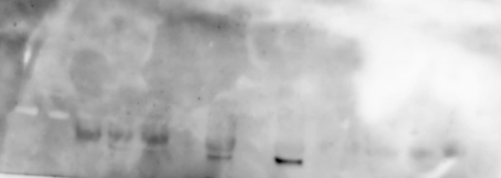
Detection of Mouse COX-2 by Western Blot
Fibrotic deposit and cardiac dysfunction correlate with decreasing CVPC presence in mdx heart. (A) Representative images of histological analysis stained using Masson trichrome technique showing myocytes (in red) and collagenous fibrotic tissue (in blue) in the left ventricle of WT and mdx hearts at 9, 24, and 52 wo. Line represents 100 µm. (B) The ratio of red and blue stained tissue was evaluated in WT hearts (open bars and black dots, n = 4–11 slices/3 animals per group) and mdx hearts (black bars and grey dots, n = 3–16 slices/3 animals per group) at the age of 24 wo and further at 52 wo. Statistical significance was calculated by Kruskal–Wallis test and Dunn‘s multiple comparison post-hoc test (*** p < 0.001, **** p < 0.0001). (C) Western blot analysis of collagen proteins and inflammatory proteins in the cardiac tissues. Left panel shows representative images of collagen 1A1 (Coll 1A1), collagen 3 (Coll 3), cyclooxygenase 2 (COX-2), and matrix metalloproteinase 9 (MMP-9) compared to the GAPDH control. The right panels show the normalized densitometry of each protein normalized by GAPDH content of WT (open bars, n = 2 animals) and mdx (black bars, n = 2 animals). Image collected and cropped by CiteAb from the following publication (https://pubmed.ncbi.nlm.nih.gov/34068508), licensed under a CC-BY license. Not internally tested by R&D Systems.Applications for COX-2 Antibody
Application
Recommended Usage
CyTOF-ready
Ready to be labeled using established conjugation methods. No BSA or other carrier proteins that could interfere with conjugation.
Immunocytochemistry
5-15 µg/mL
Sample: Immersion fixed HUVEC human umbilical vein endothelial cells and RAW 264.7 mouse monocyte/macrophage cells stimulated with LPS
Sample: Immersion fixed HUVEC human umbilical vein endothelial cells and RAW 264.7 mouse monocyte/macrophage cells stimulated with LPS
Intracellular Staining by Flow Cytometry
2.5 µg/106 cells
Sample: RAW 264.7 mouse monocyte/macrophage cell line treated LPS, fixed with paraformaldehyde and permeabilized with saponin
Sample: RAW 264.7 mouse monocyte/macrophage cell line treated LPS, fixed with paraformaldehyde and permeabilized with saponin
Western Blot
1 µg/mL
Sample: LPS-treated human peripheral blood mononuclear cell (PBMC) and RAW 264.7 mouse monocyte/macrophage cell line and PMA and LPS-treated U937 human histiocytic lymphoma cell line
Sample: LPS-treated human peripheral blood mononuclear cell (PBMC) and RAW 264.7 mouse monocyte/macrophage cell line and PMA and LPS-treated U937 human histiocytic lymphoma cell line
Reviewed Applications
Read 6 reviews rated 4.2 using AF4198 in the following applications:
Flow Cytometry Panel Builder
Bio-Techne Knows Flow Cytometry
Save time and reduce costly mistakes by quickly finding compatible reagents using the Panel Builder Tool.
Advanced Features
- Spectra Viewer - Custom analysis of spectra from multiple fluorochromes
- Spillover Popups - Visualize the spectra of individual fluorochromes
- Antigen Density Selector - Match fluorochrome brightness with antigen density

Formulation, Preparation, and Storage
Purification
Antigen Affinity-purified
Reconstitution
Reconstitute at 0.2 mg/mL in sterile PBS. For liquid material, refer to CoA for concentration.
Loading...
Formulation
Lyophilized from a 0.2 μm filtered solution in PBS with Trehalose. *Small pack size (SP) is supplied either lyophilized or as a 0.2 µm filtered solution in PBS.
Shipping
Lyophilized product is shipped at ambient temperature. Liquid small pack size (-SP) is shipped with polar packs. Upon receipt, store immediately at the temperature recommended below.
Stability & Storage
Use a manual defrost freezer and avoid repeated freeze-thaw cycles.
- 12 months from date of receipt, -20 to -70 °C as supplied.
- 1 month, 2 to 8 °C under sterile conditions after reconstitution.
- 6 months, -20 to -70 °C under sterile conditions after reconstitution.
Calculators
Background: COX-2
Long Name
Cyclooxygenase 2
Alternate Names
COX2, PGHS-2, PHS-II, PTGS2
Gene Symbol
PTGS2
UniProt
Additional COX-2 Products
Product Documents for COX-2 Antibody
Certificate of Analysis
To download a Certificate of Analysis, please enter a lot or batch number in the search box below.
Note: Certificate of Analysis not available for kit components.
Product Specific Notices for COX-2 Antibody
For research use only
Citations for COX-2 Antibody
 Powered by Bioz
Customer Reviews for COX-2 Antibody (6)
4.2 out of 5
6 Customer Ratings
Have you used COX-2 Antibody?
Submit a review and receive an Amazon gift card!
$25/€18/£15/$25CAN/¥2500 Yen for a review with an image
$10/€7/£6/$10CAN/¥1110 Yen for a review without an image
Submit a review
Customer Images
Showing
1
-
5 of
6 reviews
Showing All
Filter By:
-
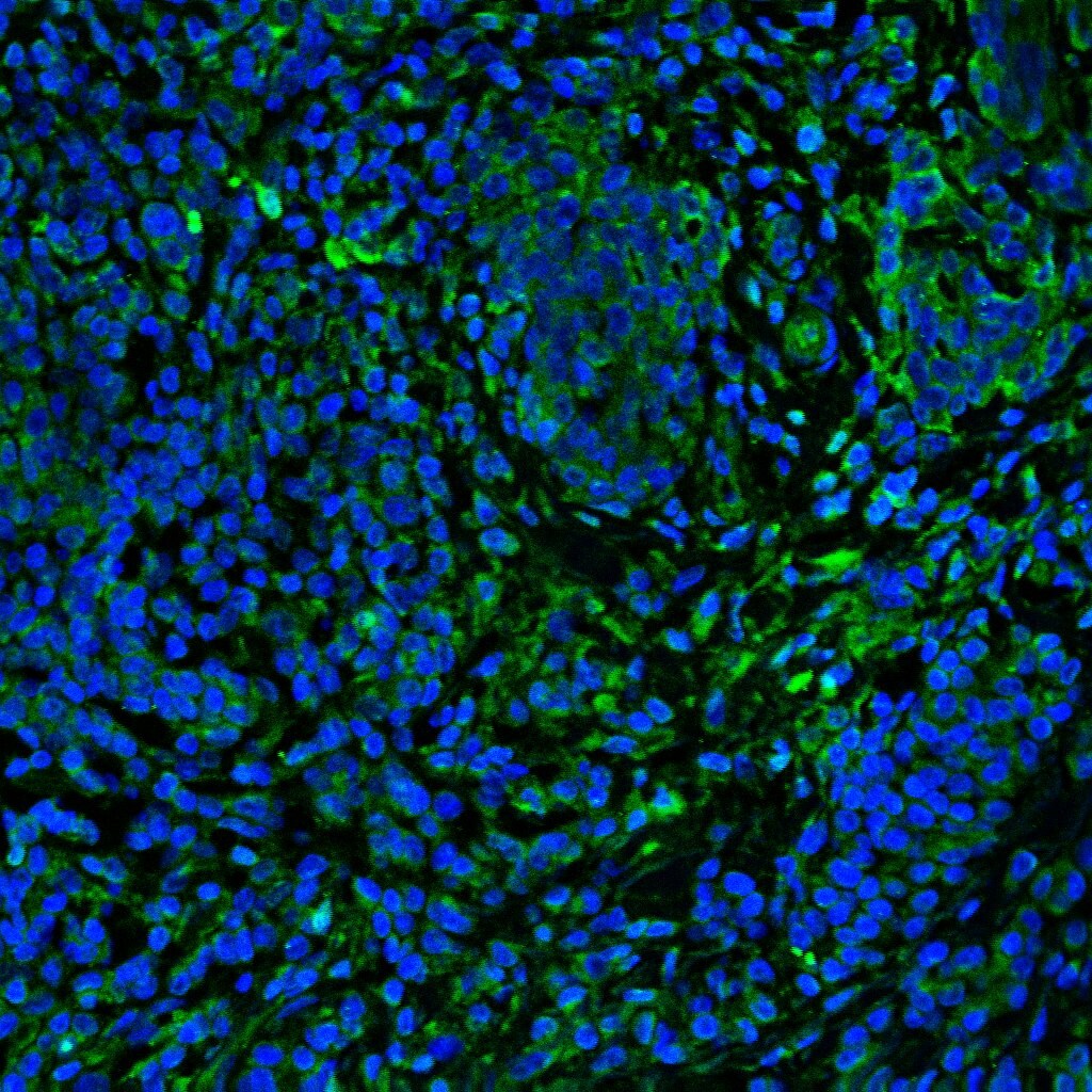
Application: Immunocytochemistry/ImmunofluorescenceSample Tested: Adult lungSpecies: MouseVerified Customer | Posted 02/14/2022Good to use!

-
Application: Immunocytochemistry/ImmunofluorescenceSample Tested: Melanoma tissueSpecies: HumanVerified Customer | Posted 11/04/2020
-
Application: Western BlotSample Tested: Adipose tissueSpecies: MouseVerified Customer | Posted 07/30/2019
-
Application: Immunocytochemistry/ImmunofluorescenceSample Tested: Spleen tissueSpecies: MouseVerified Customer | Posted 05/15/2017
-
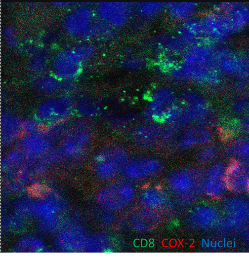
Application: Western BlotSample Tested: See PMID 24050770Species: MouseVerified Customer | Posted 01/08/2015
-
Application: Western BlotSample Tested: See PMID 24176842Species: HumanVerified Customer | Posted 01/08/2015
There are no reviews that match your criteria.
Protocols
Find general support by application which include: protocols, troubleshooting, illustrated assays, videos and webinars.
- 7-Amino Actinomycin D (7-AAD) Cell Viability Flow Cytometry Protocol
- Appropriate Fixation of IHC/ICC Samples
- Cellular Response to Hypoxia Protocols
- ClariTSA™ Fluorophore Kits
- Detection & Visualization of Antibody Binding
- Extracellular Membrane Flow Cytometry Protocol
- Flow Cytometry Protocol for Cell Surface Markers
- Flow Cytometry Protocol for Staining Membrane Associated Proteins
- Flow Cytometry Staining Protocols
- Flow Cytometry Troubleshooting Guide
- ICC Cell Smear Protocol for Suspension Cells
- ICC Immunocytochemistry Protocol Videos
- ICC for Adherent Cells
- Immunocytochemistry (ICC) Protocol
- Immunocytochemistry Troubleshooting
- Immunofluorescence of Organoids Embedded in Cultrex Basement Membrane Extract
- Immunohistochemistry (IHC) and Immunocytochemistry (ICC) Protocols
- Intracellular Flow Cytometry Protocol Using Alcohol (Methanol)
- Intracellular Flow Cytometry Protocol Using Detergents
- Intracellular Nuclear Staining Flow Cytometry Protocol Using Detergents
- Intracellular Staining Flow Cytometry Protocol Using Alcohol Permeabilization
- Intracellular Staining Flow Cytometry Protocol Using Detergents to Permeabilize Cells
- Preparing Samples for IHC/ICC Experiments
- Preventing Non-Specific Staining (Non-Specific Binding)
- Primary Antibody Selection & Optimization
- Propidium Iodide Cell Viability Flow Cytometry Protocol
- Protocol for Liperfluo
- Protocol for VisUCyte™ HRP Polymer Detection Reagent
- Protocol for the Characterization of Human Th22 Cells
- Protocol for the Characterization of Human Th9 Cells
- Protocol for the Fluorescent ICC Staining of Cell Smears - Graphic
- Protocol for the Fluorescent ICC Staining of Cultured Cells on Coverslips - Graphic
- Protocol for the Preparation and Fluorescent ICC Staining of Cells on Coverslips
- Protocol for the Preparation and Fluorescent ICC Staining of Non-adherent Cells
- Protocol for the Preparation and Fluorescent ICC Staining of Stem Cells on Coverslips
- Protocol for the Preparation of a Cell Smear for Non-adherent Cell ICC - Graphic
- Protocol: Annexin V and PI Staining by Flow Cytometry
- Protocol: Annexin V and PI Staining for Apoptosis by Flow Cytometry
- R&D Systems Quality Control Western Blot Protocol
- TUNEL and Active Caspase-3 Detection by IHC/ICC Protocol
- The Importance of IHC/ICC Controls
- Troubleshooting Guide: Fluorokine Flow Cytometry Kits
- Troubleshooting Guide: Western Blot Figures
- Western Blot Conditions
- Western Blot Protocol
- Western Blot Protocol for Cell Lysates
- Western Blot Troubleshooting
- Western Blot Troubleshooting Guide
- View all Protocols, Troubleshooting, Illustrated assays and Webinars
Loading...