Human High-mobility group box 1 protein (HMGB1), previously known as HMG-1 or amphoterin, is a member of the high mobility group box family of non-histone chromosomal proteins (1‑3). Human HMGB1 is expressed as a 30 kDa, 215 amino acid (aa) single chain polypeptide containing three domains: two N-terminal globular, 70 aa positively charged DNA-binding domains (HMG boxes A and B), and a negatively charged 30 aa C-terminal region that contains only Asp and Glu (4, 5). Residues 27‑43 and 178‑184 contain a NLS. Posttranslational modifications of the molecule have been reported, with acetylation occurring on as many as 17 lysine residues (6). HMGB1 is expressed at high levels in almost all cells (2, 4). It was originally discovered as a nuclear protein that could bend DNA. Such bending stabilizes nucleosome formation and regulates the expression of select genes upon recruitment by DNA binding proteins (1, 7, 8). It is now known that HMGB1 can also act extracellularly, both as an inflammatory mediator that promotes monocyte migration and cytokine secretion, and as a mediator of T cell-dendritic cell interaction (1, 4, 7, 9, 10). The cytokine activity of HBMG1 is restricted to the HMG B box, (3) while the A box is associated with the helix-loop-helix domain of transcription factors (11). HMBG1 is released in response to cell death and as a secretion product. Although HMBG-1 does not possess a classic signal sequence, it appears to be secreted as an acetylated form via secretory endolysosome exocytosis (6, 12). Once secreted, HMGB1 transduces cellular signals through its high affinity receptor, RAGE and, possibly, TLR2 and TLR4 (1, 3, 4). Human HMGB1 is 100% aa identical to canine HMGB1 and 99% aa identical to mouse, rat, bovine and porcine HMGB1, respectively.

Key Product Details
Species Reactivity
Human, Mouse
Applications
Immunocytochemistry
Label
Unconjugated
Antibody Source
Monoclonal Mouse IgG2B Clone # 1002147
Loading...
Product Specifications
Immunogen
Synthetic peptide containing human HMGB1
Thr85-Gly166
Accession # P09429
Thr85-Gly166
Accession # P09429
Specificity
Detects human HMGB1 in direct ELISAs. Detects human and mouse HMGB1 in Western blots.
Clonality
Monoclonal
Host
Mouse
Isotype
IgG2B
Scientific Data Images for HMGB1/HMG-1 Antibody (1002147)
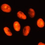
HMGB1/HMG‑1 in HeLa Human Cell Line.
HMGB1/HMG-1 was detected in immersion fixed HeLa human cervical epithelial carcinoma cell line using Mouse Anti-Human/Mouse HMGB1/HMG-1 Monoclonal Antibody (Catalog # MAB16902) at 8 µg/mL for 3 hours at room temperature. Cells were stained using the NorthernLights™ 557-conjugated Anti-Mouse IgG Secondary Antibody (red; Catalog # NL007) and counterstained with DAPI (blue). Specific staining was localized to nuclei. View our protocol for Fluorescent ICC Staining of Cells on Coverslips.Applications for HMGB1/HMG-1 Antibody (1002147)
Application
Recommended Usage
Immunocytochemistry
8-25 µg/mL
Sample: Immersion fixed HeLa human cervical epithelial carcinoma cell line
Sample: Immersion fixed HeLa human cervical epithelial carcinoma cell line
Reviewed Applications
Read 1 review rated 5 using MAB16902 in the following applications:
Formulation, Preparation, and Storage
Purification
Protein A or G purified from hybridoma culture supernatant
Reconstitution
Reconstitute at 0.5 mg/mL in sterile PBS. For liquid material, refer to CoA for concentration.
Loading...
Formulation
Lyophilized from a 0.2 μm filtered solution in PBS with Trehalose. *Small pack size (SP) is supplied either lyophilized or as a 0.2 µm filtered solution in PBS.
Shipping
Lyophilized product is shipped at ambient temperature. Liquid small pack size (-SP) is shipped with polar packs. Upon receipt, store immediately at the temperature recommended below.
Stability & Storage
Use a manual defrost freezer and avoid repeated freeze-thaw cycles.
- 12 months from date of receipt, -20 to -70 °C as supplied.
- 1 month, 2 to 8 °C under sterile conditions after reconstitution.
- 6 months, -20 to -70 °C under sterile conditions after reconstitution.
Calculators
Background: HMGB1/HMG-1
References
- Lotze, M.T. and K.J. Tracey (2005) Nat. Rev. Immunol. 5:331.
- Yang, H. et al. (2005) J. Leukoc. Biol. 78:1.
- Dumitriu, I.E. et al. (2005) Trends Immunol. 26:381.
- Degryse, B. and M. de Virgilio (2003) FEBS Lett. 553:11.
- Wen, L. et al. (1989) Nucleic Acids Res. 17:1197.
- Bonaldi, T. et al. (2003) EMBO J. 22:5551.
- Muller, S. et al. (2001) EMBO J. 20:4337.
- Bustin, M. (1999) Mol. Cell. Biol. 19:5237.
- Wang, H. et al. (1999) Science. 285:248.
- Dimitriu, I.E. et al. (2005) J. Immunol. 174:7506.
- Najima, Y. et al. (2005) J. Biol. Chem. 280:27523.
- Gardella, S. et al. (2002) EMBO Rep. 3:995.
Long Name
High Mobility Group Protein 1
Alternate Names
HMG-1, HMG1
Entrez Gene IDs
3146 (Human)
Gene Symbol
HMGB1
UniProt
Additional HMGB1/HMG-1 Products
Product Documents for HMGB1/HMG-1 Antibody (1002147)
Certificate of Analysis
To download a Certificate of Analysis, please enter a lot or batch number in the search box below.
Note: Certificate of Analysis not available for kit components.
Product Specific Notices for HMGB1/HMG-1 Antibody (1002147)
For research use only
Related Research Areas
Citations for HMGB1/HMG-1 Antibody (1002147)
 Powered by Bioz
Customer Reviews for HMGB1/HMG-1 Antibody (1002147) (1)
5 out of 5
1 Customer Rating
Have you used HMGB1/HMG-1 Antibody (1002147)?
Submit a review and receive an Amazon gift card!
$25/€18/£15/$25CAN/¥2500 Yen for a review with an image
$10/€7/£6/$10CAN/¥1110 Yen for a review without an image
Submit a review
Customer Images
Showing
1
-
1 of
1 review
Showing All
Filter By:
-
Application: Immunocytochemistry/ImmunofluorescenceSample Tested: glioma cellsSpecies: MouseVerified Customer | Posted 05/26/2022

There are no reviews that match your criteria.
Protocols
Find general support by application which include: protocols, troubleshooting, illustrated assays, videos and webinars.
- Appropriate Fixation of IHC/ICC Samples
- Cellular Response to Hypoxia Protocols
- ClariTSA™ Fluorophore Kits
- Detection & Visualization of Antibody Binding
- ICC Cell Smear Protocol for Suspension Cells
- ICC Immunocytochemistry Protocol Videos
- ICC for Adherent Cells
- Immunocytochemistry (ICC) Protocol
- Immunocytochemistry Troubleshooting
- Immunofluorescence of Organoids Embedded in Cultrex Basement Membrane Extract
- Immunohistochemistry (IHC) and Immunocytochemistry (ICC) Protocols
- Preparing Samples for IHC/ICC Experiments
- Preventing Non-Specific Staining (Non-Specific Binding)
- Primary Antibody Selection & Optimization
- Protocol for VisUCyte™ HRP Polymer Detection Reagent
- Protocol for the Fluorescent ICC Staining of Cell Smears - Graphic
- Protocol for the Fluorescent ICC Staining of Cultured Cells on Coverslips - Graphic
- Protocol for the Preparation and Fluorescent ICC Staining of Cells on Coverslips
- Protocol for the Preparation and Fluorescent ICC Staining of Non-adherent Cells
- Protocol for the Preparation and Fluorescent ICC Staining of Stem Cells on Coverslips
- Protocol for the Preparation of a Cell Smear for Non-adherent Cell ICC - Graphic
- TUNEL and Active Caspase-3 Detection by IHC/ICC Protocol
- The Importance of IHC/ICC Controls
- View all Protocols, Troubleshooting, Illustrated assays and Webinars
Loading...