
Key Product Details
Validated by
Biological Validation
Species Reactivity
Validated:
Human, Mouse
Cited:
Human, Mouse
Applications
Validated:
Immunohistochemistry, Western Blot
Cited:
Immunohistochemistry, Immunohistochemistry-Paraffin, Western Blot, Immunocytochemistry
Label
Unconjugated
Antibody Source
Polyclonal Goat IgG
Loading...
Product Specifications
Immunogen
E. coli-derived recombinant human MSX1
Met1-Thr165
Accession # P28360
Met1-Thr165
Accession # P28360
Specificity
Detects human and mouse MSX1 in Western blots.
Clonality
Polyclonal
Host
Goat
Isotype
IgG
Scientific Data Images for MSX1 Antibody
Detection of Human MSX1 by Western Blot.
Western blot shows lysates of HeLa human cervical epithelial carcinoma cell line. Gels were loaded with 30 µg of whole cell lysate (WCL), 20 µg of cytoplasmic (Cyto), and 10 µg of nuclear extracts (Nuc). PVDF membrane was probed with 0.1 µg/mL Goat Anti-Human/Mouse MSX1 Antigen Affinity-purified Polyclonal Antibody (Catalog # AF5045) followed by HRP-conjugated Anti-Goat IgG Secondary Antibody (Catalog # HAF017). A specific band for MSX1 was detected at approximately 40 kDa (as indicated). This experiment was conducted under reducing conditions and using Immunoblot Buffer Group 1.
MSX1 in Human Ovarian Cancer Tissue.
MSX1 was detected in immersion fixed paraffin-embedded sections of human ovarian cancer tissue using Goat Anti-Human/Mouse MSX1 Antigen Affinity-purified Polyclonal Antibody (Catalog # AF5045) at 0.3, 1.0 and 3.0 µg/mL overnight at 4 °C. Tissue was stained using the Anti-Goat HRP-DAB Cell & Tissue Staining Kit (brown; Catalog # CTS008) and counterstained with hematoxylin (blue). Specific staining was localized to nuclei. View our protocol for Chromogenic IHC Staining of Paraffin-embedded Tissue Sections.
Detection of Human MSX1 by Western Blot
Regulation of MSX1 expression is mediated by pSMAD1/5/8 in the human cap-stage tooth germs. (A–C) Immunostaining shows that MSX1 expression is abundant in dimethyl sulfoxide (DMSO)-treated human cap-stage tooth germs, dramatically reduced in dorsomorphin treated human cap-stage tooth germ, and unaltered in U0126 + SB203580 + SP600125 treated human cap-stage tooth germs. (D) A Western blot assay confirms the dramatically reduced expression of MSX1 in dorsomorphin treated human cap-stage tooth germs. Actin was used as the internal control. (E) Quantitative analysis of the Western blot assay. de, the dental epithelium; dm, the dental mesenchyme. Error bars represent SD. ***p < 0.001. Bar = 50 μm. Image collected and cropped by CiteAb from the following publication (https://pubmed.ncbi.nlm.nih.gov/35211032), licensed under a CC-BY license. Not internally tested by R&D Systems.
Detection of Mouse MSX1 by Immunohistochemistry
Msx1 expression in E12.5 Wls-knockout mutant and control cerebella. In control cerebellum (A,B), Msx1 is expressed in the RL. Expression of Msx1 in the RL is persisted in the Wls knockout cerebellum (C,D) and similar to Msx1 expression in the control RL. RL, rhombic lip. Scale bars, 100 μm. Image collected and cropped by CiteAb from the following open publication (https://www.frontiersin.org/articles/10.3389/fnmol.2024.1356544/full), licensed under a CC-BY license. Not internally tested by R&D Systems.
Detection of Mouse MSX1 by Immunohistochemistry
Msx1 and Msx3 expression in E12.5 Atoh1-null and wildtype cerebella. (A,B) The FISH double-labeling of Msx1 and Msx3 in the E12.5 Atoh1-null (a,a’) and wildtype cerebella (b,b’). (a’ and b’) illustrate that the sections from mutant and wildtype are collected at the same medio-lateral position. (C–F) Expression of Msx1 and Msx3 shifts and now sharing a boundary in the Atoh1-null cerebellum as denoted by the red arrow in (C). Panels (A,C–F) indicate Msx1 and Msx3 expression persistence in the Atoh1-null cerebellum. The shift of Msx1 and Msx3 expression, however, does not result in cells that co-express Msx1 and Msx3 (A,C–E). (G–I) A very different picture is shown in the wildtype cerebellum where there is a notable gap between cells that express Msx3 and Msx1 as noted by white arrow in (G). (F,J) DAPI (blue) used as a counterstain. RL, Rhombic Lip; VZ, ventricular zone. All scale bars, 100 μm. Image collected and cropped by CiteAb from the following open publication (https://www.frontiersin.org/articles/10.3389/fnmol.2024.1356544/full), licensed under a CC-BY license. Not internally tested by R&D Systems.
Detection of Mouse MSX1 by Immunohistochemistry
Temporal and spatial expression of Msx genes in the developing cerebellum. (A–C) Graphs show the dynamic nature of Msx expression in the cerebellum across 12 developmental time-points as observed from the RIKEN FANTOM5 transcriptome time-course data. (D–I) Sagittal sections of the RL with the right-side of panels denoting posterior and the bottom-side denoting ventral, with RNA in situ hybridization showing Msx genes expressed in the progenitor zones in (D–F) E11.5 and (G–I) E12.5. Msx1 expression is limited to the RL (white arrows in D,G) whereas Msx3 is limited to the VZ (black arrows in E,H). Msx2 expression is detected in the neuroepithelium but the boundary of the expression is not clear (F,I). See supplementary Figure S2 for negative control staining. RL, Rhombic Lip; VZ, Ventricular Zone. Scale bar, 100 μm. Image collected and cropped by CiteAb from the following open publication (https://www.frontiersin.org/articles/10.3389/fnmol.2024.1356544/full), licensed under a CC-BY license. Not internally tested by R&D Systems.Applications for MSX1 Antibody
Application
Recommended Usage
Immunohistochemistry
5-15 µg/mL
Sample: Immersion fixed paraffin-embedded sections of human ovarian cancer tissue
Sample: Immersion fixed paraffin-embedded sections of human ovarian cancer tissue
Western Blot
0.1 µg/mL
Sample: HeLa human cervical epithelial carcinoma cell line
Sample: HeLa human cervical epithelial carcinoma cell line
Reviewed Applications
Read 4 reviews rated 4.8 using AF5045 in the following applications:
Formulation, Preparation, and Storage
Purification
Antigen Affinity-purified
Reconstitution
Reconstitute at 0.2 mg/mL in sterile PBS. For liquid material, refer to CoA for concentration.
Loading...
Formulation
Lyophilized from a 0.2 μm filtered solution in PBS with Trehalose. *Small pack size (SP) is supplied either lyophilized or as a 0.2 µm filtered solution in PBS.
Shipping
Lyophilized product is shipped at ambient temperature. Liquid small pack size (-SP) is shipped with polar packs. Upon receipt, store immediately at the temperature recommended below.
Stability & Storage
Use a manual defrost freezer and avoid repeated freeze-thaw cycles.
- 12 months from date of receipt, -20 to -70 °C as supplied.
- 1 month, 2 to 8 °C under sterile conditions after reconstitution.
- 6 months, -20 to -70 °C under sterile conditions after reconstitution.
Calculators
Background: MSX1
Long Name
Msh Homeobox 1
Alternate Names
HOX7, HYD1, OFC5
Gene Symbol
MSX1
UniProt
Additional MSX1 Products
Product Documents for MSX1 Antibody
Certificate of Analysis
To download a Certificate of Analysis, please enter a lot or batch number in the search box below.
Note: Certificate of Analysis not available for kit components.
Product Specific Notices for MSX1 Antibody
For research use only
Citations for MSX1 Antibody
 Powered by Bioz
Customer Reviews for MSX1 Antibody (4)
4.8 out of 5
4 Customer Ratings
Have you used MSX1 Antibody?
Submit a review and receive an Amazon gift card!
$25/€18/£15/$25CAN/¥2500 Yen for a review with an image
$10/€7/£6/$10CAN/¥1110 Yen for a review without an image
Submit a review
Customer Images
Showing
1
-
4 of
4 reviews
Showing All
Filter By:
-
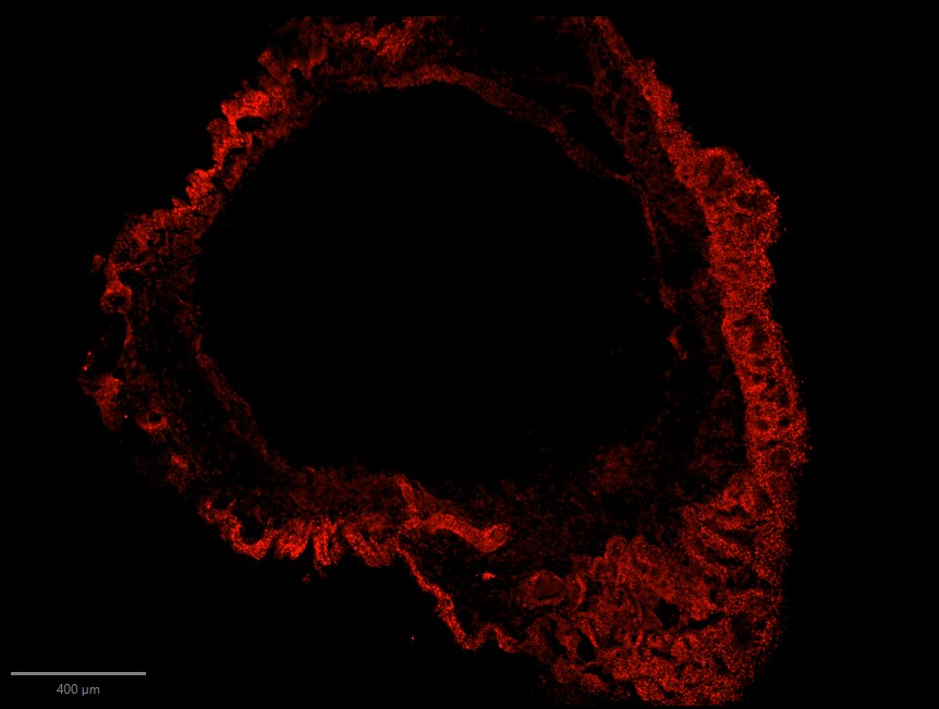
Application: Immunohistochemistry-FrozenSample Tested: choroid plexus organoidSpecies: HumanVerified Customer | Posted 04/17/2026MSX1 staining in choroid plexus organoid showing choroid plexus epitheliumfixation: 4% PFA 4° overnight blocking + permeablization: 10% BSA + 1% TX in PBS Primary antibody: MSX1 (1:200) diluted in 4% BSA + 0.1% TX in PBS 4° overnight Secondary antibody: anti-goat 568 antibody diluted in 4% BSA + 0.1% TX RT 1h

-
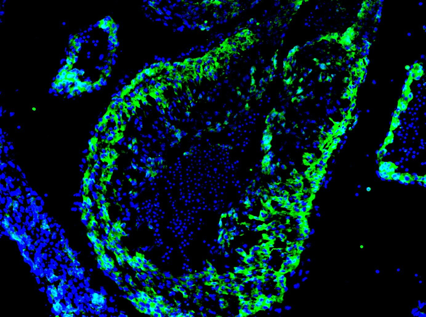
Application: Immunocytochemistry/ImmunofluorescenceSample Tested: E12.5 mouse embryo fixed in 4% PFASpecies: MouseVerified Customer | Posted 06/23/2021Fixed 4% PFA overnight. Blocked with 1% BSA Primary antibody dilution - 1:20 Secondary antibody - Invitrogen Alexa Fluor 488 Secondary antibody dilution - 1:1000 Stained on a E12.5 mouse heart section.
-
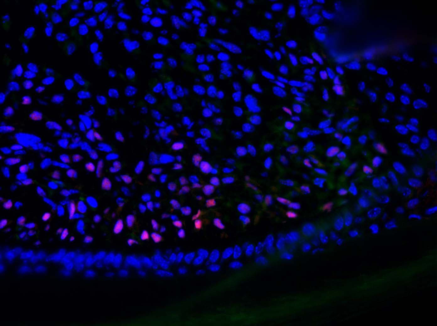
Application: Immunocytochemistry/ImmunofluorescenceSample Tested: mouse digitsSpecies: MouseVerified Customer | Posted 12/17/2018Primary antibody was used at 1:100 and incubated at 4°C overnight.
-
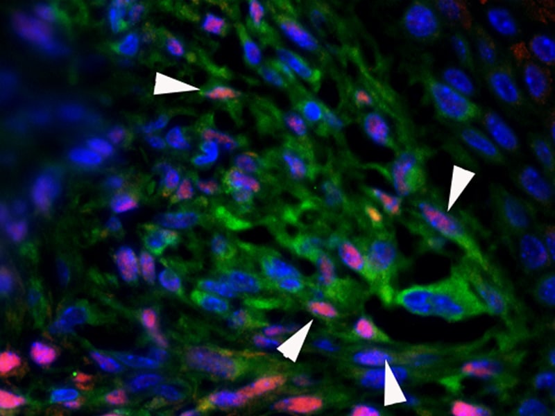
Application: Immunocytochemistry/ImmunofluorescenceSample Tested: digit sectionSpecies: MouseVerified Customer | Posted 12/17/2018Msx1 expression on frozen sections of regenerating mouse digit was detected with AF5045 (1:200) incubated overnight in fridge ( 4°C) in PBS/3% BSA/0.1% Triton-X100, secondary antibody (1:500) incubated at room temperature for 90 minutes, followed by nuclei counterstaining with DAPI.
There are no reviews that match your criteria.
Protocols
Find general support by application which include: protocols, troubleshooting, illustrated assays, videos and webinars.
- Antigen Retrieval Protocol (PIER)
- Antigen Retrieval for Frozen Sections Protocol
- Appropriate Fixation of IHC/ICC Samples
- Cellular Response to Hypoxia Protocols
- Chromogenic IHC Staining of Formalin-Fixed Paraffin-Embedded (FFPE) Tissue Protocol
- Chromogenic Immunohistochemistry Staining of Frozen Tissue
- ClariTSA™ Fluorophore Kits
- Detection & Visualization of Antibody Binding
- Fluorescent IHC Staining of Frozen Tissue Protocol
- Graphic Protocol for Heat-induced Epitope Retrieval
- Graphic Protocol for the Preparation and Fluorescent IHC Staining of Frozen Tissue Sections
- Graphic Protocol for the Preparation and Fluorescent IHC Staining of Paraffin-embedded Tissue Sections
- Graphic Protocol for the Preparation of Gelatin-coated Slides for Histological Tissue Sections
- IHC Sample Preparation (Frozen sections vs Paraffin)
- Immunofluorescent IHC Staining of Formalin-Fixed Paraffin-Embedded (FFPE) Tissue Protocol
- Immunohistochemistry (IHC) and Immunocytochemistry (ICC) Protocols
- Immunohistochemistry Frozen Troubleshooting
- Immunohistochemistry Paraffin Troubleshooting
- Preparing Samples for IHC/ICC Experiments
- Preventing Non-Specific Staining (Non-Specific Binding)
- Primary Antibody Selection & Optimization
- Protocol for Heat-Induced Epitope Retrieval (HIER)
- Protocol for Making a 4% Formaldehyde Solution in PBS
- Protocol for VisUCyte™ HRP Polymer Detection Reagent
- Protocol for the Preparation & Fixation of Cells on Coverslips
- Protocol for the Preparation and Chromogenic IHC Staining of Frozen Tissue Sections
- Protocol for the Preparation and Chromogenic IHC Staining of Frozen Tissue Sections - Graphic
- Protocol for the Preparation and Chromogenic IHC Staining of Paraffin-embedded Tissue Sections
- Protocol for the Preparation and Chromogenic IHC Staining of Paraffin-embedded Tissue Sections - Graphic
- Protocol for the Preparation and Fluorescent IHC Staining of Frozen Tissue Sections
- Protocol for the Preparation and Fluorescent IHC Staining of Paraffin-embedded Tissue Sections
- Protocol for the Preparation of Gelatin-coated Slides for Histological Tissue Sections
- R&D Systems Quality Control Western Blot Protocol
- TUNEL and Active Caspase-3 Detection by IHC/ICC Protocol
- The Importance of IHC/ICC Controls
- Troubleshooting Guide: Immunohistochemistry
- Troubleshooting Guide: Western Blot Figures
- Western Blot Conditions
- Western Blot Protocol
- Western Blot Protocol for Cell Lysates
- Western Blot Troubleshooting
- Western Blot Troubleshooting Guide
- View all Protocols, Troubleshooting, Illustrated assays and Webinars
Loading...
Associated Pathways
