
Key Product Details
Validated by
Species Reactivity
Validated:
Cited:
Applications
Validated:
Cited:
Label
Antibody Source
Product Specifications
Immunogen
Met1-Arg153
Accession # Q8R4B8
Specificity
Clonality
Host
Isotype
Scientific Data Images for NLRP3/NALP3 Antibody (768319)
Detection of Mouse NLRP3/NALP3 by Western Blot.
Western blot shows lysates of J774A.1 mouse reticulum cell sarcoma macrophage cell line and RAW 264.7 mouse monocyte/macrophage cell line. PVDF membrane was probed with 2 µg/mL of Rat Anti-Human/Mouse NLRP3/NALP3 Monoclonal Antibody (Catalog # MAB7578) followed by HRP-conjugated Anti-Rat IgG Secondary Antibody (Catalog # HAF005). A specific band was detected for NLRP3/NALP3 at approximately 117 kDa (as indicated). This experiment was conducted under reducing conditions and using Immunoblot Buffer Group 1.Catalog # AF6789is recommended for use with human lysates.
NLRP3/NALP3 in RAW 264.7 Mouse Cell Line.
NLRP3/NALP3 was detected in immersion fixed RAW 264.7 mouse monocyte/macrophage cell line using Rat Anti-Human/Mouse NLRP3/NALP3 Monoclonal Antibody (Catalog # MAB7578) at 10 µg/mL for 3 hours at room temperature. Cells were stained using the Northern-Lights™ 557-conjugated Anti-Rat IgG Secondary Antibody (red; Catalog # NL013) and counterstained with DAPI (blue). Specific staining was localized to cytoplasm. View our protocol for Fluorescent ICC Staining of Cells on Coverslips.
NLRP3/NALP3 in Human Tonsil Tissue.
NLRP3/NALP3 was detected in immersion fixed paraffin-embedded sections of human tonsil tissue using Rat Anti-Human/Mouse NLRP3/NALP3 Monoclonal Antibody (Catalog # MAB7578) at 5 µg/mL overnight at 4 °C. Before incubation with the primary antibody, tissue was subjected to heat-induced epitope retrieval using Antigen Retrieval Reagent-Basic (Catalog # CTS013). Tissue was stained using the Anti-Rat IgG VisUCyte™ HRP Polymer Antibody (brown; Catalog # VC005) and counterstained with hematoxylin (blue). Specific staining was localized to cytoplasm in lymphocytes. View our protocol for IHC Staining with VisUCyte HRP Polymer Detection Reagents.
Detection of NLRP3/NALP3 in RAW 264.7 Mouse Cell Line by Flow Cytometry.
RAW 264.7 mouse monocyte/macrophage cell line was stained with Rat Anti-Human/Mouse NLRP3/ NALP3 Monoclonal Antibody (Catalog # MAB7578, filled histo-gram) or isotype control antibody (Catalog # MAB006, open histo-gram), followed by Allophycocyanin-conjugated Anti-Rat IgG Secondary Antibody (Catalog # F0113). To facilitate intracellular staining, cells were fixed with paraformaldehyde and permeabilized with saponin.
Detection of NLRP3/NALP3 in Human Monocytes by Flow Cytometry.
Human peripheral blood monocytes was stained with Rat Anti-Human/Mouse NLRP3/NALP3 Monoclonal Antibody (Catalog # MAB7578, filled histogram) or isotype control antibody (Catalog # MAB006, open histogram), followed by Allophycocyanin-conjugated Anti-Rat IgG Secondary Antibody (Catalog # F0113). To facilitate intracellular staining, cells were fixed with paraformaldehyde and permeabilized with saponin.
Detection of Mouse NLRP3/NALP3 by Western Blot
Sodium butyrate (NaB) and hydroxybutyric acid (HBA) eyedrops improve clinical parameters and decrease inflammation in animals subjected to alkali burns (AB). (A) Digital images of corneas stained with 0.1% sodium fluorescein demonstrating intact corneal epithelium after topical NaB, HBA or balanced salt solution (BSS) treatment for 5 days quater in die (QID) in untreated mice (n = 5 animals/group). Scale bar: 1000 µm; (B) Representative color digital images used to score corneal opacity (top row) and representative fluorescein stained corneas used to create wound closure rate (bottom row) at 5 days (n = 15 animals/group) after alkali burn. Scale bar: 1000 µm; (C) Corneal opacity in corneas subjected to alkali burn and topically treated with either NaB, HBA, or BSS and compared with dexamethasone (Dex) (n = 15 animals/group); (D) Wound closure rate in corneas subjected to alkali burn and topically treated with either NaB, HBA, or BSS and compared with Dex (n = 15 animals/group); (E) Mean ± SEM of results of gene expression analysis of NLRP3, Caspase-1, and IL-1 beta in whole cornea of animals subjected to alkali burn for 2 or 5 days and topically treated with either NaB, HBA, or BSS and compared with Dex (n = 5 animals/group). * p < 0.05, *** p < 0.001, **** p < 0.0001. (F) Representative digital images of western blot of NLRP3 and beta -actin in cornea epithelium of animals subjected to alkali burn for 2 days and topically treated with either NaB, HBA, or BSS (n = 12 animals/3 samples/group). Image collected and cropped by CiteAb from the following publication (https://pubmed.ncbi.nlm.nih.gov/28273882), licensed under a CC-BY license. Not internally tested by R&D Systems.
Detection of Mouse NLRP3/NALP3 by Western Blot
Upregulation of nucleotide-binding oligomerization domain-containing protein (NOD)-like receptor family pyrin domain containing 3 (NLRP3) expression by corneal epithelium in animals subjected to alkali burn (AB) at 2 days (2 d) and 5 days (5 d) post-injury. (A) Gene expression analysis of NLRP3 messenger mRNA (mRNA) transcript in whole cornea of animals subjected to alkali burn (n = 8 animals/group). Graphs show means ± standard error of the mean (SEM). **** p < 0.0001; (B) Representative merged digital images of laser scanning confocal microscopy of corneas cryosections immunostained for NLRP3 (green) with propidium iodide nuclei counterstaining (DNA in red) in corneas subjected to alkali burn (images are representative of n = 6 animals/group). Scale bar: 50 µm; (C) Representative digital images of western blot of NLRP3 and beta -actin in cornea epithelium of animals subjected to alkali burn for 2 and 5 days. Each lane is a different sample (n = 12 animals/3 samples/group). MW: Molecular weight; UT: untreated control; 2 d and 5 d refer to mice subjected to alkali burn and euthanized 2 or 5 days post-injury, respectively (1 and 2 indicate different samples). Image collected and cropped by CiteAb from the following publication (https://pubmed.ncbi.nlm.nih.gov/28273882), licensed under a CC-BY license. Not internally tested by R&D Systems.
Detection of Mouse NLRP3/NALP3 by Immunocytochemistry/Immunofluorescence
Upregulation of nucleotide-binding oligomerization domain-containing protein (NOD)-like receptor family pyrin domain containing 3 (NLRP3) expression by corneal epithelium in animals subjected to alkali burn (AB) at 2 days (2 d) and 5 days (5 d) post-injury. (A) Gene expression analysis of NLRP3 messenger mRNA (mRNA) transcript in whole cornea of animals subjected to alkali burn (n = 8 animals/group). Graphs show means ± standard error of the mean (SEM). **** p < 0.0001; (B) Representative merged digital images of laser scanning confocal microscopy of corneas cryosections immunostained for NLRP3 (green) with propidium iodide nuclei counterstaining (DNA in red) in corneas subjected to alkali burn (images are representative of n = 6 animals/group). Scale bar: 50 µm; (C) Representative digital images of western blot of NLRP3 and beta -actin in cornea epithelium of animals subjected to alkali burn for 2 and 5 days. Each lane is a different sample (n = 12 animals/3 samples/group). MW: Molecular weight; UT: untreated control; 2 d and 5 d refer to mice subjected to alkali burn and euthanized 2 or 5 days post-injury, respectively (1 and 2 indicate different samples). Image collected and cropped by CiteAb from the following publication (https://pubmed.ncbi.nlm.nih.gov/28273882), licensed under a CC-BY license. Not internally tested by R&D Systems.
Detection of Mouse NLRP3/NALP3 by Immunohistochemistry
NLRP3 inflammasome-dependent pyroptosis occurs in liver fibrosis. (A) IHC staining for GSDMD, IL-1 beta, and IL-18 in liver sections from liver fibrosis patients and HCs. Scale bar: 40 µm. (B–D) ELISA analyses of serum levels of GSDMD (B), IL-1 beta (C), and IL-18 (D) in liver fibrosis patients (n = 89) and HCs (n = 60). (E) Representative immunofluorescence images of NLRP3 (red) and albumin (hepatocyte marker) (top), F4/80 (KC marker) (middle) or alpha -SMA (HSC marker) (bottom) (green) from the human fibrotic liver tissues. Scale bar: 40 µm. (F) Schematic diagram of the study. Liver fibrosis was induced by CCl4 injection for 8 weeks. (G) Representative mouse liver histology of H&E, Sirius Red staining, and IHC staining for alpha -SMA, GSDMD, and IL-1 beta. Black scale bar: 100 µm; Red scale bar: 50 µm. (H–J) ELISA analyses for serum levels of GSDMD (H), IL-1 beta (I), and IL-18 (J) in CCl4 group mouse (n = 5) and vehicle group mouse (n = 5). (K) Representative immunofluorescence images of NLRP3 (red) and albumin (hepatocyte marker) (top), F4/80 (KC marker) (middle) or alpha -SMA (HSC marker) (bottom) (green) from the 8-week CCl4-treated mouse liver. The vehicle group mouse liver was used as a control. Scale bar: 40 µm. (L) The qRT-PCR analysis for mRNA levels of IL-1 beta in THP-1 macrophages treated with LPS to induce pyroptosis. (M) ELISA analysis for IL-1 beta expression in supernatants from THP-1. (N) Western blot analysis of COL1A1, alpha -SMA, and TGF-beta expression in LX-2 cells which were exposed to CM from LPS-treated THP-1 macrophages. The protein expression was quantified by densitometry and normalized to beta -actin and are shown as fold changes relative to the control group (right panel). ** p < 0.01, *** p < 0.001. Image collected and cropped by CiteAb from the following open publication (https://pubmed.ncbi.nlm.nih.gov/36429008), licensed under a CC-BY license. Not internally tested by R&D Systems.
Detection of Human NLRP3/NALP3 by Immunohistochemistry
NLRP3 inflammasome-dependent pyroptosis occurs in liver fibrosis. (A) IHC staining for GSDMD, IL-1 beta, and IL-18 in liver sections from liver fibrosis patients and HCs. Scale bar: 40 µm. (B–D) ELISA analyses of serum levels of GSDMD (B), IL-1 beta (C), and IL-18 (D) in liver fibrosis patients (n = 89) and HCs (n = 60). (E) Representative immunofluorescence images of NLRP3 (red) and albumin (hepatocyte marker) (top), F4/80 (KC marker) (middle) or alpha -SMA (HSC marker) (bottom) (green) from the human fibrotic liver tissues. Scale bar: 40 µm. (F) Schematic diagram of the study. Liver fibrosis was induced by CCl4 injection for 8 weeks. (G) Representative mouse liver histology of H&E, Sirius Red staining, and IHC staining for alpha -SMA, GSDMD, and IL-1 beta. Black scale bar: 100 µm; Red scale bar: 50 µm. (H–J) ELISA analyses for serum levels of GSDMD (H), IL-1 beta (I), and IL-18 (J) in CCl4 group mouse (n = 5) and vehicle group mouse (n = 5). (K) Representative immunofluorescence images of NLRP3 (red) and albumin (hepatocyte marker) (top), F4/80 (KC marker) (middle) or alpha -SMA (HSC marker) (bottom) (green) from the 8-week CCl4-treated mouse liver. The vehicle group mouse liver was used as a control. Scale bar: 40 µm. (L) The qRT-PCR analysis for mRNA levels of IL-1 beta in THP-1 macrophages treated with LPS to induce pyroptosis. (M) ELISA analysis for IL-1 beta expression in supernatants from THP-1. (N) Western blot analysis of COL1A1, alpha -SMA, and TGF-beta expression in LX-2 cells which were exposed to CM from LPS-treated THP-1 macrophages. The protein expression was quantified by densitometry and normalized to beta -actin and are shown as fold changes relative to the control group (right panel). ** p < 0.01, *** p < 0.001. Image collected and cropped by CiteAb from the following open publication (https://pubmed.ncbi.nlm.nih.gov/36429008), licensed under a CC-BY license. Not internally tested by R&D Systems.
Detection of Mouse NLRP3/NALP3 by Immunohistochemistry
DAMP S100A8 along with NLRP3 inflammasome-dependent pyroptosis is positively related to the progression of liver fibrosis. (A) Representative IHC images for S100A8 and S100A9 in liver sections from liver fibrosis patients and HCs. (B,C) ELISA analyses for serum levels of S100A8 and S100A9 in liver fibrosis patients and HCs. (D) Comparison of serum S100A8 and S100A9 levels in liver fibrosis patients with different phases. (E–G) Distribution of serum GSDMD (E), IL-1 beta (F), and IL-18 (G) levels in liver fibrosis patients with different phases (F0–4). (H–J) Correlation between serum S100A8 levels and GSDMD (H), IL-1 beta (I) or IL-18 (J) levels in liver fibrosis patients. (K) Representative mouse liver morphology and staining with H&E and Sirius Red. (L–O) IHC staining of mouse liver sections for NLRP3, S100A8, and S100A9. Black scale bar: 100 µm; Red scale bar: 50 µm. ELISA analyses for serum levels of S100A8 (L), GSDMD (M), IL-1 beta (n), and IL-18 (O) in 4-, 6-, and 8 week-mouse models of liver fibrosis. *** p < 0.001. Image collected and cropped by CiteAb from the following open publication (https://pubmed.ncbi.nlm.nih.gov/36429008), licensed under a CC-BY license. Not internally tested by R&D Systems.
Detection of Mouse NLRP3/NALP3 by Immunohistochemistry
Inhibition of NLRP3 inflammasome-dependent pyroptosis alleviates liver fibrosis progression. (A) Experimental protocol of NLRP3 inhibitor MCC950 or saline application based on CCl4 injection in mice. (B) Representative liver histology of H&E and Sirius Red staining. The expression of alpha -SMA, NLRP3, GSDMD, and IL-1 beta was determined by immunohistochemistry. Black scale bar: 100 µm; Red scale bar: 50 µm. (C–E) Serum levels of ALT, AST, and TP were measured. * p < 0.05. Image collected and cropped by CiteAb from the following open publication (https://pubmed.ncbi.nlm.nih.gov/36429008), licensed under a CC-BY license. Not internally tested by R&D Systems.
Detection of Human NLRP3/NALP3 by Immunohistochemistry
Expression of NLRP3, caspase‐1 and gasdermin D‐N‐terminal (GSDMD‐N) in synovial tissues from patients with osteoarthritis (OA) or rheumatoid arthritis (RA). (A) Sections were stained with antibodies against the indicated proteins. Scale bar, 100 μm. (B) Sections were stained with an antibody against the macrophage marker CD68 and another antibody against a pyroptosis‐related marker. Nuclei were counterstained with DAPI. Scale bar, 50 μm. Image collected and cropped by CiteAb from the following open publication (https://pubmed.ncbi.nlm.nih.gov/37386795), licensed under a CC-BY license. Not internally tested by R&D Systems.
Detection of NLRP3/NALP3 by Immunocytochemistry/ Immunofluorescence
Effect of ginsenoside Rh4 on NLRP3 expression in LPS and nigericin-induced microglia cells. (A): NLRP3 (red) and nuclei (blue) using a laser confocal microscope; (B): Quantitative analysis of NLRP3 fluorescence intensity. Scale bars, 70 μm. The bar represents the mean ± standard error of the mean (n = 3). The Student’s t-test determined significant differences (### p < 0.001 compared with normal cells, *** p < 0.001 with LPS and nigericin-treated cells). Image collected and cropped by CiteAb from the following open publication (https://pubmed.ncbi.nlm.nih.gov/37239965), licensed under a CC-BY license. Not internally tested by R&D Systems.
Detection of Human NLRP3/NALP3 by Immunohistochemistry
Expression of NLRP3, caspase‐1 and gasdermin D‐N‐terminal (GSDMD‐N) in synovial tissues from patients with osteoarthritis (OA) or rheumatoid arthritis (RA). (A) Sections were stained with antibodies against the indicated proteins. Scale bar, 100 μm. (B) Sections were stained with an antibody against the macrophage marker CD68 and another antibody against a pyroptosis‐related marker. Nuclei were counterstained with DAPI. Scale bar, 50 μm. Image collected and cropped by CiteAb from the following open publication (https://pubmed.ncbi.nlm.nih.gov/37386795), licensed under a CC-BY license. Not internally tested by R&D Systems.
Detection of NLRP3/NALP3 by Immunocytochemistry/ Immunofluorescence
Effect of ginsenoside Rh4 on NLRP3 expression in LPS and nigericin-induced microglia cells. (A): NLRP3 (red) and nuclei (blue) using a laser confocal microscope; (B): Quantitative analysis of NLRP3 fluorescence intensity. Scale bars, 70 μm. The bar represents the mean ± standard error of the mean (n = 3). The Student’s t-test determined significant differences (### p < 0.001 compared with normal cells, *** p < 0.001 with LPS and nigericin-treated cells). Image collected and cropped by CiteAb from the following open publication (https://pubmed.ncbi.nlm.nih.gov/37239965), licensed under a CC-BY license. Not internally tested by R&D Systems.Applications for NLRP3/NALP3 Antibody (768319)
CyTOF-ready
Immunocytochemistry
Sample: Immersion fixed RAW 264.7 mouse monocyte/macrophage cell line
Immunohistochemistry
Sample: Immersion fixed paraffin-embedded sections of human tonsil tissue
Intracellular Staining by Flow Cytometry
Sample: RAW 264.7 mouse monocyte/macrophage cell line and human peripheral blood monocytes fixed with paraformaldehyde and permeabilized with saponin.
Western Blot
Sample:
J774A.1 mouse reticulum cell sarcoma macrophage cell line and RAW 264.7 mouse monocyte/macrophage cell line
Catalog # AF6789 is recommended for use with human lysates.
Reviewed Applications
Read 6 reviews rated 4.7 using MAB7578 in the following applications:
Flow Cytometry Panel Builder
Bio-Techne Knows Flow Cytometry
Save time and reduce costly mistakes by quickly finding compatible reagents using the Panel Builder Tool.
Advanced Features
- Spectra Viewer - Custom analysis of spectra from multiple fluorochromes
- Spillover Popups - Visualize the spectra of individual fluorochromes
- Antigen Density Selector - Match fluorochrome brightness with antigen density

Formulation, Preparation, and Storage
Purification
Reconstitution
Sterile PBS to a final concentration of 0.5 mg/mL. For liquid material, refer to CoA for concentration.
Formulation
Shipping
Stability & Storage
- 12 months from date of receipt, -20 to -70 °C as supplied.
- 1 month, 2 to 8 °C under sterile conditions after reconstitution.
- 6 months, -20 to -70 °C under sterile conditions after reconstitution.
Calculators
Background: NLRP3/NALP3
Long Name
Alternate Names
Gene Symbol
UniProt
Additional NLRP3/NALP3 Products
Product Documents for NLRP3/NALP3 Antibody (768319)
Certificate of Analysis
To download a Certificate of Analysis, please enter a lot or batch number in the search box below.
Note: Certificate of Analysis not available for kit components.
Product Specific Notices for NLRP3/NALP3 Antibody (768319)
For research use only
Related Research Areas
Citations for NLRP3/NALP3 Antibody (768319)
 Powered by Bioz
Customer Reviews for NLRP3/NALP3 Antibody (768319) (6)
Have you used NLRP3/NALP3 Antibody (768319)?
Submit a review and receive an Amazon gift card!
$25/€18/£15/$25CAN/¥2500 Yen for a review with an image
$10/€7/£6/$10CAN/¥1110 Yen for a review without an image
Submit a review
Customer Images


-
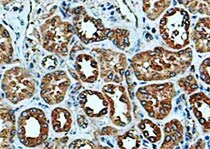
Application: ImmunohistochemistrySample Tested: Kidney tissueSpecies: MouseVerified Customer | Posted 03/17/2022

-
Application: ImmunohistochemistrySample Tested: Colon tissueSpecies: miceVerified Customer | Posted 12/16/2021
-
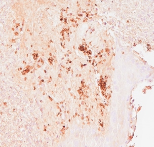
Application: ImmunohistochemistrySample Tested: Dental pulp tissueSpecies: HumanVerified Customer | Posted 08/18/2021
-
Application: Immunocytochemistry/ImmunofluorescenceSample Tested: Connective tissueSpecies: RatVerified Customer | Posted 07/06/2021
-
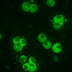
Application: Immunocytochemistry/ImmunofluorescenceSample Tested: J774A.1 mouse reticulum cell sarcoma macrophage cell lineSpecies: MouseVerified Customer | Posted 07/21/2017
-
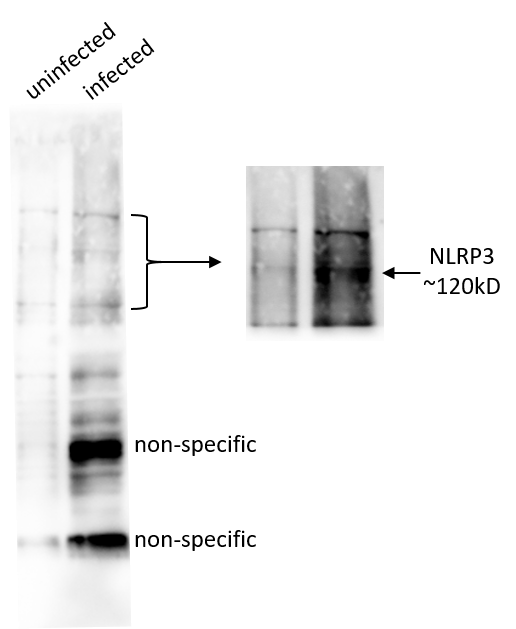
Application: Western BlotSample Tested: primary macrophagesSpecies: MouseVerified Customer | Posted 01/11/2017
There are no reviews that match your criteria.
Protocols
Find general support by application which include: protocols, troubleshooting, illustrated assays, videos and webinars.
- 7-Amino Actinomycin D (7-AAD) Cell Viability Flow Cytometry Protocol
- Antigen Retrieval Protocol (PIER)
- Antigen Retrieval for Frozen Sections Protocol
- Appropriate Fixation of IHC/ICC Samples
- Cellular Response to Hypoxia Protocols
- Chromogenic IHC Staining of Formalin-Fixed Paraffin-Embedded (FFPE) Tissue Protocol
- Chromogenic Immunohistochemistry Staining of Frozen Tissue
- ClariTSA™ Fluorophore Kits
- Detection & Visualization of Antibody Binding
- Extracellular Membrane Flow Cytometry Protocol
- Flow Cytometry Protocol for Cell Surface Markers
- Flow Cytometry Protocol for Staining Membrane Associated Proteins
- Flow Cytometry Staining Protocols
- Flow Cytometry Troubleshooting Guide
- Fluorescent IHC Staining of Frozen Tissue Protocol
- Graphic Protocol for Heat-induced Epitope Retrieval
- Graphic Protocol for the Preparation and Fluorescent IHC Staining of Frozen Tissue Sections
- Graphic Protocol for the Preparation and Fluorescent IHC Staining of Paraffin-embedded Tissue Sections
- Graphic Protocol for the Preparation of Gelatin-coated Slides for Histological Tissue Sections
- ICC Cell Smear Protocol for Suspension Cells
- ICC Immunocytochemistry Protocol Videos
- ICC for Adherent Cells
- IHC Sample Preparation (Frozen sections vs Paraffin)
- Immunocytochemistry (ICC) Protocol
- Immunocytochemistry Troubleshooting
- Immunofluorescence of Organoids Embedded in Cultrex Basement Membrane Extract
- Immunofluorescent IHC Staining of Formalin-Fixed Paraffin-Embedded (FFPE) Tissue Protocol
- Immunohistochemistry (IHC) and Immunocytochemistry (ICC) Protocols
- Immunohistochemistry Frozen Troubleshooting
- Immunohistochemistry Paraffin Troubleshooting
- Intracellular Flow Cytometry Protocol Using Alcohol (Methanol)
- Intracellular Flow Cytometry Protocol Using Detergents
- Intracellular Nuclear Staining Flow Cytometry Protocol Using Detergents
- Intracellular Staining Flow Cytometry Protocol Using Alcohol Permeabilization
- Intracellular Staining Flow Cytometry Protocol Using Detergents to Permeabilize Cells
- Preparing Samples for IHC/ICC Experiments
- Preventing Non-Specific Staining (Non-Specific Binding)
- Primary Antibody Selection & Optimization
- Propidium Iodide Cell Viability Flow Cytometry Protocol
- Protocol for Heat-Induced Epitope Retrieval (HIER)
- Protocol for Liperfluo
- Protocol for Making a 4% Formaldehyde Solution in PBS
- Protocol for VisUCyte™ HRP Polymer Detection Reagent
- Protocol for the Characterization of Human Th22 Cells
- Protocol for the Characterization of Human Th9 Cells
- Protocol for the Fluorescent ICC Staining of Cell Smears - Graphic
- Protocol for the Fluorescent ICC Staining of Cultured Cells on Coverslips - Graphic
- Protocol for the Preparation & Fixation of Cells on Coverslips
- Protocol for the Preparation and Chromogenic IHC Staining of Frozen Tissue Sections
- Protocol for the Preparation and Chromogenic IHC Staining of Frozen Tissue Sections - Graphic
- Protocol for the Preparation and Chromogenic IHC Staining of Paraffin-embedded Tissue Sections
- Protocol for the Preparation and Chromogenic IHC Staining of Paraffin-embedded Tissue Sections - Graphic
- Protocol for the Preparation and Fluorescent ICC Staining of Cells on Coverslips
- Protocol for the Preparation and Fluorescent ICC Staining of Non-adherent Cells
- Protocol for the Preparation and Fluorescent ICC Staining of Stem Cells on Coverslips
- Protocol for the Preparation and Fluorescent IHC Staining of Frozen Tissue Sections
- Protocol for the Preparation and Fluorescent IHC Staining of Paraffin-embedded Tissue Sections
- Protocol for the Preparation of Gelatin-coated Slides for Histological Tissue Sections
- Protocol for the Preparation of a Cell Smear for Non-adherent Cell ICC - Graphic
- Protocol: Annexin V and PI Staining by Flow Cytometry
- Protocol: Annexin V and PI Staining for Apoptosis by Flow Cytometry
- R&D Systems Quality Control Western Blot Protocol
- TUNEL and Active Caspase-3 Detection by IHC/ICC Protocol
- The Importance of IHC/ICC Controls
- Troubleshooting Guide: Fluorokine Flow Cytometry Kits
- Troubleshooting Guide: Immunohistochemistry
- Troubleshooting Guide: Western Blot Figures
- Western Blot Conditions
- Western Blot Protocol
- Western Blot Protocol for Cell Lysates
- Western Blot Troubleshooting
- Western Blot Troubleshooting Guide
- View all Protocols, Troubleshooting, Illustrated assays and Webinars
FAQs for NLRP3/NALP3 Antibody (768319)
-
Q: Does this NLRP3/NALP3 antibody come in lyophilized form?
A: Yes, we carry this NLRP2 antibody in lyophilized format.
-
Q: What is the immunogen sequence of this NLRP3/NALP3 antibodies antibody?
A: An E. coli derived sequence corresponding to Met1 - Arg153 of mouse NLRP3/NALP3.
-
Q: Does this NLRP3/NALP3 antibody come in lyophilized form?
A: Yes, we carry this NLRP2 antibody in lyophilized format.
-
Q: What is the immunogen sequence of this NLRP3/NALP3 antibodies antibody?
A: An E. coli derived sequence corresponding to Met1 - Arg153 of mouse NLRP3/NALP3.
Associated Pathways

