Pax3/Pax7 /Pax7 Antibody (274212)
R&D Systems | Catalog # MAB2457

Key Product Details
Species Reactivity
Validated:
Human, Mouse
Cited:
Human, Mouse, Porcine
Applications
Validated:
Western Blot, Intracellular Staining by Flow Cytometry, Immunocytochemistry, CyTOF-ready
Cited:
Immunohistochemistry, Western Blot, Flow Cytometry, Immunocytochemistry, Chromatin Immunoprecipitation (ChIP), Functional Assay
Label
Unconjugated
Antibody Source
Monoclonal Mouse IgG2A Clone # 274212
Loading...
Product Specifications
Immunogen
E. coli-derived recombinant human Pax3 (isoform Pax3a)
Met1-Ser215
Accession # NP_000429
Met1-Ser215
Accession # NP_000429
Specificity
Detects human and mouse Pax3 and Pax7 in direct ELISAs and Western blots. In direct ELISAs, approximately 20% cross-reactivity with recombinant human (rh) Pax1 is observed, approximately 10% with rhPax6, and less than 2% with rhPax2, rhPax4, rhPax5, and rhPax9.
Clonality
Monoclonal
Host
Mouse
Isotype
IgG2A
Scientific Data Images for Pax3/Pax7 /Pax7 Antibody (274212)
Detection of Pax3/Pax7 in B16-F1 cells by Flow Cytometry
B16-F1 cells were stained with Mouse Anti-Human/Mouse Pax3/Pax7 /Pax7 Monoclonal Antibody (Catalog # MAB2457, filled histogram) or isotype control antibody (Catalog # MAB003, open histogram) followed by Allophycocyanin-conjugated Anti-Mouse IgG Secondary Antibody (Catalog # F0101B). To facilitate intracellular staining, cells were fixed with Flow Cytometry Fixation Buffer (Catalog # FC004) and permeabilized with Flow Cytometry Permeabilization/Wash Buffer I (Catalog # FC005). View our protocol for Staining Intracellular Molecules.
Pax3/Pax7 in B16‑F1 Mouse Cell Line.
Pax3/Pax7 was detected in immersion fixed B16-F1 mouse melanoma cell line using Mouse Anti-Human/Mouse Pax3/Pax7 Monoclonal Antibody (Catalog # MAB2457) at 2 µg/mL for 3 hours at room temperature. Cells were stained using the NorthernLights™ 557-conjugated Anti-Mouse IgG Secondary Antibody (red; Catalog # NL007) and counterstained with DAPI (blue). Specific staining was localized to nuclei. View our protocol for Fluorescent ICC Staining of Cells on Coverslips.
Detection of Human Pax3/Pax7 by Immunocytochemistry/Immunofluorescence
Characterization of iMPCs during monolayer differentiation. a–e Representative immunostaining of Pax3 (a), Myf5 (b), MyoD (c), and MyoG (d), and corresponding quantification (e) during iMPC expansion. Scale bar=100 µm. f Representative FACS analysis for CD56 in H9 and TRiPSC derived iMPCs. g Representative immunostaining (top) and quantification (bottom) of Pax7+ and MyoG+ cell populations for H9 and TRiPS derived myotubes at 2 weeks of monolayer differentiation. (n = 6 samples from 2 differentiations for each cell line). h Representative immunostaining and quantification of GFP+/Pax7+ and GFP-/Pax7+ cell pools at 2 weeks of monolayer differentiation. Scale bar=50 µm. (n = 4 samples from 2 differentiations for each cell line). i Representative immunostaining and quantification of myotube diameter at 1, 2, and 4 weeks of monolayer differentiation. (*P < 0.05 vs. 1 week, #P < 0.05 vs. 4 week, Tukey–Kramer HSD test; n = 6 samples from 2 differentiations for each cell line). Scale bars=50 µm. Data are presented as mean ± SEM Image collected and cropped by CiteAb from the following publication (https://pubmed.ncbi.nlm.nih.gov/29317646), licensed under a CC-BY license. Not internally tested by R&D Systems.
Detection of Pax3/Pax7/Pax7 by Immunohistochemistry
Immunofluorescent analysis of Pax3 expression in the developing mouse cerebellum.(A) Bar plot showing the percentage of Pax3 + cells co-stained (y-axis) with cerebellar cell markers Pax2, Foxp2 and Calbindin at E12, E15, and P3 (x-axis). (B) Top: Immunofluorescent co-staining of Pax3 (red) and Foxp2 (green) in embryonic cerebellum at E15. Merged image is a composite image of the Pax3, Foxp2, and DAPI. Bottom: Immunofluorescent co-staining of Pax3 (red) and Calb (green) in the postnatal cerebellum at P0. Merged image is a composite image of the Pax3, Foxp2, and DAPI. Labels: VZ: Ventricular zone, EGL: External granular layer, PCL: Purkinje cell layer, ML: Molecular layer, Scalebars = 100 µm. (C) Immunofluorescent staining of Pax3 (red) in the developing cerebellum at P9. Image collected and cropped by CiteAb from the following open publication (https://pubmed.ncbi.nlm.nih.gov/35942939), licensed under a CC-BY license. Not internally tested by R&D Systems.
Detection of Pax3/Pax7/Pax7 by Immunohistochemistry
Immunofluorescent analysis of Pax3 expression in the developing mouse cerebellum.(A) Bar plot showing the percentage of Pax3 + cells co-stained (y-axis) with cerebellar cell markers Pax2, Foxp2 and Calbindin at E12, E15, and P3 (x-axis). (B) Top: Immunofluorescent co-staining of Pax3 (red) and Foxp2 (green) in embryonic cerebellum at E15. Merged image is a composite image of the Pax3, Foxp2, and DAPI. Bottom: Immunofluorescent co-staining of Pax3 (red) and Calb (green) in the postnatal cerebellum at P0. Merged image is a composite image of the Pax3, Foxp2, and DAPI. Labels: VZ: Ventricular zone, EGL: External granular layer, PCL: Purkinje cell layer, ML: Molecular layer, Scalebars = 100 µm. (C) Immunofluorescent staining of Pax3 (red) in the developing cerebellum at P9. Image collected and cropped by CiteAb from the following open publication (https://pubmed.ncbi.nlm.nih.gov/35942939), licensed under a CC-BY license. Not internally tested by R&D Systems.Applications for Pax3/Pax7 /Pax7 Antibody (274212)
Application
Recommended Usage
CyTOF-ready
Ready to be labeled using established conjugation methods. No BSA or other carrier proteins that could interfere with conjugation.
Immunocytochemistry
8-25 µg/mL
Sample: Immersion fixed B16-F1 mouse melanoma cell line
Sample: Immersion fixed B16-F1 mouse melanoma cell line
Intracellular Staining by Flow Cytometry
0.25 µg/106 cells
Sample: B16‑F1 mouse melanoma cell line fixed with paraformaldehyde and permeabilized with saponin
Sample: B16‑F1 mouse melanoma cell line fixed with paraformaldehyde and permeabilized with saponin
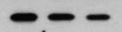
Western Blot
1 µg/mL
Sample: Recombinant Human Pax3
Sample: Recombinant Human Pax3
Reviewed Applications
Read 2 reviews rated 4.5 using MAB2457 in the following applications:
Flow Cytometry Panel Builder
Bio-Techne Knows Flow Cytometry
Save time and reduce costly mistakes by quickly finding compatible reagents using the Panel Builder Tool.
Advanced Features
- Spectra Viewer - Custom analysis of spectra from multiple fluorochromes
- Spillover Popups - Visualize the spectra of individual fluorochromes
- Antigen Density Selector - Match fluorochrome brightness with antigen density

Formulation, Preparation, and Storage
Purification
Protein A or G purified from hybridoma culture supernatant
Reconstitution
Reconstitute at 0.5 mg/mL in sterile PBS. For liquid material, refer to CoA for concentration.
Loading...
Formulation
Lyophilized from a 0.2 μm filtered solution in PBS with Trehalose. See Certificate of Analysis for details.
*Small pack size (-SP) is supplied either lyophilized or as a 0.2 µm filtered solution in PBS.
*Small pack size (-SP) is supplied either lyophilized or as a 0.2 µm filtered solution in PBS.
Shipping
Lyophilized product is shipped at ambient temperature. Liquid small pack size (-SP) is shipped with polar packs. Upon receipt, store immediately at the temperature recommended below.
Stability & Storage
Use a manual defrost freezer and avoid repeated freeze-thaw cycles.
- 12 months from date of receipt, -20 to -70 °C as supplied.
- 1 month, 2 to 8 °C under sterile conditions after reconstitution.
- 6 months, -20 to -70 °C under sterile conditions after reconstitution.
Calculators
Background: Pax3/Pax7
Long Name
Paired Box Gene 3 /Paired Box Gene 7
UniProt
Additional Pax3/Pax7 Products
Product Documents for Pax3/Pax7 /Pax7 Antibody (274212)
Certificate of Analysis
To download a Certificate of Analysis, please enter a lot or batch number in the search box below.
Note: Certificate of Analysis not available for kit components.
Product Specific Notices for Pax3/Pax7 /Pax7 Antibody (274212)
For research use only
Related Research Areas
Citations for Pax3/Pax7 /Pax7 Antibody (274212)
 Powered by Bioz
Customer Reviews for Pax3/Pax7 /Pax7 Antibody (274212) (2)
4.5 out of 5
2 Customer Ratings
Have you used Pax3/Pax7 /Pax7 Antibody (274212)?
Submit a review and receive an Amazon gift card!
$25/€18/£15/$25CAN/¥2500 Yen for a review with an image
$10/€7/£6/$10CAN/¥1110 Yen for a review without an image
Submit a review
Customer Images
Showing
1
-
2 of
2 reviews
Showing All
Filter By:
-
Application: Western BlotSample Tested: B16-F1 mouse melanoma cell lineSpecies: MouseVerified Customer | Posted 09/13/2021

-
Application: Western BlotSample Tested: See PMID 21078671Species: OtherVerified Customer | Posted 02/19/2015
There are no reviews that match your criteria.
Protocols
Find general support by application which include: protocols, troubleshooting, illustrated assays, videos and webinars.
- 7-Amino Actinomycin D (7-AAD) Cell Viability Flow Cytometry Protocol
- Appropriate Fixation of IHC/ICC Samples
- Cellular Response to Hypoxia Protocols
- ClariTSA™ Fluorophore Kits
- Detection & Visualization of Antibody Binding
- Extracellular Membrane Flow Cytometry Protocol
- Flow Cytometry Protocol for Cell Surface Markers
- Flow Cytometry Protocol for Staining Membrane Associated Proteins
- Flow Cytometry Staining Protocols
- Flow Cytometry Troubleshooting Guide
- ICC Cell Smear Protocol for Suspension Cells
- ICC Immunocytochemistry Protocol Videos
- ICC for Adherent Cells
- Immunocytochemistry (ICC) Protocol
- Immunocytochemistry Troubleshooting
- Immunofluorescence of Organoids Embedded in Cultrex Basement Membrane Extract
- Immunohistochemistry (IHC) and Immunocytochemistry (ICC) Protocols
- Intracellular Flow Cytometry Protocol Using Alcohol (Methanol)
- Intracellular Flow Cytometry Protocol Using Detergents
- Intracellular Nuclear Staining Flow Cytometry Protocol Using Detergents
- Intracellular Staining Flow Cytometry Protocol Using Alcohol Permeabilization
- Intracellular Staining Flow Cytometry Protocol Using Detergents to Permeabilize Cells
- Preparing Samples for IHC/ICC Experiments
- Preventing Non-Specific Staining (Non-Specific Binding)
- Primary Antibody Selection & Optimization
- Propidium Iodide Cell Viability Flow Cytometry Protocol
- Protocol for Liperfluo
- Protocol for VisUCyte™ HRP Polymer Detection Reagent
- Protocol for the Characterization of Human Th22 Cells
- Protocol for the Characterization of Human Th9 Cells
- Protocol for the Fluorescent ICC Staining of Cell Smears - Graphic
- Protocol for the Fluorescent ICC Staining of Cultured Cells on Coverslips - Graphic
- Protocol for the Preparation and Fluorescent ICC Staining of Cells on Coverslips
- Protocol for the Preparation and Fluorescent ICC Staining of Non-adherent Cells
- Protocol for the Preparation and Fluorescent ICC Staining of Stem Cells on Coverslips
- Protocol for the Preparation of a Cell Smear for Non-adherent Cell ICC - Graphic
- Protocol: Annexin V and PI Staining by Flow Cytometry
- Protocol: Annexin V and PI Staining for Apoptosis by Flow Cytometry
- R&D Systems Quality Control Western Blot Protocol
- TUNEL and Active Caspase-3 Detection by IHC/ICC Protocol
- The Importance of IHC/ICC Controls
- Troubleshooting Guide: Fluorokine Flow Cytometry Kits
- Troubleshooting Guide: Western Blot Figures
- Western Blot Conditions
- Western Blot Protocol
- Western Blot Protocol for Cell Lysates
- Western Blot Troubleshooting
- Western Blot Troubleshooting Guide
- View all Protocols, Troubleshooting, Illustrated assays and Webinars
Loading...