Bcl-2/adenovirus E1B 19 kDa protein-interacting protein 3-like (BNIP3L) also known as BNIP3 alpha and NIP3-like protein X (Nix), is a proapoptotic member of BNIP3 protein family. BNIP3L is a functional homolog of BNIP3 and both proteins contain a single Bcl-2 homology 3 (BH3) domain. BNIP3L is a 219 amino acid (aa), 24 kDa (predicted) protein that contains a C-terminal transmembrane domain required for mitochondrial localization, homodimerisation, and regulation of its proapoptotic function. BNIP3L may antagonize the activity of BCL2 family antiapoptic proteins by directly interactacting with proteins such as viral E1B-19K and cellular Bcl-2 and Bcl-xL. BNIP3L shares 56% amino acid sequence identity with BNIP3 and 98% with mouse and rat BNIP3L.

Key Product Details
Species Reactivity
Human, Mouse, Rat
Applications
Western Blot, Immunocytochemistry
Label
Unconjugated
Antibody Source
Polyclonal Goat IgG
Loading...
Product Specifications
Immunogen
E. coli-derived recombinant human BNIP3L
Ser2-Glu184
Accession # O60238
Ser2-Glu184
Accession # O60238
Specificity
Detects endogenous human, mouse and rat BNIP3L in Western blots. In Western blots, less than 10% cross-reactivity to recombinant human BNIP3L is observed.
Clonality
Polyclonal
Host
Goat
Isotype
IgG
Scientific Data Images for BNIP3L Antibody
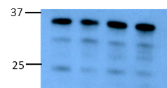
Detection of Human/Mouse/Rat BNIP3L by Western Blot.
Western blot shows lysates of Raji human Burkitt's lymphoma cell line, A20 mouse B cell lymphoma cell line, and Rat-2 rat embryonic fibroblast cell line. PVDF membrane was probed with 1 µg/mL of Goat Anti-Human/Mouse/Rat BNIP3L Antigen Affinity-purified Polyclonal Antibody (Catalog # AF4030) followed by HRP-conjugated Anti-Goat IgG Secondary Antibody (Catalog # HAF109). A specific band was detected for BNIP3L at approximately 35kDa (as indicated). This experiment was conducted under reducing conditions and using Immunoblot Buffer Group 2.
BNIP3L in HeLa Human Cell Line.
BNIP3L was detected in immersion fixed HeLa human cervical epithelial carcinoma cell line using Goat Anti-Human/Mouse/Rat BNIP3L Antigen Affinity-purified Polyclonal Antibody (Catalog # AF4030) at 10 µg/mL for 3 hours at room temperature. Cells were stained using the NorthernLights™ 557-conjugated Anti-Goat IgG Secondary Antibody (red; Catalog # NL001) and counterstained with DAPI (blue). Specific staining was localized to cytoplasm. View our protocol for Fluorescent ICC Staining of Cells on Coverslips.Applications for BNIP3L Antibody
Application
Recommended Usage
Immunocytochemistry
5-15 µg/mL
Sample: Immersion fixed HeLa human cervical epithelial carcinoma cell line
Sample: Immersion fixed HeLa human cervical epithelial carcinoma cell line
Western Blot
1 µg/mL
Sample: Raji human Burkitt's lymphoma cell line, A20 mouse B cell lymphoma cell line, and Rat-2 rat embryonic fibroblast cell line
Sample: Raji human Burkitt's lymphoma cell line, A20 mouse B cell lymphoma cell line, and Rat-2 rat embryonic fibroblast cell line
Reviewed Applications
Read 1 review rated 4 using AF4030 in the following applications:
Formulation, Preparation, and Storage
Purification
Antigen Affinity-purified
Reconstitution
Reconstitute at 0.2 mg/mL in sterile PBS. For liquid material, refer to CoA for concentration.
Loading...
Formulation
Lyophilized from a 0.2 μm filtered solution in PBS with Trehalose. *Small pack size (SP) is supplied either lyophilized or as a 0.2 µm filtered solution in PBS.
Shipping
Lyophilized product is shipped at ambient temperature. Liquid small pack size (-SP) is shipped with polar packs. Upon receipt, store immediately at the temperature recommended below.
Stability & Storage
Use a manual defrost freezer and avoid repeated freeze-thaw cycles.
- 12 months from date of receipt, -20 to -70 °C as supplied.
- 1 month, 2 to 8 °C under sterile conditions after reconstitution.
- 6 months, -20 to -70 °C under sterile conditions after reconstitution.
Calculators
Background: BNIP3L
Long Name
BCL2/Adenovirus E1B 19 Kda Protein-Interacting Protein 3-Like
Alternate Names
BNIP3a, Nip3L, Nix
Gene Symbol
BNIP3L
UniProt
Additional BNIP3L Products
Product Documents for BNIP3L Antibody
Certificate of Analysis
To download a Certificate of Analysis, please enter a lot or batch number in the search box below.
Note: Certificate of Analysis not available for kit components.
Product Specific Notices for BNIP3L Antibody
For research use only
Related Research Areas
Citations for BNIP3L Antibody
 Powered by Bioz
Customer Reviews for BNIP3L Antibody (1)
4 out of 5
1 Customer Rating
Have you used BNIP3L Antibody?
Submit a review and receive an Amazon gift card!
$25/€18/£15/$25CAN/¥2500 Yen for a review with an image
$10/€7/£6/$10CAN/¥1110 Yen for a review without an image
Submit a review
Customer Images
Showing
1
-
1 of
1 review
Showing All
Filter By:
-
Application: Western BlotSample Tested: Bone Marrrow Derived MacrophagesSpecies: MouseVerified Customer | Posted 10/15/2020Western blot of BMDM cell lysates incubated with 1:800 dilution of the antibody and detected with anti-goat HRP.

There are no reviews that match your criteria.
Protocols
Find general support by application which include: protocols, troubleshooting, illustrated assays, videos and webinars.
- Appropriate Fixation of IHC/ICC Samples
- Cellular Response to Hypoxia Protocols
- ClariTSA™ Fluorophore Kits
- Detection & Visualization of Antibody Binding
- ICC Cell Smear Protocol for Suspension Cells
- ICC Immunocytochemistry Protocol Videos
- ICC for Adherent Cells
- Immunocytochemistry (ICC) Protocol
- Immunocytochemistry Troubleshooting
- Immunofluorescence of Organoids Embedded in Cultrex Basement Membrane Extract
- Immunohistochemistry (IHC) and Immunocytochemistry (ICC) Protocols
- Preparing Samples for IHC/ICC Experiments
- Preventing Non-Specific Staining (Non-Specific Binding)
- Primary Antibody Selection & Optimization
- Protocol for VisUCyte™ HRP Polymer Detection Reagent
- Protocol for the Fluorescent ICC Staining of Cell Smears - Graphic
- Protocol for the Fluorescent ICC Staining of Cultured Cells on Coverslips - Graphic
- Protocol for the Preparation and Fluorescent ICC Staining of Cells on Coverslips
- Protocol for the Preparation and Fluorescent ICC Staining of Non-adherent Cells
- Protocol for the Preparation and Fluorescent ICC Staining of Stem Cells on Coverslips
- Protocol for the Preparation of a Cell Smear for Non-adherent Cell ICC - Graphic
- R&D Systems Quality Control Western Blot Protocol
- TUNEL and Active Caspase-3 Detection by IHC/ICC Protocol
- The Importance of IHC/ICC Controls
- Troubleshooting Guide: Western Blot Figures
- Western Blot Conditions
- Western Blot Protocol
- Western Blot Protocol for Cell Lysates
- Western Blot Troubleshooting
- Western Blot Troubleshooting Guide
- View all Protocols, Troubleshooting, Illustrated assays and Webinars
Loading...