
Key Product Details
Species Reactivity
Validated:
Cited:
Applications
Validated:
Cited:
Label
Antibody Source
Product Specifications
Immunogen
aa 352-523
Specificity
Clonality
Host
Isotype
Scientific Data Images for Pax7 Antibody (PAX7)
Pax7 in C2C12 Mouse Cell Line.
Pax7 was detected in immersion fixed C2C12 mouse myoblast cell line using 10 µg/mL Mouse Anti-Human/Mouse/Rat/Chicken Pax7 Monoclonal Antibody (Catalog # MAB1675) for 3 hours at room temperature. Cells were stained (red). View our protocol for Fluorescent ICC Staining of Cells on Coverslips.
Pax7 in iBJ6 iPS Cell Line.
Pax7 was detected in immersion fixed iBJ6 iPS cell line differentiated into myoblasts using Mouse Anti-Human/Mouse/Rat/Chicken Pax7 Monoclonal Antibody (Catalog # MAB1675) at 10 µg/mL for 3 hours at room temperature. Cells were stained using the NorthernLights™ 557-conjugated Anti-Mouse IgG Secondary Antibody (red; Catalog # NL007) and counterstained with DAPI (blue). Specific staining was localized to nuclei. View our protocol for Fluorescent ICC Staining of Stem Cells on Coverslips.
Pax7 in Human Skeletal Muscle.
Pax7 was detected in immersion fixed paraffin-embedded sections of human skeletal muscle using Mouse Anti-Human/Mouse/Rat/Chicken Pax7 Monoclonal Antibody (Catalog # MAB1675) at 5 µg/mL for 1 hour at room temperature followed by incubation with the Anti-Mouse IgG VisUCyte™ HRP Polymer Antibody (VC001). Before incubation with the primary antibody, tissue was subjected to heat-induced epitope retrieval using Antigen Retrieval Reagent-Basic (CTS013). Tissue was stained using DAB (brown) and counterstained with hematoxylin (blue). Specific staining was localized to cell nuclei. Staining was performed using our protocol for IHC Staining with VisUCyte HRP Polymer Detection Reagents.
Detection of Pax7 by Western Blot
Protein expression analyses during differentiation and ID1 expression analyses. a Protein expression profiles of PAX7, MYOD, and Desmin were compared between regular pCM (rpCM) cells and pCM cells overexpressing miR-146b (pCM-146b OE) after 4 days of myotube differentiation. The graph represents a density comparison of Western blotting results (n = 3; *p < 0.05). b Expression profiles of ID1 were compared between the rpCM cells and pCM-146b OE cells (n = 3; ***p < 0.001) Image collected and cropped by CiteAb from the following open publication (https://pubmed.ncbi.nlm.nih.gov/32471354), licensed under a CC-BY license. Not internally tested by R&D Systems.Applications for Pax7 Antibody (PAX7)
Immunocytochemistry
Sample: Immersion fixed C2C12 mouse myoblast cell line and iBJ6 iPS cell line differentiated into myoblasts
Immunohistochemistry
Sample: Immersion fixed paraffin-embedded sections of human skeletal muscle
Reviewed Applications
Read 4 reviews rated 4.8 using MAB1675 in the following applications:
Formulation, Preparation, and Storage
Purification
Reconstitution
Reconstitute at 0.5 mg/mL in sterile PBS. For liquid material, refer to CoA for concentration.
Formulation
Shipping
Stability & Storage
- 12 months from date of receipt, -20 to -70 °C as supplied.
- 1 month, 2 to 8 °C under sterile conditions after reconstitution.
- 6 months, -20 to -70 °C under sterile conditions after reconstitution.
Calculators
Background: Pax7
References
- Jostes, B. et al. (1990) Mech. Dev. 33(1):27.
- Asakura, A. et al. (2001) Differentiation 68(4-5):245.
- Kawakami, A. et al. (1997) Mech. Dev. 66:119.
- Ericson, J. et al. (1996) Cell 87:661.
- Berggren, K. et al. (2001) Dev. Dyn. 222:1.
- Takebayashi, H. et al. (2002) Mech. Dev. 113:169.
- McLoon, L.K. and J. Wirtschafter (2003) Invest. Opthamal. Vis. Sci. 44:1927.
- Yamamoto, S. et al. (2001) J. Neurosci. 21:9814.
Long Name
Alternate Names
Gene Symbol
Additional Pax7 Products
Product Documents for Pax7 Antibody (PAX7)
Certificate of Analysis
To download a Certificate of Analysis, please enter a lot or batch number in the search box below.
Note: Certificate of Analysis not available for kit components.
Product Specific Notices for Pax7 Antibody (PAX7)
For research use only
Related Research Areas
Citations for Pax7 Antibody (PAX7)
 Powered by Bioz
Customer Reviews for Pax7 Antibody (PAX7) (4)
Have you used Pax7 Antibody (PAX7)?
Submit a review and receive an Amazon gift card!
$25/€18/£15/$25CAN/¥2500 Yen for a review with an image
$10/€7/£6/$10CAN/¥1110 Yen for a review without an image
Submit a review
Customer Images
-
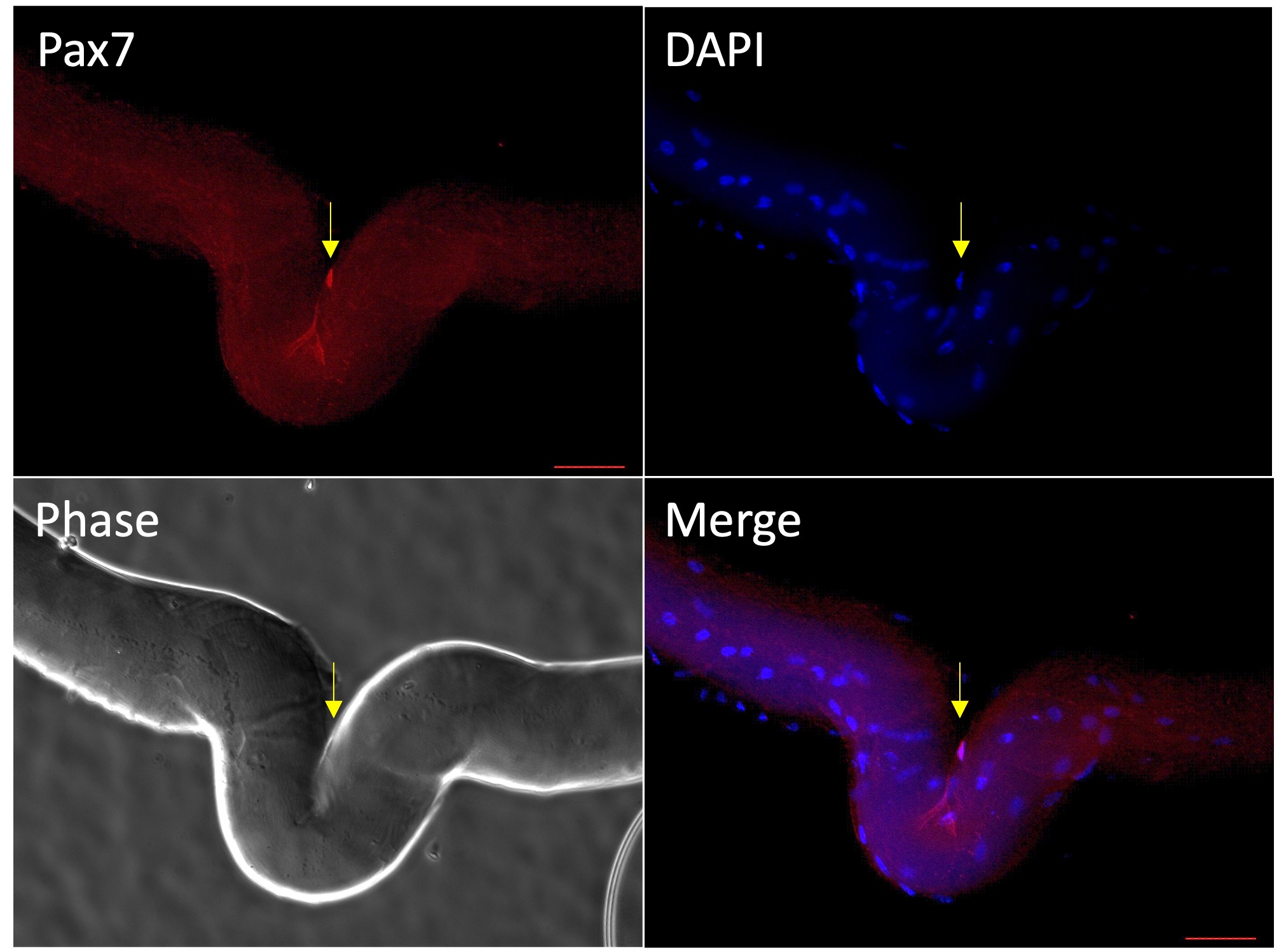
Application: Immunocytochemistry/ImmunofluorescenceSample Tested: single muscle fiberSpecies: MouseVerified Customer | Posted 07/03/2022Fixed with PFA.Block with BSA.1:500 primary ab.

-
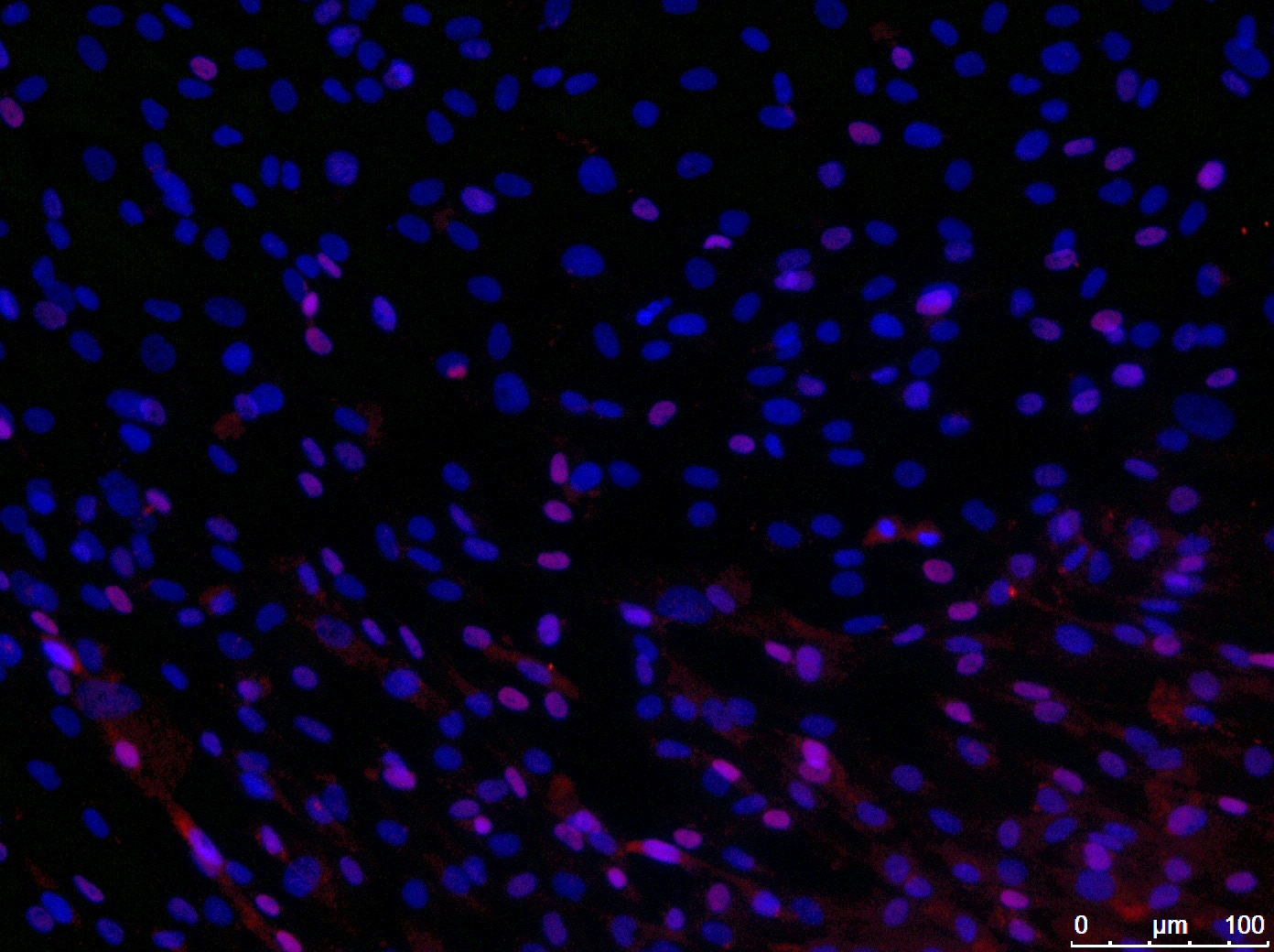
Application: Immunocytochemistry/ImmunofluorescenceSample Tested: human muscle precursor cells and Muscle precursor cellsSpecies: HumanVerified Customer | Posted 03/29/2022
-
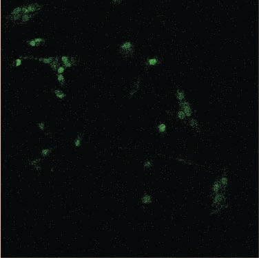
Application: Immunocytochemistry/ImmunofluorescenceSample Tested: Primary myoblastsSpecies: Horse and MouseVerified Customer | Posted 12/04/2020
-
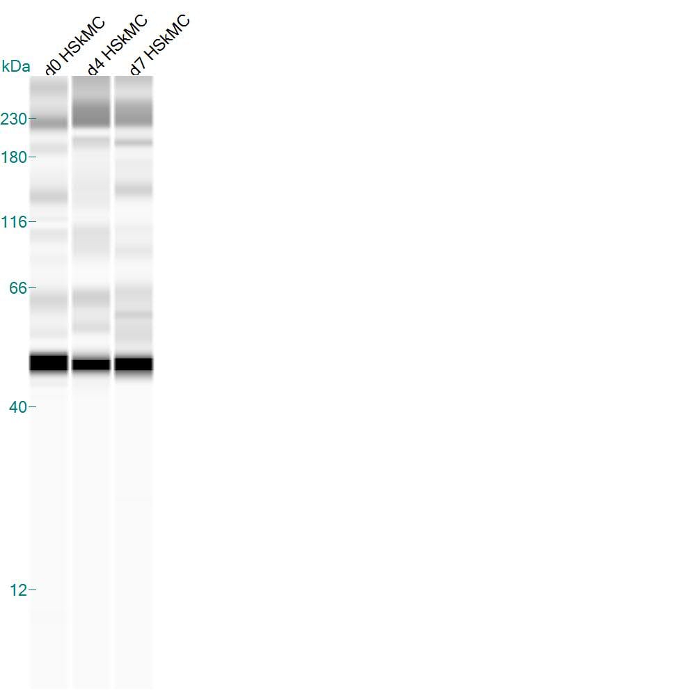
Application: Simple WesternSample Tested: Human skeletal muscle satellite-derived cell lysateSpecies: HumanVerified Customer | Posted 02/25/2015Human Skeletal muscle satellite derived cells at 0, 4, and 7 days of differentiation. Specific band at 49 kDa.
There are no reviews that match your criteria.
Protocols
Find general support by application which include: protocols, troubleshooting, illustrated assays, videos and webinars.
- Antigen Retrieval Protocol (PIER)
- Antigen Retrieval for Frozen Sections Protocol
- Appropriate Fixation of IHC/ICC Samples
- Cellular Response to Hypoxia Protocols
- Chromogenic IHC Staining of Formalin-Fixed Paraffin-Embedded (FFPE) Tissue Protocol
- Chromogenic Immunohistochemistry Staining of Frozen Tissue
- ClariTSA™ Fluorophore Kits
- Detection & Visualization of Antibody Binding
- Fluorescent IHC Staining of Frozen Tissue Protocol
- Graphic Protocol for Heat-induced Epitope Retrieval
- Graphic Protocol for the Preparation and Fluorescent IHC Staining of Frozen Tissue Sections
- Graphic Protocol for the Preparation and Fluorescent IHC Staining of Paraffin-embedded Tissue Sections
- Graphic Protocol for the Preparation of Gelatin-coated Slides for Histological Tissue Sections
- ICC Cell Smear Protocol for Suspension Cells
- ICC Immunocytochemistry Protocol Videos
- ICC for Adherent Cells
- IHC Sample Preparation (Frozen sections vs Paraffin)
- Immunocytochemistry (ICC) Protocol
- Immunocytochemistry Troubleshooting
- Immunofluorescence of Organoids Embedded in Cultrex Basement Membrane Extract
- Immunofluorescent IHC Staining of Formalin-Fixed Paraffin-Embedded (FFPE) Tissue Protocol
- Immunohistochemistry (IHC) and Immunocytochemistry (ICC) Protocols
- Immunohistochemistry Frozen Troubleshooting
- Immunohistochemistry Paraffin Troubleshooting
- Preparing Samples for IHC/ICC Experiments
- Preventing Non-Specific Staining (Non-Specific Binding)
- Primary Antibody Selection & Optimization
- Protocol for Heat-Induced Epitope Retrieval (HIER)
- Protocol for Making a 4% Formaldehyde Solution in PBS
- Protocol for VisUCyte™ HRP Polymer Detection Reagent
- Protocol for the Fluorescent ICC Staining of Cell Smears - Graphic
- Protocol for the Fluorescent ICC Staining of Cultured Cells on Coverslips - Graphic
- Protocol for the Preparation & Fixation of Cells on Coverslips
- Protocol for the Preparation and Chromogenic IHC Staining of Frozen Tissue Sections
- Protocol for the Preparation and Chromogenic IHC Staining of Frozen Tissue Sections - Graphic
- Protocol for the Preparation and Chromogenic IHC Staining of Paraffin-embedded Tissue Sections
- Protocol for the Preparation and Chromogenic IHC Staining of Paraffin-embedded Tissue Sections - Graphic
- Protocol for the Preparation and Fluorescent ICC Staining of Cells on Coverslips
- Protocol for the Preparation and Fluorescent ICC Staining of Non-adherent Cells
- Protocol for the Preparation and Fluorescent ICC Staining of Stem Cells on Coverslips
- Protocol for the Preparation and Fluorescent IHC Staining of Frozen Tissue Sections
- Protocol for the Preparation and Fluorescent IHC Staining of Paraffin-embedded Tissue Sections
- Protocol for the Preparation of Gelatin-coated Slides for Histological Tissue Sections
- Protocol for the Preparation of a Cell Smear for Non-adherent Cell ICC - Graphic
- TUNEL and Active Caspase-3 Detection by IHC/ICC Protocol
- The Importance of IHC/ICC Controls
- Troubleshooting Guide: Immunohistochemistry
- View all Protocols, Troubleshooting, Illustrated assays and Webinars
FAQs for Pax7 Antibody (PAX7)
-
Q: Does MAB1675 recognize Bovine PAX7?
A: MAB1675 binds to the C-terminal region of the Chicken PAX7 sequence, a region that is not present in the bovine PAX7 sequence. We would not expect MAB1675 to recognize Bovine PAX7 for this reason.
Associated Pathways
