G-protein beta -subunit-like (G beta L), also known as mLST8, is a subunit of the Target of Rapamycin (TOR) complex. G beta L contains 7 WD-40 repeats, and binds to the kinase domain of TOR in both the Raptor (Rapamycin-sensitive)-and Rictor (Rapamycin-insensitive)-containing complexes. G beta L is necessary for the full activation of the TOR kinase, and phosphorylation of many downstream proteins, including 4E-BP1 and p70S6 kinase.

Key Product Details
Species Reactivity
Human, Mouse, Rat
Applications
Immunohistochemistry, Western Blot
Label
Unconjugated
Antibody Source
Polyclonal Goat IgG
Loading...
Product Specifications
Immunogen
E. coli-derived recombinant human G beta L
Met1-Gly327
Accession # Q9BVC4
Met1-Gly327
Accession # Q9BVC4
Specificity
Detects human, mouse, and rat G beta L in Western blots.
Clonality
Polyclonal
Host
Goat
Isotype
IgG
Scientific Data Images for GBL Antibody
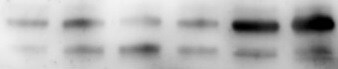
Detection of Human/Mouse/Rat G beta L by Western Blot.
Western blot shows lysates of HeLa human cervical epithelial carcinoma cell line, MCF-7 human breast cancer cell line, Balb/3T3 mouse embryonic fibroblast cell line, and PC-12 rat adrenal pheochromocytoma cell line. PVDF membrane was probed with 1 µg/mL of Human/Mouse/Rat G beta L Antigen Affinity-purified Polyclonal Antibody (Catalog # AF4004) followed by HRP-conjugated Anti-Goat IgG Secondary Antibody (Catalog # HAF109). A specific band was detected for G beta L at approximately 40 kDa (as indicated). This experiment was conducted under reducing conditions and using Immunoblot Buffer Group 1.
G beta L in Human Prostate.
G beta L was detected in immersion fixed paraffin-embedded sections of human prostate using Goat Anti-Human/Mouse/Rat G beta L Antigen Affinity-purified Polyclonal Antibody (Catalog # AF4004) at 10 µg/mL overnight at 4 °C. Tissue was stained using the Anti-Goat HRP-DAB Cell & Tissue Staining Kit (brown; Catalog # CTS008) and counterstained with hematoxylin (blue). Specific staining was localized to neuronal cell bodies. View our protocol for Chromogenic IHC Staining of Paraffin-embedded Tissue Sections.Applications for GBL Antibody
Application
Recommended Usage
Immunohistochemistry
5-15 µg/mL
Sample: Immersion fixed paraffin-embedded sections of human prostate
Sample: Immersion fixed paraffin-embedded sections of human prostate
Western Blot
1 µg/mL
Sample: HeLa human cervical epithelial carcinoma cell line, MCF-7 human breast cancer cell line, Balb/3T3 mouse embryonic fibroblast cell line, and PC-12 rat adrenal pheochromocytoma cell line
Sample: HeLa human cervical epithelial carcinoma cell line, MCF-7 human breast cancer cell line, Balb/3T3 mouse embryonic fibroblast cell line, and PC-12 rat adrenal pheochromocytoma cell line
Reviewed Applications
Read 1 review rated 5 using AF4004 in the following applications:
Formulation, Preparation, and Storage
Purification
Antigen Affinity-purified
Reconstitution
Reconstitute at 0.2 mg/mL in sterile PBS. For liquid material, refer to CoA for concentration.
Loading...
Formulation
Lyophilized from a 0.2 μm filtered solution in PBS with Trehalose. *Small pack size (SP) is supplied either lyophilized or as a 0.2 µm filtered solution in PBS.
Shipping
Lyophilized product is shipped at ambient temperature. Liquid small pack size (-SP) is shipped with polar packs. Upon receipt, store immediately at the temperature recommended below.
Stability & Storage
Use a manual defrost freezer and avoid repeated freeze-thaw cycles.
- 12 months from date of receipt, -20 to -70 °C as supplied.
- 1 month, 2 to 8 °C under sterile conditions after reconstitution.
- 6 months, -20 to -70 °C under sterile conditions after reconstitution.
Calculators
Background: GBL
Long Name
G protein beta subunit-like
Alternate Names
mLST8
Entrez Gene IDs
64223 (Human)
Gene Symbol
MLST8
UniProt
Additional GBL Products
Product Documents for GBL Antibody
Certificate of Analysis
To download a Certificate of Analysis, please enter a lot or batch number in the search box below.
Note: Certificate of Analysis not available for kit components.
Product Specific Notices for GBL Antibody
For research use only
Related Research Areas
Citations for GBL Antibody
 Powered by Bioz
Customer Reviews for GBL Antibody (1)
5 out of 5
1 Customer Rating
Have you used GBL Antibody?
Submit a review and receive an Amazon gift card!
$25/€18/£15/$25CAN/¥2500 Yen for a review with an image
$10/€7/£6/$10CAN/¥1110 Yen for a review without an image
Submit a review
Customer Images
Showing
1
-
1 of
1 review
Showing All
Filter By:
-
Application: Western BlotSample Tested: Adipose tissueSpecies: MouseVerified Customer | Posted 10/23/2019Mouse white adipose tissue was homogenised and protein content was quantified by a BCA assay. Twenty micrograms of protein were resolved on a 4-12% Bis-Tris gel and transferred to nitrocellulose membranes. Membranes were probed with primary antibody gbl diluted 1:1000 in 5% BSA 2, before incubation with anti goat secondary horseradish peroxidase-conjugated antibody 1:5000. Blots were visualised with Immobilon Western Chemiluminescence HRP Substrate and imaged with Syngene chemiluminescence imaging system.

There are no reviews that match your criteria.
Protocols
Find general support by application which include: protocols, troubleshooting, illustrated assays, videos and webinars.
- Antigen Retrieval Protocol (PIER)
- Antigen Retrieval for Frozen Sections Protocol
- Appropriate Fixation of IHC/ICC Samples
- Cellular Response to Hypoxia Protocols
- Chromogenic IHC Staining of Formalin-Fixed Paraffin-Embedded (FFPE) Tissue Protocol
- Chromogenic Immunohistochemistry Staining of Frozen Tissue
- ClariTSA™ Fluorophore Kits
- Detection & Visualization of Antibody Binding
- Fluorescent IHC Staining of Frozen Tissue Protocol
- Graphic Protocol for Heat-induced Epitope Retrieval
- Graphic Protocol for the Preparation and Fluorescent IHC Staining of Frozen Tissue Sections
- Graphic Protocol for the Preparation and Fluorescent IHC Staining of Paraffin-embedded Tissue Sections
- Graphic Protocol for the Preparation of Gelatin-coated Slides for Histological Tissue Sections
- IHC Sample Preparation (Frozen sections vs Paraffin)
- Immunofluorescent IHC Staining of Formalin-Fixed Paraffin-Embedded (FFPE) Tissue Protocol
- Immunohistochemistry (IHC) and Immunocytochemistry (ICC) Protocols
- Immunohistochemistry Frozen Troubleshooting
- Immunohistochemistry Paraffin Troubleshooting
- Preparing Samples for IHC/ICC Experiments
- Preventing Non-Specific Staining (Non-Specific Binding)
- Primary Antibody Selection & Optimization
- Protocol for Heat-Induced Epitope Retrieval (HIER)
- Protocol for Making a 4% Formaldehyde Solution in PBS
- Protocol for VisUCyte™ HRP Polymer Detection Reagent
- Protocol for the Preparation & Fixation of Cells on Coverslips
- Protocol for the Preparation and Chromogenic IHC Staining of Frozen Tissue Sections
- Protocol for the Preparation and Chromogenic IHC Staining of Frozen Tissue Sections - Graphic
- Protocol for the Preparation and Chromogenic IHC Staining of Paraffin-embedded Tissue Sections
- Protocol for the Preparation and Chromogenic IHC Staining of Paraffin-embedded Tissue Sections - Graphic
- Protocol for the Preparation and Fluorescent IHC Staining of Frozen Tissue Sections
- Protocol for the Preparation and Fluorescent IHC Staining of Paraffin-embedded Tissue Sections
- Protocol for the Preparation of Gelatin-coated Slides for Histological Tissue Sections
- R&D Systems Quality Control Western Blot Protocol
- TUNEL and Active Caspase-3 Detection by IHC/ICC Protocol
- The Importance of IHC/ICC Controls
- Troubleshooting Guide: Immunohistochemistry
- Troubleshooting Guide: Western Blot Figures
- Western Blot Conditions
- Western Blot Protocol
- Western Blot Protocol for Cell Lysates
- Western Blot Troubleshooting
- Western Blot Troubleshooting Guide
- View all Protocols, Troubleshooting, Illustrated assays and Webinars
Loading...
Associated Pathways


