Glutathione Peroxidase 3 (GPX3), also known as Plasma Glutathione Peroxidase, is a 226 amino acid member of the glutathione peroxidase antioxidant enzyme family. The Glutathione Peroxidase family protects cell surfaces, extracellular fluid components, and enzymes from oxidative stress by catalysing the reduction of hydrogen peroxide, lipid peroxides, and organic hydroperoxide using reduced glutathione. GPX3 is secreted into plasma and like GPX1, is an ~100 kDa homotetramer consisting of four identical 23 kDa subunits, each containing a selenocysteine residue at the active site. GPX3 exhibits highest levels of mRNA expression in liver (human only), kidney, heart and lung tissues. Mouse GPX3 shares 89% and 99% amino acid identity with human and rat GPX3, respectively.
Glutathione Peroxidase 3/GPX3 Antibody
R&D Systems | Catalog # AF4199

Key Product Details
Species Reactivity
Validated:
Human, Mouse, Rat
Cited:
Human, Mouse, Transgenic Mouse
Applications
Validated:
Immunohistochemistry, Western Blot, Simple Western
Cited:
Immunohistochemistry, Western Blot, Immunocytochemistry, Simple Western, IF/IHC, Simple Plex Automated Immunoassay
Label
Unconjugated
Antibody Source
Polyclonal Goat IgG
Loading...
Product Specifications
Immunogen
E. coli-derived recombinant mouse Glutathione Peroxidase 3/GPX3
Gly74-Lys226
Accession # P46412
Gly74-Lys226
Accession # P46412
Specificity
Detects human, mouse, and rat Glutathione Peroxidase 3/GPX3 in Western blots and detects recombinant human and recombinant mouse Glutathione Peroxidase 3/GPX3 in direct ELISAs. In direct ELISAs, approximately 15% cross-reactivity with recombinant human (rh) GPX5 and recombinant mouse (rm) GPX1 is observed, and less than 4% cross-reactivity with rhGPX4, rhGPX6, and rmGPX2 is observed.
Clonality
Polyclonal
Host
Goat
Isotype
IgG
Scientific Data Images for Glutathione Peroxidase 3/GPX3 Antibody
Detection of Human/Mouse/Rat Glutathione Peroxidase 3 by Western Blot.
Western blot shows lysates of human plasma, mouse kidney tissue, and rat kidney tissue. PVDF membrane was probed with 0.5 µg/mL of Goat Anti-Human/Mouse/Rat Glutathione Peroxidase 3 Antigen Affinity-purified Polyclonal Antibody (Catalog # AF4199) followed by HRP-conjugated Anti-Goat IgG Secondary Antibody (Catalog # HAF017). A specific band was detected for Glutathione Peroxidase 3 at approximately 24kDa (as indicated). This experiment was conducted under reducing conditions and using Immunoblot Buffer Group 1.
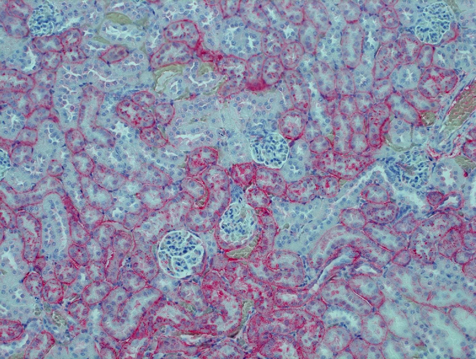
Glutathione Peroxidase 3/GPX3 in Human Kidney.
Glutathione Peroxidase 3/GPX3 was detected in immersion fixed paraffin-embedded sections of human kidney using 15 µg/mL Goat Anti-Human/Mouse/Rat Glutathione Peroxidase 3/GPX3 Antigen Affinity-purified Polyclonal Antibody (Catalog # AF4199) overnight at 4 °C. Tissue was stained with the Anti-Goat HRP-DAB Cell & Tissue Staining Kit (brown; Catalog # CTS008) and counterstained with hematoxylin (blue). Specific labeling was localized to the cytoplasm in tubules. View our protocol for Chromogenic IHC Staining of Paraffin-embedded Tissue Sections.
Detection of Human Glutathione Peroxidase 3/GPX3 by Simple WesternTM.
Simple Western lane view shows human serum, loaded at 0.2 mg/mL. A specific band was detected for Glutathione Peroxidase 3/GPX3 at approximately 29 kDa (as indicated) using 25 µg/mL of Goat Anti-Human/Mouse/Rat Glutathione Peroxidase 3/GPX3 Antigen Affinity-purified Polyclonal Antibody (Catalog # AF4199) followed by 1:50 dilution of HRP-conjugated Anti-Goat IgG Secondary Antibody (Catalog # HAF109). This experiment was conducted under reducing conditions and using the 12-230 kDa separation system.
Detection of Glutathione Peroxidase 3/GPX3 by Immunohistochemistry
Bioinformatics analysis showed that GPX3 expression was significantly increased during adipogenic differentiation of hBMSCs. A Heatmap of adipogenic marker genes and GPx family genes. B Volcano plot of adipogenic marker genes and GPx family genes. C The gene expression pattern of GPx family and oxidative stress-related enzymes during osteogenic and adipogenic differentiation of hBMSCs. Oxidative stress-related enzymes genes: PLA2G2A, LOX, CAT; Adipogenic marker genes: PPARG, CEBPA, FABP4; osteogenic marker genes: ALPL, BGLAP Image collected and cropped by CiteAb from the following open publication (https://pubmed.ncbi.nlm.nih.gov/40410842), licensed under a CC-BY license. Not internally tested by R&D Systems.
Detection of Glutathione Peroxidase 3/GPX3 by Western Blot
GPx3 expression during adipogenic differentiation of mBMSC in vitro. A Representative images of Oil Red O staining at different differentiation time points and (B) corresponding quantification. C Representative Western blot images showing the protein expressions of GPx3, along with adipogenic markers PPAR gamma and C/EBP alpha, at various differentiation stages. D-F Quantifications of band intensities in (C). G-J Relative mRNA expression levels of Gpx3 and adipogenic-specific genes Pparg, Cebp alpha, and Adipoq normalized to Actb during adipogenic differentiation. Data are presented as mean ± SD. *P < 0.05, ***P < 0.001, ****P < 0.0001 compared to the control group; ns indicates P > 0.05 Image collected and cropped by CiteAb from the following open publication (https://pubmed.ncbi.nlm.nih.gov/40410842), licensed under a CC-BY license. Not internally tested by R&D Systems.
Detection of Glutathione Peroxidase 3/GPX3 by Flow Cytometry
scRNA-seq data indicates that increased BMAds is associated with diminished Gpx3 expression during ageing. A The proportion of bone marrow cells in the young and aged bone marrow. B The expression levels of Adipoq or Lpl in the young and aged bone marrow. C-E UMAP visualization of Adipoq (adipocyte marker), Aspn (BMSC marker) and Gpx3 expression in scRNA-seq data from bone marrow cells of mice at different ages. F The expression levels of Gpx3 in BMSCs and adipocytes in the young and aged mice. 3 M, three-month-old mice; 16 M, sixteen-month-old mice; EC, endothelial cell Image collected and cropped by CiteAb from the following open publication (https://pubmed.ncbi.nlm.nih.gov/40410842), licensed under a CC-BY license. Not internally tested by R&D Systems.Applications for Glutathione Peroxidase 3/GPX3 Antibody
Application
Recommended Usage
Immunohistochemistry
5-15 µg/mL
Sample: Immersion fixed paraffin-embedded sections of human kidney
Sample: Immersion fixed paraffin-embedded sections of human kidney
Simple Western
25 µg/mL
Sample: Human serum
Sample: Human serum
Western Blot
0.5 µg/mL
Sample: Human plasma, mouse kidney tissue, and rat kidney tissue
Sample: Human plasma, mouse kidney tissue, and rat kidney tissue
Reviewed Applications
Read 1 review rated 5 using AF4199 in the following applications:
Formulation, Preparation, and Storage
Purification
Antigen Affinity-purified
Reconstitution
Reconstitute at 0.2 mg/mL in sterile PBS. For liquid material, refer to CoA for concentration.
Loading...
Formulation
Lyophilized from a 0.2 μm filtered solution in PBS with Trehalose. *Small pack size (SP) is supplied either lyophilized or as a 0.2 µm filtered solution in PBS.
Shipping
Lyophilized product is shipped at ambient temperature. Liquid small pack size (-SP) is shipped with polar packs. Upon receipt, store immediately at the temperature recommended below.
Stability & Storage
Use a manual defrost freezer and avoid repeated freeze-thaw cycles.
- 12 months from date of receipt, -20 to -70 °C as supplied.
- 1 month, 2 to 8 °C under sterile conditions after reconstitution.
- 6 months, -20 to -70 °C under sterile conditions after reconstitution.
Calculators
Background: Glutathione Peroxidase 3/GPX3
Alternate Names
GPX-P, GPX3
Gene Symbol
GPX3
UniProt
Additional Glutathione Peroxidase 3/GPX3 Products
Product Documents for Glutathione Peroxidase 3/GPX3 Antibody
Certificate of Analysis
To download a Certificate of Analysis, please enter a lot or batch number in the search box below.
Note: Certificate of Analysis not available for kit components.
Product Specific Notices for Glutathione Peroxidase 3/GPX3 Antibody
For research use only
Related Research Areas
Citations for Glutathione Peroxidase 3/GPX3 Antibody
 Powered by Bioz
Customer Reviews for Glutathione Peroxidase 3/GPX3 Antibody (1)
5 out of 5
1 Customer Rating
Have you used Glutathione Peroxidase 3/GPX3 Antibody?
Submit a review and receive an Amazon gift card!
$25/€18/£15/$25CAN/¥2500 Yen for a review with an image
$10/€7/£6/$10CAN/¥1110 Yen for a review without an image
Submit a review
Customer Images
Showing
1
-
1 of
1 review
Showing All
Filter By:
-
Application: ImmunohistochemistrySample Tested: Kidney tissueSpecies: MouseVerified Customer | Posted 04/21/2017Heat-induced antigen retrieval with citrate buffer antibody concentration 1:400

There are no reviews that match your criteria.
Protocols
Find general support by application which include: protocols, troubleshooting, illustrated assays, videos and webinars.
- Antigen Retrieval Protocol (PIER)
- Antigen Retrieval for Frozen Sections Protocol
- Appropriate Fixation of IHC/ICC Samples
- Cellular Response to Hypoxia Protocols
- Chromogenic IHC Staining of Formalin-Fixed Paraffin-Embedded (FFPE) Tissue Protocol
- Chromogenic Immunohistochemistry Staining of Frozen Tissue
- ClariTSA™ Fluorophore Kits
- Detection & Visualization of Antibody Binding
- Fluorescent IHC Staining of Frozen Tissue Protocol
- Graphic Protocol for Heat-induced Epitope Retrieval
- Graphic Protocol for the Preparation and Fluorescent IHC Staining of Frozen Tissue Sections
- Graphic Protocol for the Preparation and Fluorescent IHC Staining of Paraffin-embedded Tissue Sections
- Graphic Protocol for the Preparation of Gelatin-coated Slides for Histological Tissue Sections
- IHC Sample Preparation (Frozen sections vs Paraffin)
- Immunofluorescent IHC Staining of Formalin-Fixed Paraffin-Embedded (FFPE) Tissue Protocol
- Immunohistochemistry (IHC) and Immunocytochemistry (ICC) Protocols
- Immunohistochemistry Frozen Troubleshooting
- Immunohistochemistry Paraffin Troubleshooting
- Preparing Samples for IHC/ICC Experiments
- Preventing Non-Specific Staining (Non-Specific Binding)
- Primary Antibody Selection & Optimization
- Protocol for Heat-Induced Epitope Retrieval (HIER)
- Protocol for Making a 4% Formaldehyde Solution in PBS
- Protocol for VisUCyte™ HRP Polymer Detection Reagent
- Protocol for the Preparation & Fixation of Cells on Coverslips
- Protocol for the Preparation and Chromogenic IHC Staining of Frozen Tissue Sections
- Protocol for the Preparation and Chromogenic IHC Staining of Frozen Tissue Sections - Graphic
- Protocol for the Preparation and Chromogenic IHC Staining of Paraffin-embedded Tissue Sections
- Protocol for the Preparation and Chromogenic IHC Staining of Paraffin-embedded Tissue Sections - Graphic
- Protocol for the Preparation and Fluorescent IHC Staining of Frozen Tissue Sections
- Protocol for the Preparation and Fluorescent IHC Staining of Paraffin-embedded Tissue Sections
- Protocol for the Preparation of Gelatin-coated Slides for Histological Tissue Sections
- R&D Systems Quality Control Western Blot Protocol
- TUNEL and Active Caspase-3 Detection by IHC/ICC Protocol
- The Importance of IHC/ICC Controls
- Troubleshooting Guide: Immunohistochemistry
- Troubleshooting Guide: Western Blot Figures
- Western Blot Conditions
- Western Blot Protocol
- Western Blot Protocol for Cell Lysates
- Western Blot Troubleshooting
- Western Blot Troubleshooting Guide
- View all Protocols, Troubleshooting, Illustrated assays and Webinars
Loading...