Neuropilin-2 (Npn-2) is a 120 kDa, type I transmembrane (TM) glycoprotein that is related to the semaphorin receptor now known as Neuropilin-1 (1). Npn-2 is a complex molecule with multiple splice forms. Five transmembrane forms are known, and one 62 kDa soluble form has been identified (2). Based on the originally reported precursor size of 909 amino acids (aa), the “standard” precursor in human will have a 20 aa signal sequence, an 842 aa extracellular region, a 25 aa TM segment, and a 42 aa cytoplasmic tail (1). The extracellular region contains two N-terminal CUB (C1r/Ugef/BMP-1) domains, two jellyroll-shaped coagulation factor V type C domains, and a juxtamembrane MAM (meprin/A-5 protein/tyrosine phosphatase μ) domain (1, 3). The CUB and factor V domain are involved in VEGF and semaphorin binding. The MAM domain appears necessary for signaling through plexin-1 (4). The five transmembrane isoforms all share the same CUB, factor V and MAM domains. Splicing begins at aa 809, seven amino acids after the end of the MAM domain, and it involves the end of the extracellular region, the TM segment, and the cytoplasmic domain (a total of 101 aa). Two of the four variants show a complete replacement of these 101 aa with a totally unrelated stretch of approximately 90 aa. This creates a new TM and cytoplasmic tail. These forms are called “Npn-2b” forms. Two other isoforms (plus the standard 909 aa form) retain the 101 aa stretch, and add either 17 or 22 aa to the end of the extracellular region. These forms are called “Npn-2a” forms. The isoform offered by R&D Systems is the “a” form with the 17 aa addition. This isoform shows 94% aa identity to the equivalent regions in mouse and rat Npn-2. The soluble form of Npn-2 is 555 aa in precursor length, and contains the two CUB domains plus the first 1½ factor V type C domains (1). Npn-2 binds Sema3B through F, and VEGF isoforms 165, 145, PlGF-2, and VEGF-C (5). It is known to form homodimers and heterodimers with Npn-1, and it forms receptor complexes with plexin-1 and VEGF R1 (4, 5). Npn-2 is found on a variety of cell types including neurons (motor, autonomic, sensory), vascular endothelial cells, Schwann cells and pancreatic acinar cells.
Human/Mouse/Rat Neuropilin‑2 Antibody
R&D Systems | Catalog # AF2215

Key Product Details
Species Reactivity
Validated:
Cited:
Applications
Validated:
Cited:
Label
Antibody Source
Product Specifications
Immunogen
Gln23-Tyr855
Accession # Q7LBX6
Specificity
Clonality
Host
Isotype
Endotoxin Level
Scientific Data Images for Human/Mouse/Rat Neuropilin‑2 Antibody
Detection of Human Neuropilin‑2 by Western Blot.
Western blot shows lysates of HUVEC human umbilical vein endothelial cells. PVDF membrane was probed with 0.5 µg/mL of Goat Anti-Human/Mouse/Rat Neuropilin-2 Antigen Affinity-purified Polyclonal Antibody (Catalog # AF2215) followed by HRP-conjugated Anti-Goat IgG Secondary Antibody HAF109). A specific band was detected for Neuropilin-2 at approximately 120 kDa (as indicated). This experiment was conducted under reducing conditions and using Immunoblot Buffer Group 1.
Neuropilin‑2 in Human Pancreatic Cancer Tissue.
Neuropilin-2 was detected in immersion fixed paraffin-embedded sections of human pancreatic cancer tissue using Goat Anti-Human/Mouse/Rat Neuropilin-2 Antigen Affinity-purified Poly-clonal Antibody (Catalog # AF2215) at 5 µg/mL overnight at 4 °C. Before incubation with the primary antibody, tissue was subjected to heat-induced epitope retrieval using Antigen Retrieval Reagent-Basic (CTS013). Tissue was stained using the Anti-Goat HRP-DAB Cell & Tissue Staining Kit (brown; CTS008) and counterstained with hematoxylin (blue). View our protocol for Chromogenic IHC Staining of Paraffin-embedded Tissue Sections.
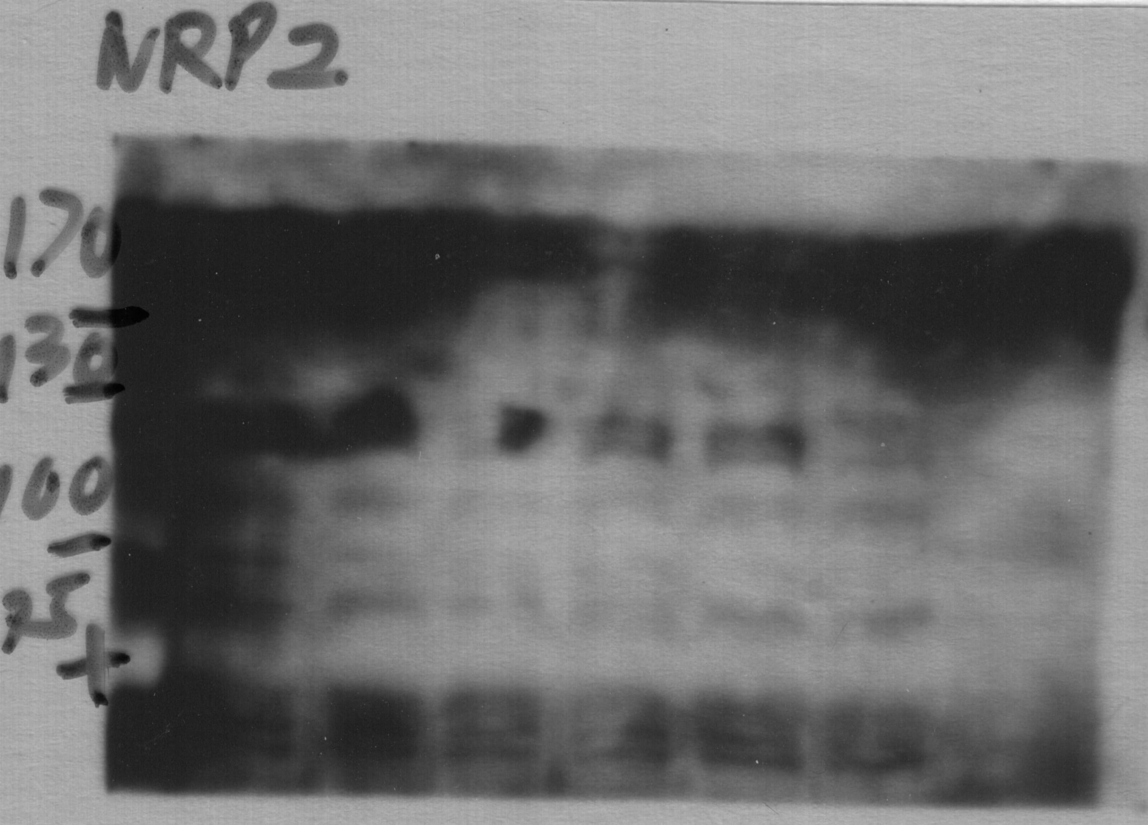
Detection of Human, Mouse, and Rat Neuropilin‑2 by Simple WesternTM.
Simple Western lane view shows lysates of HUVEC human umbilical vein endothelial cells, C6 rat glioma cell line, LL/2 mouse Lewis lung carcinoma cell line, and bEnd.3 mouse endothelioma cell line, loaded at 0.2 mg/mL. A specific band was detected for Neuropilin-2 at approximately 140 kDa (as indicated) using 10 µg/mL of Goat Anti-Human/Mouse/Rat Neuropilin-2 Antigen Affinity-purified Polyclonal Antibody (Catalog # AF2215) followed by 1:50 dilution of HRP-conjugated Anti-Goat IgG Secondary Antibody HAF109). This experiment was conducted under reducing conditions and using the 12-230 kDa separation system.
Detection of Neuropillin-2 in HUVEC Human Cell Line by Flow Cytometry.
HUVEC human umbilical vein endothelial cells were stained with Goat Anti-Human/Mouse/Rat Neuropillin-2 Polyclonal Antibody (Catalog # AF2215, filled histogram) or Goat IgG control antibody (AB-108-C, open histogram), followed by Phycoerythrin-conjugated anti-Goat IgG (F0107). Staining was performed using our Staining Surface Molecules protocol.Applications for Human/Mouse/Rat Neuropilin‑2 Antibody
Blockade of Receptor-ligand Interaction
CyTOF-ready
Flow Cytometry
Sample: HUVEC human umbilical vein endothelial cells
Immunohistochemistry
Sample: Immersion fixed paraffin-embedded sections of human pancreatic cancer tissue subjected to Antigen Retrieval Reagent-Basic (Catalog # CTS013)
Simple Western
Sample: HUVEC human umbilical vein endothelial cells, C6 rat glioma cell line, LL/2 mouse Lewis lung carcinoma cell line, and bEnd.3 mouse endothelioma cell line
Western Blot
Sample: HUVEC human umbilical vein endothelial cells
Reviewed Applications
Read 2 reviews rated 3 using AF2215 in the following applications:
Flow Cytometry Panel Builder
Bio-Techne Knows Flow Cytometry
Save time and reduce costly mistakes by quickly finding compatible reagents using the Panel Builder Tool.
Advanced Features
- Spectra Viewer - Custom analysis of spectra from multiple fluorochromes
- Spillover Popups - Visualize the spectra of individual fluorochromes
- Antigen Density Selector - Match fluorochrome brightness with antigen density

Formulation, Preparation, and Storage
Purification
Reconstitution
Reconstitute at 0.2 mg/mL in sterile PBS. For liquid material, refer to CoA for concentration.
Formulation
Shipping
Stability & Storage
- 12 months from date of receipt, -20 to -70 °C as supplied.
- 1 month, 2 to 8 °C under sterile conditions after reconstitution.
- 6 months, -20 to -70 °C under sterile conditions after reconstitution.
Calculators
Background: Neuropilin-2
References
- Chen, H. et al. (1997) Neuron 19:547.
- Rossignol, M. et al. (2000) Genomics 70:211.
- He, Z. and M. Tessier-lavigne (1997) Cell 90:739.
- Nakamura, F. and Y. Goshima (2002) Adv. Exp. Med. Biol. 515:55.
- Neufeld, G. et al. (2002) Adv. Exp. Med. Biol. 515:81.
Alternate Names
Gene Symbol
UniProt
Additional Neuropilin-2 Products
Product Documents for Human/Mouse/Rat Neuropilin‑2 Antibody
Certificate of Analysis
To download a Certificate of Analysis, please enter a lot or batch number in the search box below.
Note: Certificate of Analysis not available for kit components.
Product Specific Notices for Human/Mouse/Rat Neuropilin‑2 Antibody
This product or the use of this product is covered by U.S. Patents owned by The Regents of the University of California. This product is for research use only and is not to be used for commercial purposes. Use of this product to produce products for sale or for diagnostic, therapeutic or drug discovery purposes is prohibited. In order to obtain a license to use this product for such purposes, contact The Regents of the University of California.
U.S. Patent # 6,054,293, 6,623,738, and other U.S. and international patents pending.
For research use only
Citations for Human/Mouse/Rat Neuropilin‑2 Antibody
 Powered by Bioz
Customer Reviews for Human/Mouse/Rat Neuropilin‑2 Antibody (2)
Have you used Human/Mouse/Rat Neuropilin‑2 Antibody?
Submit a review and receive an Amazon gift card!
$25/€18/£15/$25CAN/¥2500 Yen for a review with an image
$10/€7/£6/$10CAN/¥1110 Yen for a review without an image
Submit a review
Customer Images
-
Verified Customer | Posted 04/21/2017

-
Application: Immunocytochemistry/ImmunofluorescenceSample Tested: Neonatal dorsal root ganglia cellsSpecies: MouseVerified Customer | Posted 09/01/2016
There are no reviews that match your criteria.
Protocols
Find general support by application which include: protocols, troubleshooting, illustrated assays, videos and webinars.
- 7-Amino Actinomycin D (7-AAD) Cell Viability Flow Cytometry Protocol
- Antigen Retrieval Protocol (PIER)
- Antigen Retrieval for Frozen Sections Protocol
- Appropriate Fixation of IHC/ICC Samples
- Cellular Response to Hypoxia Protocols
- Chromogenic IHC Staining of Formalin-Fixed Paraffin-Embedded (FFPE) Tissue Protocol
- Chromogenic Immunohistochemistry Staining of Frozen Tissue
- ClariTSA™ Fluorophore Kits
- Detection & Visualization of Antibody Binding
- Extracellular Membrane Flow Cytometry Protocol
- Flow Cytometry Protocol for Cell Surface Markers
- Flow Cytometry Protocol for Staining Membrane Associated Proteins
- Flow Cytometry Staining Protocols
- Flow Cytometry Troubleshooting Guide
- Fluorescent IHC Staining of Frozen Tissue Protocol
- Graphic Protocol for Heat-induced Epitope Retrieval
- Graphic Protocol for the Preparation and Fluorescent IHC Staining of Frozen Tissue Sections
- Graphic Protocol for the Preparation and Fluorescent IHC Staining of Paraffin-embedded Tissue Sections
- Graphic Protocol for the Preparation of Gelatin-coated Slides for Histological Tissue Sections
- IHC Sample Preparation (Frozen sections vs Paraffin)
- Immunofluorescent IHC Staining of Formalin-Fixed Paraffin-Embedded (FFPE) Tissue Protocol
- Immunohistochemistry (IHC) and Immunocytochemistry (ICC) Protocols
- Immunohistochemistry Frozen Troubleshooting
- Immunohistochemistry Paraffin Troubleshooting
- Intracellular Flow Cytometry Protocol Using Alcohol (Methanol)
- Intracellular Flow Cytometry Protocol Using Detergents
- Intracellular Nuclear Staining Flow Cytometry Protocol Using Detergents
- Intracellular Staining Flow Cytometry Protocol Using Alcohol Permeabilization
- Intracellular Staining Flow Cytometry Protocol Using Detergents to Permeabilize Cells
- Preparing Samples for IHC/ICC Experiments
- Preventing Non-Specific Staining (Non-Specific Binding)
- Primary Antibody Selection & Optimization
- Propidium Iodide Cell Viability Flow Cytometry Protocol
- Protocol for Heat-Induced Epitope Retrieval (HIER)
- Protocol for Liperfluo
- Protocol for Making a 4% Formaldehyde Solution in PBS
- Protocol for VisUCyte™ HRP Polymer Detection Reagent
- Protocol for the Characterization of Human Th22 Cells
- Protocol for the Characterization of Human Th9 Cells
- Protocol for the Preparation & Fixation of Cells on Coverslips
- Protocol for the Preparation and Chromogenic IHC Staining of Frozen Tissue Sections
- Protocol for the Preparation and Chromogenic IHC Staining of Frozen Tissue Sections - Graphic
- Protocol for the Preparation and Chromogenic IHC Staining of Paraffin-embedded Tissue Sections
- Protocol for the Preparation and Chromogenic IHC Staining of Paraffin-embedded Tissue Sections - Graphic
- Protocol for the Preparation and Fluorescent IHC Staining of Frozen Tissue Sections
- Protocol for the Preparation and Fluorescent IHC Staining of Paraffin-embedded Tissue Sections
- Protocol for the Preparation of Gelatin-coated Slides for Histological Tissue Sections
- Protocol: Annexin V and PI Staining by Flow Cytometry
- Protocol: Annexin V and PI Staining for Apoptosis by Flow Cytometry
- R&D Systems Quality Control Western Blot Protocol
- TUNEL and Active Caspase-3 Detection by IHC/ICC Protocol
- The Importance of IHC/ICC Controls
- Troubleshooting Guide: Fluorokine Flow Cytometry Kits
- Troubleshooting Guide: Immunohistochemistry
- Troubleshooting Guide: Western Blot Figures
- Western Blot Conditions
- Western Blot Protocol
- Western Blot Protocol for Cell Lysates
- Western Blot Troubleshooting
- Western Blot Troubleshooting Guide
- View all Protocols, Troubleshooting, Illustrated assays and Webinars
Associated Pathways
