
phospho-ERK1/ERK2 (ERK1 T202/Y204, ERK2 T185/Y187) Antibody (269434)
R&D Systems | Catalog # MAB1018


Scientific images may be resized, cropped, or adjusted for brightness or contrast for presentation purposes. These updates are limited to presentation and do not affect the underlying scientific interpretation of the data. Where available, additional source data may be provided upon request.
Key Product Details
Validated by
Species Reactivity
Validated:
Cited:
Applications
Validated:
Cited:
Label
Antibody Source
Product Specifications
Immunogen
Specificity
Clonality
Host
Isotype
Scientific Data Images for phospho-ERK1/ERK2 (ERK1 T202/Y204, ERK2 T185/Y187) Antibody (269434)
Detection of Mouse, Rat, and Human Phospho-ERK1 (T202/Y204) and ERK2 (T185/Y187) by Western Blot.
Western blot shows lysates of NIH-3T3 mouse embryonic fibroblast cell line untreated (-) or treated (+) with 100 ng/mL Rabbit Anti-Human PDGF (Catalog # 120-HD) and PC-12 rat adrenal pheochromocytoma cell line untreated or treated with 100 ng/mL Recombinant Rat beta -NGF (Catalog # 556-NG) and HeLa human cervical epithelial carcinoma cell line untreated or treated with 200 nM PMA. PVDF membrane was probed with 0.5 µg/mL of Human/Mouse/Rat Phospho-ERK1 (T202/Y204)/ERK2 (T185/Y187) Monoclonal Antibody (MAB1018), followed by HRP-conjugated Anti-Rabbit IgG Secondary Antibody (Catalog # HAF008). Specific bands were detected for Phospho-ERK1 (T202/Y204)/ ERK2 (T185/Y187) at approximately 40-45 kDa (as indicated). This experiment was conducted under reducing conditions and using Immunoblot Buffer Group 3.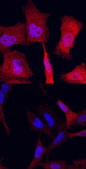
Phospho-ERK1 (T202/Y204)/ERK2 (T185/Y187) in HeLa Human Cell Line.
ERK1/ERK2 phosphorylated at T202/Y204 (ERK1) and T185/Y187 (ERK2) was detected in immersion fixed Hela human cervical epithelial carcinoma cells, unstimulated (lower panel) and stimulated (upper panel) with PMA using Rabbit Anti-Human/Mouse/Rat Phospho-ERK1/ERK2 (ERK1 T202/Y204, ERK2 T185/Y187) Monoclonal Antibody (Catalog # MAB1018) at 10 µg/mL for 3 hours at room temperature. Cells were stained using the NorthernLights™ 557-conjugated Anti-Rabbit IgG Secondary Antibody (red; Catalog # NL004) and counterstained with DAPI (blue). Specific staining was localized to cytoplasm. View our protocol for Fluorescent ICC Staining of Cells on Coverslips.
Detection of Phospho-ERK1/ERK2 in PMA-treated Jurkat Human Cell Line by Flow Cytometry.
Jurkat human acute T cell leukemia cell line were unstimulated (light orange open histogram) or treated with 50 ng/mL PMA for 10 minutes (dark orange filled histogram), then stained with Rabbit Anti-Human/Mouse/Rat Phospho-ERK1/ERK2 (ERK1 T202/Y204, ERK2 T185/Y187) Monoclonal Antibody (Catalog # MAB1018) or control antibody (Catalog # AB-105-C, blue open histogram), followed by Fluorescein-conjugated Anti-Rabbit IgG Secondary Antibody (Catalog # F0112). To facilitate intracellular staining, cells were fixed with paraformaldehyde and permeabilized with ice-cold methanol.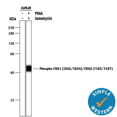
Detection of Human Phospho-ERK1 (T202/Y204)/ERK2 (T185/Y187) by Simple WesternTM.
Simple Western lane view shows lysates of Jurkat human acute T cell leukemia cell line untreated (-) or treated (+) with 200 nM PMA and Ionomycin for 20 minutes, loaded at 0.2 mg/mL. A specific band was detected for Phospho-ERK1 (T202/Y204)/ERK2 (T185/Y187) at approximately 42 kDa (as indicated) using 5 µg/mL of Rabbit Anti-Human/Mouse/Rat Phospho-ERK1/ERK2 (ERK1 T202/Y204, ERK2 T185/Y187) Monoclonal Antibody (Catalog # MAB1018). This experiment was conducted under reducing conditions and using the 12-230 kDa separation system.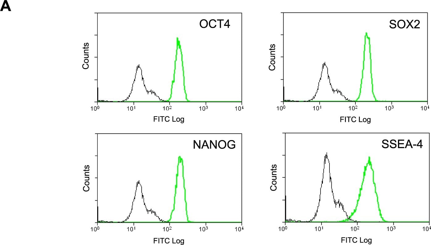
Detection of Human ERK1/ERK2 by Flow Cytometry
Differentiation Characteristics of Individual Stem Cells.(A) Flow cytometric analysis of undifferentiated stem cell markers in the clonal NTERA2.Tom cell line. Black line depicts P3X negative control and green depicts antigen specific expression (OCT4, SOX2, NANOG and SSEA4). (B) Graph depicting the percentage of TUJ1 positive neurons within differentiated colonies derived from individual RA treated NTERA2.Tom stem cells. (C) Images of differentiated NTERA2.Tom colonies derived from individual NTERA2.Tom cells on a background of wildtype NTERA2 cells. Cell were exposed to RA for 21 days (Green - beta III tubulin, Blue - Hoechst) White arrows depict beta III tubulin positive NTERA2.Tom neurons. Scale bar represents 50 µM. Image collected and cropped by CiteAb from the following publication (https://pubmed.ncbi.nlm.nih.gov/20531938), licensed under a CC-BY license. Not internally tested by R&D Systems.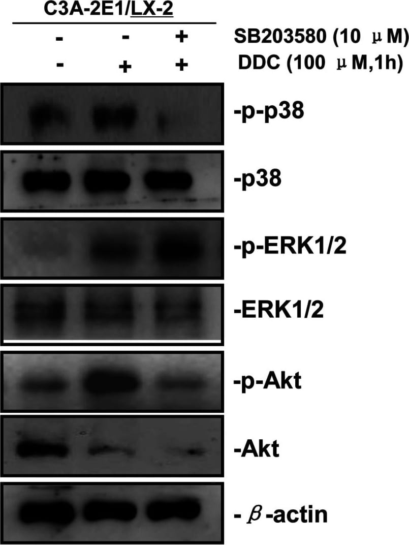
Detection of Human ERK1/ERK2 by Western Blot
p38 inhibitor SB203580 improves the up-regulation of MMP-1 by DDC through stimulating ERK1/2Co-cultures were treated with or without 100 μM DDC for 1 h in the presence or absence of p38 inhibitor (SB203580, 10 μM). Phosphorylation of ERK1/2, p38 and Akt in LX-2 cells of co-cultures were determined by Western blotting analysis. Image collected and cropped by CiteAb from the following publication (https://pubmed.ncbi.nlm.nih.gov/23577625), licensed under a CC-BY license. Not internally tested by R&D Systems.
Detection of Human ERK1/ERK2 by Western Blot
Phospho-proteome profiling results of LX-2 cells co-cultured with C3A–CYP2E1 cells with or without DDC treatment and detection of the effect of DDC on intracellular kinases in LX-2 cells of co-cultures200 μg of total cell lysates from LX-2 cells co-cultured with C3A-2E1 cells with or without 100 μM DDC for 1 h were incubated with membranes of the human phospho-MAPK Array Kit according to the manufacturer's instructions. Phospho MAPK Array data were developed on X-ray films following exposure to chemiluminescent reagents. 20 μg aliquots of total cell lysates from LX-2 cells were subjected to Western blotting analysis. (A) Template showing the location of MAPK antibodies spotted onto the human phospho-MAPK Array Kit. (B) The activation status of ERK1/2 in LX-2 cells co-cultured with C3A-2E1 cells after DDC treatment. (C) The activation status of p38 in LX-2 cells co-cultured with C3A-2E1 cells after DDC treatment. (D) The activation status of Akt in LX-2 cells co-cultured with C3A-2E1 cells after DDC treatment. (E) The activation status of ERK1/2, p38 and Akt in LX-2 cells co-cultured with C3A cells after DDC treatment. Image collected and cropped by CiteAb from the following publication (https://pubmed.ncbi.nlm.nih.gov/23577625), licensed under a CC-BY license. Not internally tested by R&D Systems.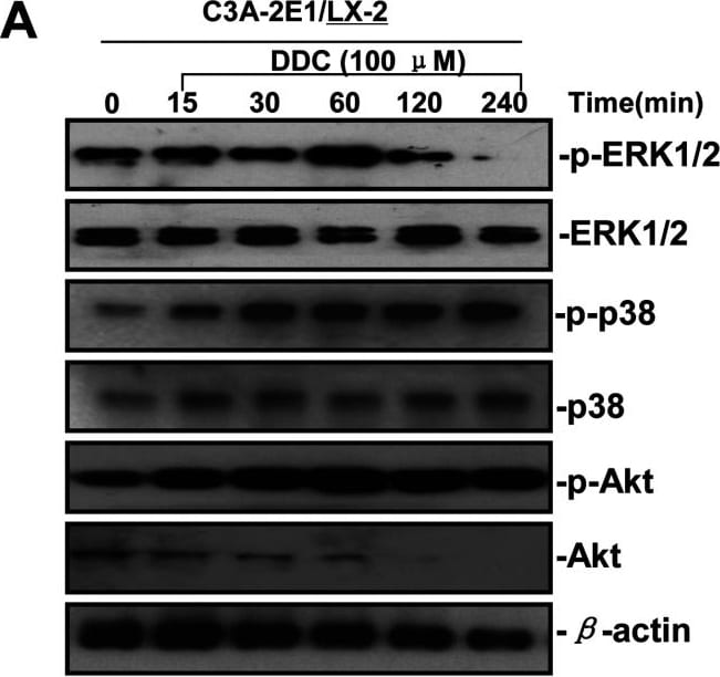
Detection of Human ERK1/ERK2 by Western Blot
DDC up-regulates MMP-1 through ERK1/2 and Akt activation(A) Co-cultures were treated with 100 μM DDC for the indicated time. Phosphorylation of ERK1/2, p38 and Akt were determined by Western blotting analysis. The corresponding non-phosphorylated ERK1/2, p38, Akt and beta -actin were used for protein loading control. (B) Co-cultures were treated with or without 100 μM DDC for 24 h in the presence or absence of ERK1/2 inhibitor (U0126, 10 μM). MMP-1 protein levels were analysed by Western blotting. (C) Co-cultures were treated with or without 100 μM DDC for 24 h in the presence or absence of p38 inhibitor (SB203580, 10 μM). MMP-1 protein levels were analysed by Western blotting. (D) Co-cultures were treated with or without 100 μM DDC for 24 h in the presence or absence of Akt inhibitor (T3830, 50 μM). MMP-1 protein levels were analysed by Western blotting. Image collected and cropped by CiteAb from the following publication (https://pubmed.ncbi.nlm.nih.gov/23577625), licensed under a CC-BY license. Not internally tested by R&D Systems.
Detection of Human ERK1/ERK2 by Western Blot
Phospho-proteome profiling results of LX-2 cells co-cultured with C3A–CYP2E1 cells with or without DDC treatment and detection of the effect of DDC on intracellular kinases in LX-2 cells of co-cultures200 μg of total cell lysates from LX-2 cells co-cultured with C3A-2E1 cells with or without 100 μM DDC for 1 h were incubated with membranes of the human phospho-MAPK Array Kit according to the manufacturer's instructions. Phospho MAPK Array data were developed on X-ray films following exposure to chemiluminescent reagents. 20 μg aliquots of total cell lysates from LX-2 cells were subjected to Western blotting analysis. (A) Template showing the location of MAPK antibodies spotted onto the human phospho-MAPK Array Kit. (B) The activation status of ERK1/2 in LX-2 cells co-cultured with C3A-2E1 cells after DDC treatment. (C) The activation status of p38 in LX-2 cells co-cultured with C3A-2E1 cells after DDC treatment. (D) The activation status of Akt in LX-2 cells co-cultured with C3A-2E1 cells after DDC treatment. (E) The activation status of ERK1/2, p38 and Akt in LX-2 cells co-cultured with C3A cells after DDC treatment. Image collected and cropped by CiteAb from the following publication (https://pubmed.ncbi.nlm.nih.gov/23577625), licensed under a CC-BY license. Not internally tested by R&D Systems.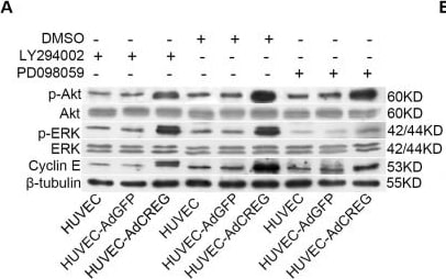
Detection of Human ERK1/ERK2 by Western Blot
ERK, but not PI3K/Akt, mediates the increase of cyclin E induced by CREG overexpression in HUVEC. (A) Expression of p-ERK, ERK, p-Akt, Akt, cyclin E and beta -tubulin was detected by Western blotting in the three groups of cells with LY294002 (50 μM) or PD098059 (20 μM) treatment. Vehicle (DMSO)-treated cells were used as control, and beta -tubulin was used as a loading control; (B,C) Pooled analysis of the levels of Akt (B) and ERK (C) signaling activation accessed by the grayscale ratios of p-Akt/Akt and p-ERK/ERK, respectively. Data are given as the mean ± SD (n = 3). *p < 0.05; **p < 0.01. NS: no significant difference. (D) After normalization to beta -tubulin, the difference in the expression of cyclin E between the HUVEC-AdGFP and HUVEC-AdCREG group was reduced by the treatment of PD098059, but not LY294002. Data are given as the mean ± SD (n = 3). **p < 0.01. NS: no significant difference. Image collected and cropped by CiteAb from the following open publication (https://pubmed.ncbi.nlm.nih.gov/24018888), licensed under a CC-BY license. Not internally tested by R&D Systems.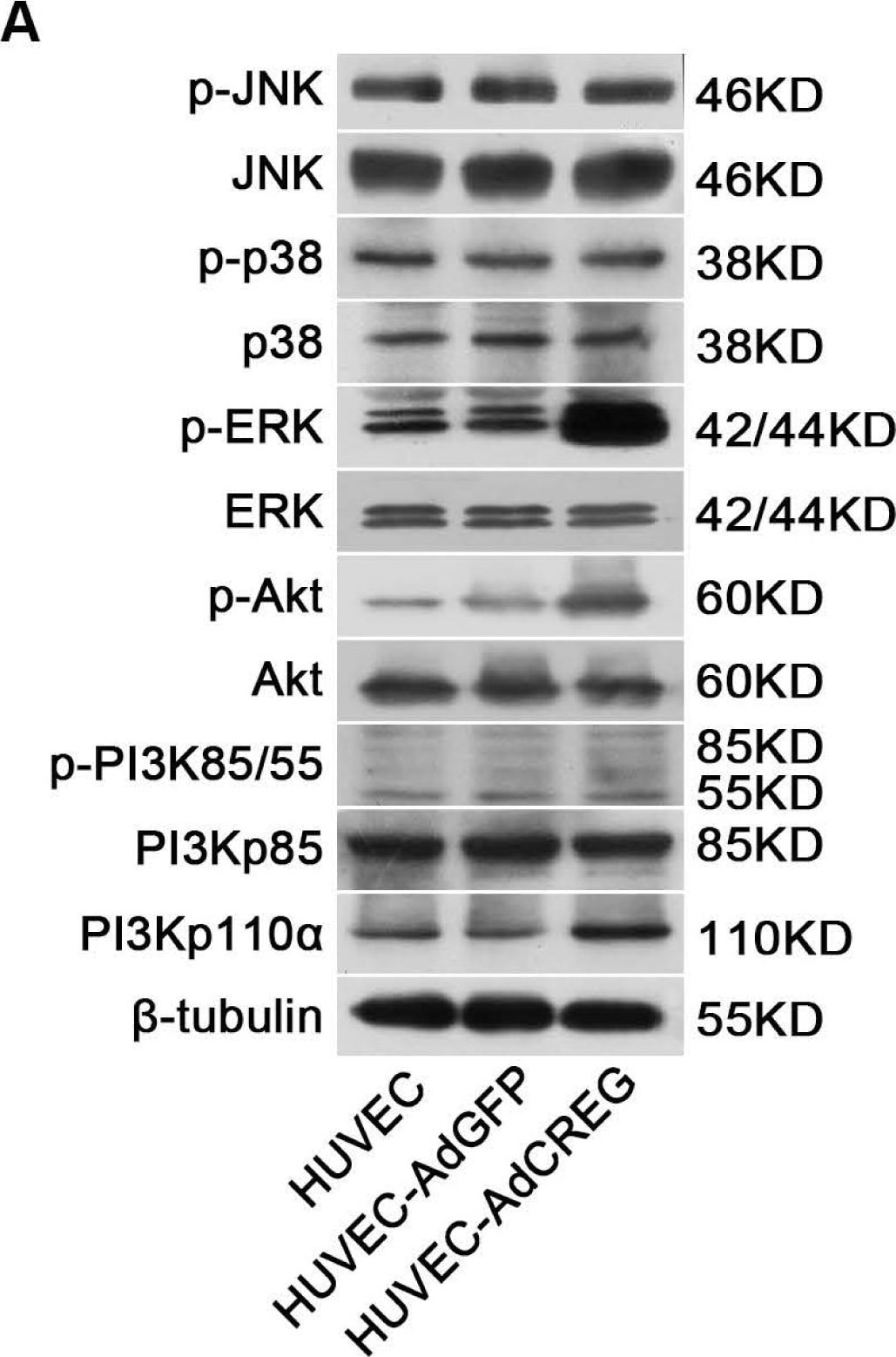
Detection of Human ERK1/ERK2 by Western Blot
CREG overexpression activates both ERK&PI3K/Akt signaling pathways,&ERK, but not PI3K/Akt, mediates CREG effects on HUVEC proliferation. (A) In the three groups of cells (HUVEC, AdGFP&AdCREG), expression of JNK, p38, ERK, PI3K&Akt signaling molecules&their phosphorylated (p-) forms detected by WB, with beta -tubulin as the loading control; Image collected & cropped by CiteAb from the following open publication (https://pubmed.ncbi.nlm.nih.gov/24018888), licensed under a CC-BY license. Not internally tested by R&D Systems.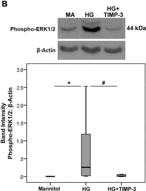
Detection of Phospho-ERK1 (T202/Y204)/ERK2 (T185/Y187) by Western Blot
Müller cells were pre-incubated with TIMP-3 (100 ng/ml) or dilution medium for 1 h before increasing the sugar content of the cultures [25 mM of mannitol as control or 25 mM of glucose (HG)]. After 24 h levels of the p65 subunit of NF-kappa B [panel (A)], phospho-ERK1/2 [panel (B)], caspase-3 [panel (C)], and ADAM17 [panel (D)] in cell lysates was determined by Western blot analysis. Results are expressed as median (interquartile range) from three different experiments performed in triplicate. Kruskal-Wallis test and Mann-Whitney test were used for comparisons between three groups and two groups, respectively. *p < 0.05 compared with values obtained from cells treated with mannitol. #p < 0.05 compared with values obtained from cells treated with HG. Image collected and cropped by CiteAb from the following open publication (https://pubmed.ncbi.nlm.nih.gov/35082694), licensed under a CC-BY license. Not internally tested by R&D Systems.Applications for phospho-ERK1/ERK2 (ERK1 T202/Y204, ERK2 T185/Y187) Antibody (269434)
CyTOF-ready
Immunocytochemistry
Sample: Immersion fixed HeLa human cervical epithelial carcinoma cell line stimulated with PMA
Intracellular Staining by Flow Cytometry
Sample: Jurkat human acute T cell leukemia cell line treated with PMA, fixed with paraformaldehyde and permeabilized with ice-cold methanol
Simple Western
Sample: Jurkat human acute T cell leukemia cell line treated with PMA and Ionomycin
Western Blot
Sample: NIH‑3T3 mouse embryonic fibroblast cell line treated with Human PDGF (Catalog # 120-HD) and PC‑12 rat adrenal pheochromocytoma cell line treated with Recombinant Rat beta -NGF (Catalog # 556-NG)
Reviewed Applications
Read 2 reviews rated 5 using MAB1018 in the following applications:
Flow Cytometry Panel Builder
Bio-Techne Knows Flow Cytometry
Save time and reduce costly mistakes by quickly finding compatible reagents using the Panel Builder Tool.
Advanced Features
- Spectra Viewer - Custom analysis of spectra from multiple fluorochromes
- Spillover Popups - Visualize the spectra of individual fluorochromes
- Antigen Density Selector - Match fluorochrome brightness with antigen density

Formulation, Preparation, and Storage
Purification
Reconstitution
Reconstitute at 0.5 mg/mL in sterile PBS. For liquid material, refer to CoA for concentration.
Formulation
Shipping
Stability & Storage
- 12 months from date of receipt, -20 to -70 °C as supplied.
- 1 month, 2 to 8 °C under sterile conditions after reconstitution.
- 6 months, -20 to -70 °C under sterile conditions after reconstitution.
Calculators
Background: ERK1/ERK2
Long Name
Alternate Names
Additional ERK1/ERK2 Products
Product Documents for phospho-ERK1/ERK2 (ERK1 T202/Y204, ERK2 T185/Y187) Antibody (269434)
Certificate of Analysis
To download a Certificate of Analysis, please enter a lot or batch number in the search box below.
Note: Certificate of Analysis not available for kit components.
Product Specific Notices for phospho-ERK1/ERK2 (ERK1 T202/Y204, ERK2 T185/Y187) Antibody (269434)
For research use only
Citations for phospho-ERK1/ERK2 (ERK1 T202/Y204, ERK2 T185/Y187) Antibody (269434)
 Powered by Bioz
Customer Reviews for phospho-ERK1/ERK2 (ERK1 T202/Y204, ERK2 T185/Y187) Antibody (269434) (2)
Have you used phospho-ERK1/ERK2 (ERK1 T202/Y204, ERK2 T185/Y187) Antibody (269434)?
Submit a review and receive an Amazon gift card!
$25/€18/£15/$25CAN/¥2500 for a review with an image
$10/€7/£6/$10CAN/¥1110 for a review without an image
Submit a review
Customer Images
-
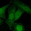
Application: Immunocytochemistry/ImmunofluorescenceSample Tested: HeLa cellsSpecies: HumanVerified Customer | Posted 10/11/2021

-
Application: Western BlotSample Tested: Cell ExtractsSpecies: HumanVerified Customer | Posted 06/29/2016
There are no reviews that match your criteria.
Protocols
Find general support by application which include: protocols, troubleshooting, illustrated assays, videos and webinars.
- 7-Amino Actinomycin D (7-AAD) Cell Viability Flow Cytometry Protocol
- Appropriate Fixation of IHC/ICC Samples
- Cellular Response to Hypoxia Protocols
- ClariTSA™ Fluorophore Kits
- Detection & Visualization of Antibody Binding
- Extracellular Membrane Flow Cytometry Protocol
- Flow Cytometry Protocol for Cell Surface Markers
- Flow Cytometry Protocol for Staining Membrane Associated Proteins
- Flow Cytometry Staining Protocols
- Flow Cytometry Troubleshooting Guide
- ICC Cell Smear Protocol for Suspension Cells
- ICC Immunocytochemistry Protocol Videos
- ICC for Adherent Cells
- Immunocytochemistry (ICC) Protocol
- Immunocytochemistry Troubleshooting
- Immunofluorescence of Organoids Embedded in Cultrex Basement Membrane Extract
- Immunohistochemistry (IHC) and Immunocytochemistry (ICC) Protocols
- Intracellular Flow Cytometry Protocol Using Alcohol (Methanol)
- Intracellular Flow Cytometry Protocol Using Detergents
- Intracellular Nuclear Staining Flow Cytometry Protocol Using Detergents
- Intracellular Staining Flow Cytometry Protocol Using Alcohol Permeabilization
- Intracellular Staining Flow Cytometry Protocol Using Detergents to Permeabilize Cells
- Preparing Samples for IHC/ICC Experiments
- Preventing Non-Specific Staining (Non-Specific Binding)
- Primary Antibody Selection & Optimization
- Propidium Iodide Cell Viability Flow Cytometry Protocol
- Protocol for Liperfluo
- Protocol for VisUCyte™ HRP Polymer Detection Reagent
- Protocol for the Characterization of Human Th22 Cells
- Protocol for the Characterization of Human Th9 Cells
- Protocol for the Fluorescent ICC Staining of Cell Smears - Graphic
- Protocol for the Fluorescent ICC Staining of Cultured Cells on Coverslips - Graphic
- Protocol for the Preparation and Fluorescent ICC Staining of Cells on Coverslips
- Protocol for the Preparation and Fluorescent ICC Staining of Non-adherent Cells
- Protocol for the Preparation and Fluorescent ICC Staining of Stem Cells on Coverslips
- Protocol for the Preparation of a Cell Smear for Non-adherent Cell ICC - Graphic
- Protocol: Annexin V and PI Staining by Flow Cytometry
- Protocol: Annexin V and PI Staining for Apoptosis by Flow Cytometry
- R&D Systems Quality Control Western Blot Protocol
- TUNEL and Active Caspase-3 Detection by IHC/ICC Protocol
- The Importance of IHC/ICC Controls
- Troubleshooting Guide: Fluorokine Flow Cytometry Kits
- Troubleshooting Guide: Western Blot Figures
- Western Blot Conditions
- Western Blot Protocol
- Western Blot Protocol for Cell Lysates
- Western Blot Troubleshooting
- Western Blot Troubleshooting Guide
- View all Protocols, Troubleshooting, Illustrated assays and Webinars
Associated Pathways

