The tumor suppressor gene PTEN (phosphatase and tensin homolog deleted on chromosome 10), also known as MMAC1 (mutated in multiple advanced cancers 1), encodes a phosphatase that contains the catalytic signature motif (HCXXGXXRS/T) found in all members of the protein tyrosine phosphatase family. In vitro, the recombinant PTEN has both lipid phosphatase and protein phosphatase activities (1, 2). Interestingly, accumulating evidence has shown that the tumor suppressor activity of PTEN relies on its ability to dephosphorylate phosphatidylinositol (3, 4, 5)-triphosphate specifically at position 3 of the inositol ring (3). This activity reduces the levels of phosphatidylinositol (3, 4, 5)-triphosphate which is specifically produced from phosphatidylinositol (4, 5)-diphosphate by PI 3-kinase upon activation by a variety of stimuli. Therefore, PTEN antagonizes PI 3-kinase-induced downstream signaling events and cellular processes including cell growth, apoptosis and cell motility. In vivo, the importance of PTEN catalytic activity in its tumor suppressor functions is underscored by the fact that the majority of PTEN missense mutations detected in tumor specimens target the phosphatase domain and cause a loss in PTEN phosphatase activity (4).

Key Product Details
Validated by
Species Reactivity
Validated:
Cited:
Applications
Validated:
Cited:
Label
Antibody Source
Product Specifications
Immunogen
Ser385-Val403
Accession # P60484
Specificity
Clonality
Host
Isotype
Scientific Data Images for PTEN Antibody
Detection of Human/Mouse/Rat PTEN by Western Blot.
Western blot shows lysates of mouse and rat brain tissue, A431 human epithelial carcinoma cell line, and MRC-5 human embryonic lung fibroblast cell line. PVDF membrane was probed with 0.1 µg/mL Rabbit Anti-Human/Mouse/Rat PTEN Antigen Affinity-purified Polyclonal Antibody (Catalog # AF847) followed by HRP-conjugated Anti-Rabbit IgG Secondary Antibody (Catalog # HAF008). For additional reference, recombinant human PTEN (5 ng) was included. A specific band for PTEN was detected at approximately 54 kDa (as indicated). This experiment was conducted under reducing conditions and using Immunoblot Buffer Group 4.
PTEN in Human Pancreas.
PTEN was detected in immersion fixed paraffin-embedded sections of human pancreas using Rabbit Anti-Human/Mouse/Rat PTEN Antigen Affinity-purified Polyclonal Antibody (Catalog # AF847) at 3 µg/mL for 1 hour at room temperature followed by incubation with the Anti-Rabbit IgG VisUCyte™ HRP Polymer Antibody (VC003). Before incubation with the primary antibody, tissue was subjected to heat-induced epitope retrieval using Antigen Retrieval Reagent-Basic (CTS013). Tissue was stained using DAB (brown) and counterstained with hematoxylin (blue). Specific staining was localized to cytoplasm in islet cells. Staining was performed using our protocol for IHC Staining with VisUCyte HRP Polymer Detection Reagents.
PTEN in Mouse Pancreas.
PTEN was detected in immersion fixed paraffin-embedded sections of mouse pancreas using Rabbit Anti-Human/Mouse/Rat PTEN Antigen Affinity-purified Polyclonal Antibody (Catalog # AF847) at 3 µg/mL for 1 hour at room temperature followed by incubation with the Anti-Rabbit IgG VisUCyte™ HRP Polymer Antibody (VC003). Before incubation with the primary antibody, tissue was subjected to heat-induced epitope retrieval using Antigen Retrieval Reagent-Basic (CTS013). Tissue was stained using DAB (brown) and counterstained with hematoxylin (blue). Specific staining was localized to cytoplasm in islet cells. Staining was performed using our protocol for IHC Staining with VisUCyte HRP Polymer Detection Reagents.
Detection of PTEN in Human PBMC lymphocytes by Flow Cytometry.
Human peripheral blood lymphocytes were stained with Rabbit Anti-Human/Mouse/Rat PTEN Antigen Affinity-purified Polyclonal Antibody (Catalog # AF847, filled histogram) or control antibody (Catalog # AB-105-C, open histogram), followed by Phycoerythrin-conjugated Anti-Rabbit IgG Secondary Antibody (Catalog # F0110). To facilitate intracellular staining, cells were fixed with paraformaldehyde and permeabilized with saponin.
Detection of Human PTEN by Simple WesternTM.
Simple Western lane view shows lysates of A431 human epithelial carcinoma cell line and HeLa human cervical epithelial carcinoma cell line, loaded at 0.2 mg/mL. A specific band was detected for PTEN at approximately 60 kDa (as indicated) using 1 µg/mL of Rabbit Anti-Human/Mouse/Rat PTEN Antigen Affinity-purified Polyclonal Antibody (Catalog # AF847). This experiment was conducted under reducing conditions and using the 12-230 kDa separation system.
Western Blot Shows Human PTEN Specificity by Using Knockout Cell Line.
Western blot shows lysates of HeLa human cervical epithelial carcinoma parental cell line and PTEN knockout HeLa cell line (KO). PVDF membrane was probed with 0.1 µg/mL of Rabbit Anti-Human/Mouse/Rat PTEN Antigen Affinity-purified Polyclonal Antibody (Catalog # AF847) followed by HRP-conjugated Anti-Rabbit IgG Secondary Antibody (Catalog # HAF008). A specific band was detected for PTEN at approximately 55 kDa (as indicated) in the parental HeLa cell line, but is not detectable in knockout HeLa cell line. GAPDH (Catalog # AF5718) is shown as a loading control. This experiment was conducted under reducing conditions and using Immunoblot Buffer Group 1.
Detection of Human PTEN by Western Blot
FoxM1 inhibitor induced RASSF1A and PTEN expression and YAP phosphorylation in mCRC cells. T84 and Colo 205 cells were treated with FoxM1 inhibitor, thiostrepton (Th) with different doses (0–8 µM) for 48 h. (A,B) mRNA was extracted after 24 h for detection of RASSF1A by qRT-PCR. Bar graphs show quantitative results normalized to GAPDH mRNA levels. Cell lysates were monitored by immunoblot for FoxM1(C), RASSF1A (D), p-YAP and total YAP (E), PTEN (F), and GAPDH as indicated. Immunoblots were quantified by scanning densitometry and normalized against GAPDH expression (lower panels of (C–E) for FoxM1, RASSF1A and p-YAP, respectively. Right panel of (F) represents the quantification of PTEN). (G) Patient derived organoids were treated with (8 µM) and without thiostrepton for 24 h and 48 h. Organoid cell lysates were analyzed by immunoblot for RASSF1A expression and quantified by densitometry ((G), lower panel). The results are from three independent experiments. (* p < 0.05, ** p < 0.01, *** p < 0.001). Image collected and cropped by CiteAb from the following open publication (https://pubmed.ncbi.nlm.nih.gov/30744076), licensed under a CC-BY license. Not internally tested by R&D Systems.Applications for PTEN Antibody
CyTOF-ready
Immunohistochemistry
Sample: Immersion fixed paraffin-embedded sections of human pancreas and immersion fixed paraffin-embedded sections of mouse pancreas
Intracellular Staining by Flow Cytometry
Sample: Human peripheral blood lymphocytes fixed with paraformaldehyde and permeabilized with saponin
Knockout Validated
Simple Western
Sample: A431 human epithelial carcinoma cell line and HeLa human cervical epithelial carcinoma cell line
Western Blot
Sample: Mouse and rat brain tissue, A431 human epithelial carcinoma cell line, and MRC-5 human embryonic lung fibroblast cell line
Reviewed Applications
Read 2 reviews rated 5 using AF847 in the following applications:
Flow Cytometry Panel Builder
Bio-Techne Knows Flow Cytometry
Save time and reduce costly mistakes by quickly finding compatible reagents using the Panel Builder Tool.
Advanced Features
- Spectra Viewer - Custom analysis of spectra from multiple fluorochromes
- Spillover Popups - Visualize the spectra of individual fluorochromes
- Antigen Density Selector - Match fluorochrome brightness with antigen density

Formulation, Preparation, and Storage
Purification
Reconstitution
Reconstitute at 0.2 mg/mL in sterile PBS. For liquid material, refer to CoA for concentration.
Formulation
Shipping
Stability & Storage
- 12 months from date of receipt, -20 to -70 °C as supplied.
- 1 month, 2 to 8 °C under sterile conditions after reconstitution.
- 6 months, -20 to -70 °C under sterile conditions after reconstitution.
Calculators
Background: PTEN
References
- Maehama, T. and J. Dixon (1998) J. Biol. Chem. 273:13375.
- Das, S. et al. (2003) Proc. Natl. Acad. Sci. USA 100:7491.
- Myers, M. et al. (1998) Proc. Natl. Acad. Sci. USA 95:13513.
- Waite, K. and C. Eng (2002) Am. J. Hum. Genet. 70:829.
Long Name
Alternate Names
Gene Symbol
UniProt
Additional PTEN Products
Product Documents for PTEN Antibody
Certificate of Analysis
To download a Certificate of Analysis, please enter a lot or batch number in the search box below.
Note: Certificate of Analysis not available for kit components.
Product Specific Notices for PTEN Antibody
This product is covered by the following U.S. patent: USSN # 10/299,003.
For research use only
Related Research Areas
Citations for PTEN Antibody
 Powered by Bioz
Customer Reviews for PTEN Antibody (2)
Have you used PTEN Antibody?
Submit a review and receive an Amazon gift card!
$25/€18/£15/$25CAN/¥2500 Yen for a review with an image
$10/€7/£6/$10CAN/¥1110 Yen for a review without an image
Submit a review
Customer Images

-
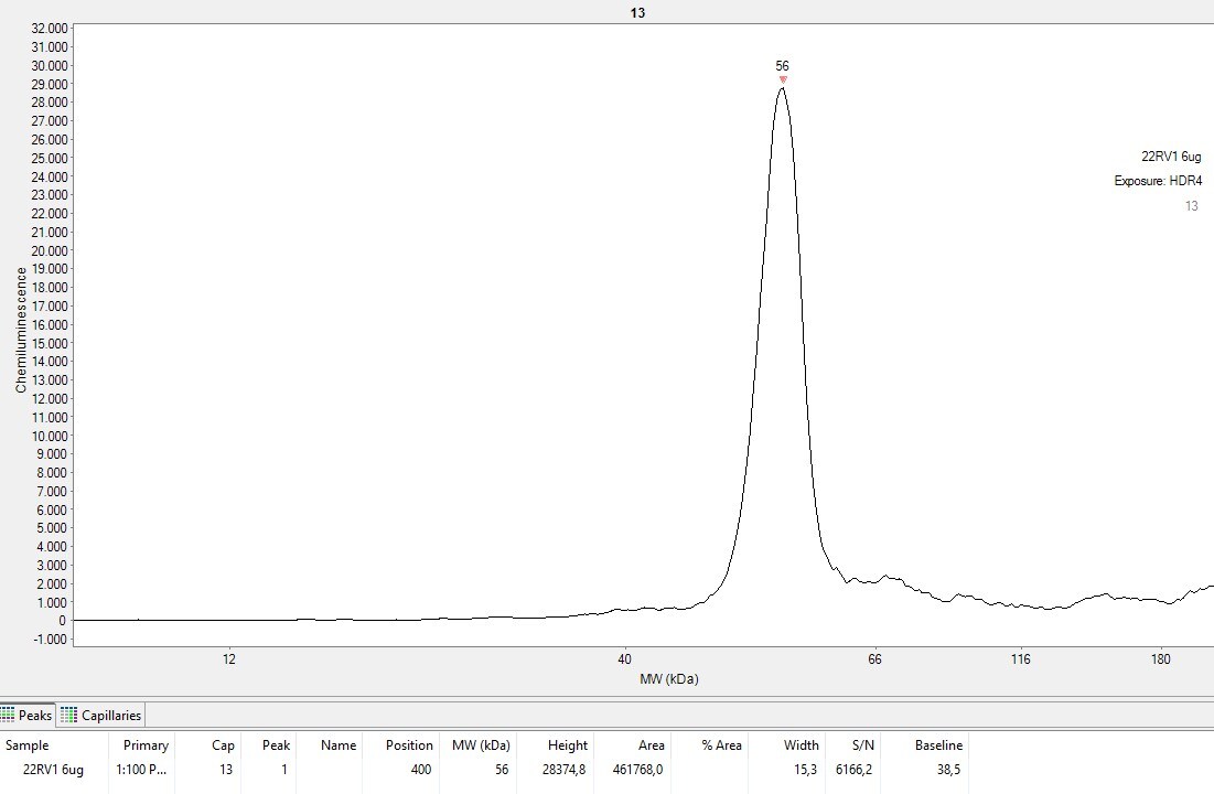
Application: Simple WesternSample Tested: HeLa human cervical epithelial carcinoma cell line and 22RV1 Prostate CancerSpecies: HumanVerified Customer | Posted 02/23/2026This was the 22RV1 peak at 1 to 100 and 2 ug/uL protein concentration.Examined 2 cell lines at 1 to 50 and 1 to 100 primary antibody dilution with 12-230 kDa separation module and anti-rabbit RTU secondary antibody for Simple-Western

-
Application: Western BlotSample Tested: PANC-1 human pancreatic carcinoma cell lineSpecies: HumanVerified Customer | Posted 01/12/2018
There are no reviews that match your criteria.
Protocols
Find general support by application which include: protocols, troubleshooting, illustrated assays, videos and webinars.
- 7-Amino Actinomycin D (7-AAD) Cell Viability Flow Cytometry Protocol
- Antigen Retrieval Protocol (PIER)
- Antigen Retrieval for Frozen Sections Protocol
- Appropriate Fixation of IHC/ICC Samples
- Cellular Response to Hypoxia Protocols
- Chromogenic IHC Staining of Formalin-Fixed Paraffin-Embedded (FFPE) Tissue Protocol
- Chromogenic Immunohistochemistry Staining of Frozen Tissue
- ClariTSA™ Fluorophore Kits
- Detection & Visualization of Antibody Binding
- Extracellular Membrane Flow Cytometry Protocol
- Flow Cytometry Protocol for Cell Surface Markers
- Flow Cytometry Protocol for Staining Membrane Associated Proteins
- Flow Cytometry Staining Protocols
- Flow Cytometry Troubleshooting Guide
- Fluorescent IHC Staining of Frozen Tissue Protocol
- Graphic Protocol for Heat-induced Epitope Retrieval
- Graphic Protocol for the Preparation and Fluorescent IHC Staining of Frozen Tissue Sections
- Graphic Protocol for the Preparation and Fluorescent IHC Staining of Paraffin-embedded Tissue Sections
- Graphic Protocol for the Preparation of Gelatin-coated Slides for Histological Tissue Sections
- IHC Sample Preparation (Frozen sections vs Paraffin)
- Immunofluorescent IHC Staining of Formalin-Fixed Paraffin-Embedded (FFPE) Tissue Protocol
- Immunohistochemistry (IHC) and Immunocytochemistry (ICC) Protocols
- Immunohistochemistry Frozen Troubleshooting
- Immunohistochemistry Paraffin Troubleshooting
- Intracellular Flow Cytometry Protocol Using Alcohol (Methanol)
- Intracellular Flow Cytometry Protocol Using Detergents
- Intracellular Nuclear Staining Flow Cytometry Protocol Using Detergents
- Intracellular Staining Flow Cytometry Protocol Using Alcohol Permeabilization
- Intracellular Staining Flow Cytometry Protocol Using Detergents to Permeabilize Cells
- Preparing Samples for IHC/ICC Experiments
- Preventing Non-Specific Staining (Non-Specific Binding)
- Primary Antibody Selection & Optimization
- Propidium Iodide Cell Viability Flow Cytometry Protocol
- Protocol for Heat-Induced Epitope Retrieval (HIER)
- Protocol for Liperfluo
- Protocol for Making a 4% Formaldehyde Solution in PBS
- Protocol for VisUCyte™ HRP Polymer Detection Reagent
- Protocol for the Characterization of Human Th22 Cells
- Protocol for the Characterization of Human Th9 Cells
- Protocol for the Preparation & Fixation of Cells on Coverslips
- Protocol for the Preparation and Chromogenic IHC Staining of Frozen Tissue Sections
- Protocol for the Preparation and Chromogenic IHC Staining of Frozen Tissue Sections - Graphic
- Protocol for the Preparation and Chromogenic IHC Staining of Paraffin-embedded Tissue Sections
- Protocol for the Preparation and Chromogenic IHC Staining of Paraffin-embedded Tissue Sections - Graphic
- Protocol for the Preparation and Fluorescent IHC Staining of Frozen Tissue Sections
- Protocol for the Preparation and Fluorescent IHC Staining of Paraffin-embedded Tissue Sections
- Protocol for the Preparation of Gelatin-coated Slides for Histological Tissue Sections
- Protocol: Annexin V and PI Staining by Flow Cytometry
- Protocol: Annexin V and PI Staining for Apoptosis by Flow Cytometry
- R&D Systems Quality Control Western Blot Protocol
- TUNEL and Active Caspase-3 Detection by IHC/ICC Protocol
- The Importance of IHC/ICC Controls
- Troubleshooting Guide: Fluorokine Flow Cytometry Kits
- Troubleshooting Guide: Immunohistochemistry
- Troubleshooting Guide: Western Blot Figures
- Western Blot Conditions
- Western Blot Protocol
- Western Blot Protocol for Cell Lysates
- Western Blot Troubleshooting
- Western Blot Troubleshooting Guide
- View all Protocols, Troubleshooting, Illustrated assays and Webinars
Associated Pathways


