Receptor-Interacting Protein 1 (RIP1, also known as RIPK1) is a 671 amino acid (aa) 75 kDa protein that contains an N-terminal protein kinase domain, a C-terminal death domain, and a unique internal region called the intermediate domain. RIP1 is a serine/threonine protein kinase and is constitutively expressed in many tissues. RIP1 interacts with the cytoplasmic death domain of FAS and TNF receptors and is an important element in the signal transduction machinery that mediates apoptosis. RIP1 has been shown to interact with a number of proteins including TRADD, TRAF1, TRAF2, and TRAF3, to form larger signaling complexes. These complexes, in turn, activate specific signaling cascades, such as NF kappa B. RIP1 also interacts through the C-terminal RIP homotypic interaction motif (RHIM) of TRIF in TLR3 dependent activation of NF kappa B.

Key Product Details
Validated by
Knockout/Knockdown
Species Reactivity
Validated:
Human, Mouse, Rat
Cited:
Human, Mouse, Rat
Applications
Validated:
Knockout Validated, Western Blot, Immunocytochemistry, Simple Western
Cited:
Western Blot, Flow Cytometry
Label
Unconjugated
Antibody Source
Monoclonal Mouse IgG1 Clone # 334640
Loading...
Product Specifications
Immunogen
E. coli-derived recombinant human RIPK1/RIP1
Met1-Asn671
Accession # Q13546
Met1-Asn671
Accession # Q13546
Specificity
Detects human, mouse and rat RIPK1/RIP1 in Western blots.
Clonality
Monoclonal
Host
Mouse
Isotype
IgG1
Scientific Data Images for RIPK1/RIP1 Antibody (334640)
Detection of Human/Mouse/Rat RIPK1/RIP1 by Western Blot.
Western blot shows lysates of Raji human Burkitt's lymphoma cell line, Jurkat human acute T cell leukemia cell line, DA3 mouse myeloma cell line, and L6 rat myoblast cell line. PVDF membrane was probed with 0.5 µg/mL of Mouse Anti-Human/Mouse/Rat RIPK1/RIP1 Monoclonal Antibody (Catalog # MAB3585) followed by HRP-conjugated Anti-Mouse IgG Secondary Antibody (Catalog # HAF007). A specific band was detected for RIPK1/RIP1 at approximately 75 kDa (as indicated). This experiment was conducted under reducing conditions and using Immunoblot Buffer Group 2.
RIPK1/RIP1 in MCF‑7 Human Cell Line.
RIPK1/RIP1 was detected in immersion fixed MCF-7 human breast cancer cell line using Mouse Anti-Human/Mouse/Rat RIPK1/RIP1 Monoclonal Antibody (Catalog # MAB3585) at 25 µg/mL for 3 hours at room temperature. Cells were stained using the NorthernLights™ 557-conjugated Anti-Mouse IgG Secondary Antibody (red; Catalog # NL007) and counterstained with DAPI (blue). Specific staining was localized to cytoplasm. View our protocol for Fluorescent ICC Staining of Cells on Coverslips.
Detection of Human RIPK1/RIP1 by Simple WesternTM.
Simple Western lane view shows lysates of Jurkat human acute T cell leukemia cell line and MCF‑7 human breast cancer cell line, loaded at 0.2 mg/mL. A specific band was detected for RIPK1/RIP1 at approximately 78 kDa (as indicated) using 10 µg/mL of Mouse Anti-Human/Mouse/Rat RIPK1/RIP1 Monoclonal Antibody (Catalog # MAB3585). This experiment was conducted under reducing conditions and using the 12-230 kDa separation system.Non-specific interaction with the 230 kDa Simple Western standard may be seen with this antibody.

Western Blot Shows Human RIPK1/RIP1 Specificity by Using Knockout Cell Line.
Western blot shows lysates of MCF-7 human breast cancer parental cell line and RIPK1/RIP1 knockout MCF-7 cell line (KO). PVDF membrane was probed with 0.5 µg/mL of Mouse Anti-Human/Mouse/Rat RIPK1/RIP1 Monoclonal Antibody (Catalog # MAB3585) followed by HRP-conjugated Anti-Mouse IgG Secondary Antibody (Catalog # HAF018). A specific band was detected for RIPK1/RIP1 at approximately 75 kDa (as indicated) in the parental MCF-7 cell line, but is not detectable in knockout MCF-7 cell line. GAPDH (Catalog # MAB5718) is shown as a loading control. This experiment was conducted under reducing conditions and using Immunoblot Buffer Group 1.
Detection of RIPK1/RIP1 by Western Blot
Proteomic analysis identified proteins upregulated in the presence of pro-survival HSP70/BAG1S complex. a U2OS MYC-ER cells expressing ectopic vector, BAG1S, or BAG1 delta S depleted of endogenous BAG1 protein. MYC activity induced for 12 or 24 h with ±100 nM 4-OHT treatment. Lysates analyzed via IB to detect changes in known HSP70 chaperone client proteins GCR, XIAP and RAF1. b Schematic of experimental conditions representing endogenous BAG1 (vector - shLUC), BAG1 knockdown (vector - shBAG1), BAG1S only (BAG1S - shBAG1), or BAG1 delta S only (BAG1 delta S - shBAG1) evaluated for differences in global protein levels. Proteomics analysis outlined with exclusion criteria for significant protein differences between samples. c Efficient knockdown of endogenous BAG1 and rescue of BAG1S and BAG1 delta S shown by IB for samples subjected to proteomics analysis. d Proteomic hits assessed based on schematic of compiled proteins with ≥|1.5| fold change in knockdown compared to control and p ≤ 0.05 across all conditions. Protein expression levels obtained for each sample indexed by specific protein and clustered by UniProt biological process classification. P-values representative of experimental triplicates submitted for proteomic assessment. e Venn diagram showing proteins partially rescued with reintroduction of BAG1S or BAG1 delta S. Increase of ≥10% constitutes a partial rescue. Overlapping proteins with BAG1S or BAG1 delta S indicative of proteins rescued by either ectopic protein. f Verification of proteomics via detection of BAG1S rescued targets SLC7A6 and POLR1D by IB. Image collected and cropped by CiteAb from the following open publication (https://pubmed.ncbi.nlm.nih.gov/30902071), licensed under a CC-BY license. Not internally tested by R&D Systems.
Detection of RIPK1/RIP1 by Western Blot
RPE flatmounts are viable for at least one week after culture in vitro. (A) RPE flatmounts were prepared from 2- and 9-month-old wild-type C57BL/6J mice and cultured in complete medium with or without H2O2 for up to one week. Protein lysates were prepared from the RPE flatmounts and subjected to Western blotting analysis. The cell death marker RIP1 was used to evaluate cell viability; vinculin was used as an internal control. As a positive control: RPE flatmounts treated with either 1 mM or 10 mM H2O2 had significant expression of RIP1. Further, the RPE flatmount that was cultured for 7 days without changing the medium (mishandled sample) showed a slight amount of RIP1, whereas the RPE flatmounts cultured with frequent medium changes did not express RIP1, suggesting that the RPE cells obtained even from older mice (9 months) were viable after at least 7 days of culture. (B) RPE flatmounts were obtained from 3-month-old wild-type C57BL/6J mice and cultured in complete medium with or without H2O2 treatment for up to one week. RPE flatmounts were then fixed in 2.5% glutaraldehyde and processed for transmission electron microscopy (TEM). The TEM imaging of the RPE flatmounts cultured for 16 h, 3 days, and 7 days showed that these RPE cells have normal nuclei (yellow arrows) and organelles, such as mitochondria (yellow arrowheads), and abundant microvilli. H2O2-treated RPE flatmounts showed cell death symptoms, including chromatin condensation and fragmentation (red arrow), a disrupted cell membrane, complete loss of microvilli, a translucent cytoplasm, and severely damaged mitochondria (red arrowheads). (C) ZO-1 immunostaining on 2-month-old RPE flatmounts cultured for 3 days showed robust ZO-1 expression, indicating RPE cell integrity. Image collected and cropped by CiteAb from the following open publication (https://pubmed.ncbi.nlm.nih.gov/34769409), licensed under a CC-BY license. Not internally tested by R&D Systems.Applications for RIPK1/RIP1 Antibody (334640)
Application
Recommended Usage
Immunocytochemistry
8-25 µg/mL
Sample: Immersion fixed MCF-7 human breast cancer cell line
Sample: Immersion fixed MCF-7 human breast cancer cell line
Knockout Validated
RIPK1/RIP1
is specifically detected in MCF‑7 human breast cancer cell line parental cell line but is not detectable in
RIPK1/RIP1 knockout MCF‑7 cell line.
Simple Western
10 µg/mL
Sample: Jurkat human acute T cell leukemia cell line and MCF‑7 human breast cancer cell line
Sample: Jurkat human acute T cell leukemia cell line and MCF‑7 human breast cancer cell line
Western Blot
0.5 µg/mL
Sample: Raji human Burkitt's lymphoma cell line, Jurkat human acute T cell leukemia cell line, DA3 mouse myeloma cell line, and L6 rat myoblast cell line
Sample: Raji human Burkitt's lymphoma cell line, Jurkat human acute T cell leukemia cell line, DA3 mouse myeloma cell line, and L6 rat myoblast cell line
Reviewed Applications
Read 2 reviews rated 5 using MAB3585 in the following applications:
Formulation, Preparation, and Storage
Purification
Protein A or G purified from hybridoma culture supernatant
Reconstitution
Reconstitute at 0.5 mg/mL in sterile PBS. For liquid material, refer to CoA for concentration.
Loading...
Formulation
Lyophilized from a 0.2 μm filtered solution in PBS with Trehalose. *Small pack size (SP) is supplied either lyophilized or as a 0.2 µm filtered solution in PBS.
Shipping
Lyophilized product is shipped at ambient temperature. Liquid small pack size (-SP) is shipped with polar packs. Upon receipt, store immediately at the temperature recommended below.
Stability & Storage
Use a manual defrost freezer and avoid repeated freeze-thaw cycles.
- 12 months from date of receipt, -20 to -70 °C as supplied.
- 1 month, 2 to 8 °C under sterile conditions after reconstitution.
- 6 months, -20 to -70 °C under sterile conditions after reconstitution.
Calculators
Background: RIPK1/RIP1
Long Name
Receptor (TNFRSF)-Interacting Serine-Threonine Kinase 1
Alternate Names
RIP, RIP1
Gene Symbol
RIPK1
UniProt
Additional RIPK1/RIP1 Products
Product Documents for RIPK1/RIP1 Antibody (334640)
Certificate of Analysis
To download a Certificate of Analysis, please enter a lot or batch number in the search box below.
Note: Certificate of Analysis not available for kit components.
Product Specific Notices for RIPK1/RIP1 Antibody (334640)
For research use only
Citations for RIPK1/RIP1 Antibody (334640)
 Powered by Bioz
Customer Reviews for RIPK1/RIP1 Antibody (334640) (2)
5 out of 5
2 Customer Ratings
Have you used RIPK1/RIP1 Antibody (334640)?
Submit a review and receive an Amazon gift card!
$25/€18/£15/$25CAN/¥2500 Yen for a review with an image
$10/€7/£6/$10CAN/¥1110 Yen for a review without an image
Submit a review
Customer Images
Showing
1
-
2 of
2 reviews
Showing All
Filter By:
-
Application: Western BlotSample Tested: Liver tissueSpecies: RatVerified Customer | Posted 11/08/2023Used for rat liver tissue sample at 1:1000 dilution.

-
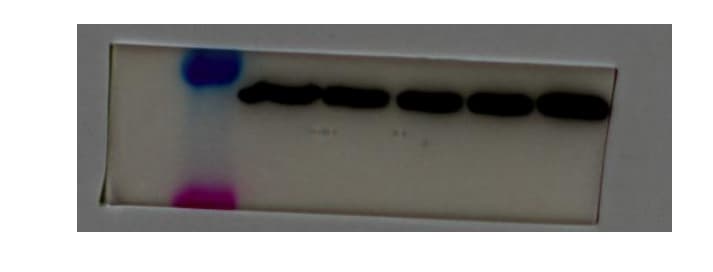
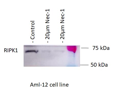
Application: Western BlotSample Tested: AML12 (alpha mouse liver 12) cellSpecies: MouseVerified Customer | Posted 05/04/2020Aml12 cell line. Cells treated with Nec-1 (RIPK1 inhibitor) for 24 hours. Primary antibody: 1:1000 Secondary antibody: 1: 200000
There are no reviews that match your criteria.
Protocols
Find general support by application which include: protocols, troubleshooting, illustrated assays, videos and webinars.
- Appropriate Fixation of IHC/ICC Samples
- Cellular Response to Hypoxia Protocols
- ClariTSA™ Fluorophore Kits
- Detection & Visualization of Antibody Binding
- ICC Cell Smear Protocol for Suspension Cells
- ICC Immunocytochemistry Protocol Videos
- ICC for Adherent Cells
- Immunocytochemistry (ICC) Protocol
- Immunocytochemistry Troubleshooting
- Immunofluorescence of Organoids Embedded in Cultrex Basement Membrane Extract
- Immunohistochemistry (IHC) and Immunocytochemistry (ICC) Protocols
- Preparing Samples for IHC/ICC Experiments
- Preventing Non-Specific Staining (Non-Specific Binding)
- Primary Antibody Selection & Optimization
- Protocol for VisUCyte™ HRP Polymer Detection Reagent
- Protocol for the Fluorescent ICC Staining of Cell Smears - Graphic
- Protocol for the Fluorescent ICC Staining of Cultured Cells on Coverslips - Graphic
- Protocol for the Preparation and Fluorescent ICC Staining of Cells on Coverslips
- Protocol for the Preparation and Fluorescent ICC Staining of Non-adherent Cells
- Protocol for the Preparation and Fluorescent ICC Staining of Stem Cells on Coverslips
- Protocol for the Preparation of a Cell Smear for Non-adherent Cell ICC - Graphic
- R&D Systems Quality Control Western Blot Protocol
- TUNEL and Active Caspase-3 Detection by IHC/ICC Protocol
- The Importance of IHC/ICC Controls
- Troubleshooting Guide: Western Blot Figures
- Western Blot Conditions
- Western Blot Protocol
- Western Blot Protocol for Cell Lysates
- Western Blot Troubleshooting
- Western Blot Troubleshooting Guide
- View all Protocols, Troubleshooting, Illustrated assays and Webinars
Loading...
Associated Pathways


