RelA belongs to a family of transcription factors (NF kappa B (nuclear factor kappa from B cells) complex) that play a fundamental role in inflammatory and immune responses. The NF kappa B complex is composed of a heterodimer of a Rel family member (RelA, c-Rel, RelB) and either NF kappa B1 or NF kappa B2 subunits. RelA and NF kappa B1 are the most common heterodimeric pair. The NF kappa B complex is sequestered in the cytoplasm by inhibitory I kappa B proteins. Upon cellular activation, the ubiquitin-proteosome pathway degrades the I kappa B proteins allowing the NF kappa B complex to translocate to the nucleus and activate gene transcription.

Key Product Details
Validated by
Knockout/Knockdown, Biological Validation
Species Reactivity
Validated:
Human, Mouse, Rat
Cited:
Human, Mouse
Applications
Validated:
Knockout Validated, Immunohistochemistry, Western Blot, Immunocytochemistry, Simple Western, Chromatin Immunoprecipitation (ChIP)
Cited:
Western Blot, Immunoprecipitation, Chromatin Immunoprecipitation (ChIP)
Label
Unconjugated
Antibody Source
Polyclonal Sheep IgG
Loading...
Product Specifications
Immunogen
E. coli-derived recombinant human RelA/NF kappa B p65 isoform 1
Asn456-Ser551
Accession # Q04206
Asn456-Ser551
Accession # Q04206
Specificity
Detects human, mouse, and rat RelA/NF kappa B p65 in Western blots.
Clonality
Polyclonal
Host
Sheep
Isotype
IgG
Scientific Data Images for RelA/NFkB p65 Antibody
Detection of Human RelA/NF kappa B p65 by Western Blot.
Western blot shows lysates of K562 human chronic myelogenous leukemia cell line, Daudi human Burkitt's lymphoma cell line, and LNCaP human prostate cancer cell line. PVDF membrane was probed with 1 µg/mL of Sheep Anti-Human/Mouse/Rat RelA/NF kappa B p65 Antigen Affinity-purified Polyclonal Antibody (Catalog # AF5078) followed by HRP-conjugated Anti-Sheep IgG Secondary Antibody (Catalog # HAF016). A specific band was detected for RelA/NF kappa B p65 at approximately 70 kDa (as indicated). This experiment was conducted under reducing conditions and using Immunoblot Buffer Group 1.
Detection of Human and Mouse RelA/NF kappa B p65 by Western Blot.
Western blot shows lysates of K562 human chronic myelogenous leukemia cell line, HeLa human cervical epithelial carcinoma cell line, and Neuro-2A mouse neuroblastoma cell line. PVDF membrane was probed with 1 µg/mL of Sheep Anti-Human/Mouse/Rat RelA/NF kappa B p65 Antigen Affinity-purified Polyclonal Antibody (Catalog # AF5078) followed by HRP-conjugated Anti-Sheep IgG Secondary Antibody (Catalog # HAF016). A specific band was detected for RelA/NF kappa B p65 at approximately 70 kDa (as indicated). This experiment was conducted under reducing conditions and using Immunoblot Buffer Group 1.
Detection of Mouse and Rat RelA/NF kappa B p65 by Western Blot.
Western blot shows lysates of NIH-3T3 mouse embryonic fibroblast cell line and C6 rat glioma cell line. PVDF membrane was probed with 1 µg/mL of Sheep Anti-Human/Mouse RelA/NF kappa B p65 Antigen Affinity-purified Polyclonal Antibody (Catalog # AF5078) followed by HRP-conjugated Anti-Sheep IgG Secondary Antibody (Catalog # HAF016). A specific band was detected for RelA/NF kappa B p65 at approximately 65 kDa (as indicated). This experiment was conducted under reducing conditions and using Immunoblot Buffer Group 1.
Detection of RelA/NF kappa B p65-regulated Genes by Chromatin Immuno-precipitation.
Jurkat human acute T cell leukemia cell line treated with 50 ng/mL PMA and 200 ng/mL calcium ionomycin for overnight was fixed using formaldehyde, resuspended in lysis buffer, and sonicated to shear chromatin. RelA/NF kappa B p65/DNA complexes were immunoprecipitated using 5 µg Sheep Anti-Human/Mouse/Rat RelA/NF kappa B p65 Antigen Affinity-purified Polyclonal Antibody (Catalog # AF5078) or control antibody (Catalog # 5-001-A) for 15 minutes in an ultrasonic bath, followed by Biotinylated Anti-Sheep IgG Secondary Antibody (Catalog # BAF016). Immunocomplexes were captured using 50 µL of MagCellect Streptavidin Ferrofluid (Catalog # MAG999) and DNA was purified using chelating resin solution. Thep21promoter was detected by standard PCR.
RelA/NF kappa B p65 in HeLa Human Cell Line.
RelA/NF kappa B p65 was detected in immersion fixed HeLa human cervical epithelial carcinoma cells untreated (upper panel) or treated (lower panel) with 20 ng/mL Recombinant Human TNF-alpha (Catalog # 210-TA) for 10 minutes using Sheep Anti-Human/Mouse/Rat RelA/NF kappa B p65 Antigen Affinity-purified Polyclonal Antibody (Catalog # AF5078) at 10 µg/mL for 3 hours at room temperature. Cells were stained using the NorthernLights™ 557-conjugated Anti-Sheep IgG Secondary Antibody (red; Catalog # NL010) and counterstained with DAPI (blue). Specific staining was localized to cytoplasm in untreated cells and nuclei in treated cells. View our protocol for Fluorescent ICC Staining of Cells on Coverslips.
RelA/NF kappa B p65 in Human Squamous Cell Carcinoma.
RelA/NF kappa B p65 was detected in immersion fixed paraffin-embedded sections of human squamous cell carcinoma using Sheep Anti-Human/Mouse/Rat RelA/NF kappa B p65 Antigen Affinity-purified Polyclonal Antibody (Catalog # AF5078) at 3 µg/mL overnight at 4 °C. Tissue was stained using the Anti-Sheep HRP-DAB Cell & Tissue Staining Kit (brown; Catalog # CTS019) and counterstained with hematoxylin (blue). Specific staining was localized to cytoplasm in cancer cells. View our protocol for Chromogenic IHC Staining of Paraffin-embedded Tissue Sections.
Detection of Human, Mouse, and Rat RelA/NF kappa B p65 by Simple WesternTM.
Simple Western lane view shows lysates of HeLa human cervical epithelial carcinoma cell line, Neuro-2A mouse neuroblastoma cell line, and C6 rat glioma cell line, loaded at 0.5 mg/mL. A specific band was detected for RelA/NF kappa B p65 at approximately 65 kDa (as indicated) using 20 µg/mL of Sheep Anti-Human/Mouse/Rat RelA/NF kappa B p65 Antigen Affinity-purified Polyclonal Antibody (Catalog # AF5078) followed by 1:50 dilution of HRP-conjugated Anti-Sheep IgG Secondary Antibody (Catalog # HAF016). This experiment was conducted under reducing conditions and using the 12-230 kDa separation system.
Western Blot Shows Human RelA/NF kappa B p65 Specificity by Using Knockout Cell Line.
Western blot shows lysates of HeLa human cervical epithelial carcinoma parental cell line and RelA/NF kappa B p65 knockout HeLa cell line (KO). PVDF membrane was probed with 1 µg/mL of Sheep Anti-Human/Mouse/Rat RelA/NF kappa B p65 Antigen Affinity-purified Polyclonal Antibody (Catalog # AF5078) followed by HRP-conjugated Anti-Sheep IgG Secondary Antibody (Catalog # HAF016). A specific band was detected for RelA/NF kappa B p65 at approximately 65 kDa (as indicated) in the parental HeLa cell line, but is not detectable in knockout HeLa cell line. GAPDH (Catalog # AF5718) is shown as a loading control. This experiment was conducted under reducing conditions and using Immunoblot Buffer Group 1.Applications for RelA/NFkB p65 Antibody
Application
Recommended Usage
Chromatin Immunoprecipitation (ChIP)
5 µg/5 x 106 cells
Sample: PMA and calcium ionomycin treated Jurkat human acute T cell leukemia cell line chromatin, p21 promoter detected by standard PCR
Sample: PMA and calcium ionomycin treated Jurkat human acute T cell leukemia cell line chromatin, p21 promoter detected by standard PCR
Immunocytochemistry
5-15 µg/mL
Sample: Immersion fixed HeLa human cervical epithelial carcinoma cells untreated or treated with 20 ng/mL Recombinant Human TNF-alpha (Catalog # 210-TA)
Sample: Immersion fixed HeLa human cervical epithelial carcinoma cells untreated or treated with 20 ng/mL Recombinant Human TNF-alpha (Catalog # 210-TA)
Immunohistochemistry
5-15 µg/mL
Sample: Immersion fixed paraffin-embedded sections of human squamous cell carcinoma
Sample: Immersion fixed paraffin-embedded sections of human squamous cell carcinoma
Knockout Validated
RelA/NF kappa B p65 is specifically detected in HeLa human cervical epithelial carcinoma parental cell line but is not detectable in RelA/NF kappa B p65 knockout HeLa cell line.
Simple Western
20 µg/mL
Sample: HeLa human cervical epithelial carcinoma cell line, Neuro‑2A mouse neuroblastoma cell line, and C6 rat glioma cell line
Sample: HeLa human cervical epithelial carcinoma cell line, Neuro‑2A mouse neuroblastoma cell line, and C6 rat glioma cell line
Western Blot
1 µg/mL
Sample: K562 human chronic myelogenous leukemia cell line, Daudi human Burkitt's lymphoma cell line, LNCaP human prostate cancer cell line, HeLa human cervical epithelial carcinoma cell line, Neuro‑2A mouse neuroblastoma cell line, NIH‑3T3 mouse embryonic fibroblast cell line, and C6 rat glioma cell line
Sample: K562 human chronic myelogenous leukemia cell line, Daudi human Burkitt's lymphoma cell line, LNCaP human prostate cancer cell line, HeLa human cervical epithelial carcinoma cell line, Neuro‑2A mouse neuroblastoma cell line, NIH‑3T3 mouse embryonic fibroblast cell line, and C6 rat glioma cell line
Reviewed Applications
Read 2 reviews rated 5 using AF5078 in the following applications:
Formulation, Preparation, and Storage
Purification
Antigen Affinity-purified
Reconstitution
Reconstitute at 0.2 mg/mL in sterile PBS. For liquid material, refer to CoA for concentration.
Loading...
Formulation
Lyophilized from a 0.2 μm filtered solution in PBS with Trehalose. *Small pack size (SP) is supplied either lyophilized or as a 0.2 µm filtered solution in PBS.
Shipping
Lyophilized product is shipped at ambient temperature. Liquid small pack size (-SP) is shipped with polar packs. Upon receipt, store immediately at the temperature recommended below.
Stability & Storage
Use a manual defrost freezer and avoid repeated freeze-thaw cycles.
- 12 months from date of receipt, -20 to -70 °C as supplied.
- 1 month, 2 to 8 °C under sterile conditions after reconstitution.
- 6 months, -20 to -70 °C under sterile conditions after reconstitution.
Calculators
Background: RelA/NFkB p65
Long Name
v-rel Reticuloendotheliosis Viral Oncogene Homolog A
Alternate Names
NFkB p65, NFKB3, p65RelA
Entrez Gene IDs
5970 (Human)
Gene Symbol
RELA
UniProt
Additional RelA/NFkB p65 Products
Product Documents for RelA/NFkB p65 Antibody
Certificate of Analysis
To download a Certificate of Analysis, please enter a lot or batch number in the search box below.
Note: Certificate of Analysis not available for kit components.
Product Specific Notices for RelA/NFkB p65 Antibody
For research use only
Related Research Areas
Citations for RelA/NFkB p65 Antibody
 Powered by Bioz
Customer Reviews for RelA/NFkB p65 Antibody (2)
5 out of 5
2 Customer Ratings
Have you used RelA/NFkB p65 Antibody?
Submit a review and receive an Amazon gift card!
$25/€18/£15/$25CAN/¥2500 Yen for a review with an image
$10/€7/£6/$10CAN/¥1110 Yen for a review without an image
Submit a review
Customer Images

Showing
1
-
2 of
2 reviews
Showing All
Filter By:
-
Application: ELISASample Tested: xenograft PD sampleSpecies: HumanVerified Customer | Posted 10/24/2019

-
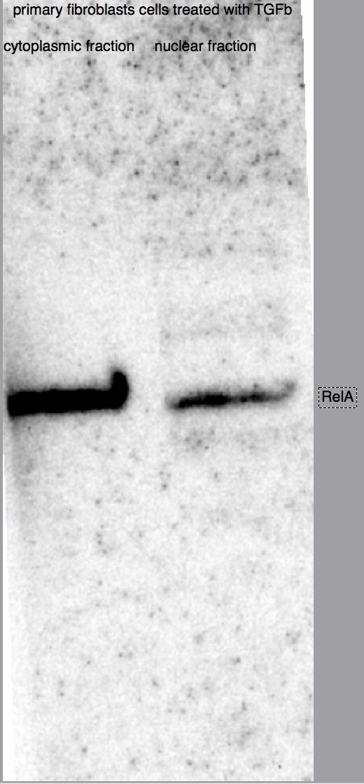
Application: Western BlotSample Tested: Human fibroblastSpecies: HumanVerified Customer | Posted 08/23/2018
There are no reviews that match your criteria.
Protocols
Find general support by application which include: protocols, troubleshooting, illustrated assays, videos and webinars.
- Antigen Retrieval Protocol (PIER)
- Antigen Retrieval for Frozen Sections Protocol
- Appropriate Fixation of IHC/ICC Samples
- Cellular Response to Hypoxia Protocols
- ChIP Protocol Video
- Chromatin Immunoprecipitation (ChIP) Protocol
- Chromatin Immunoprecipitation Protocol
- Chromogenic IHC Staining of Formalin-Fixed Paraffin-Embedded (FFPE) Tissue Protocol
- Chromogenic Immunohistochemistry Staining of Frozen Tissue
- ClariTSA™ Fluorophore Kits
- Detection & Visualization of Antibody Binding
- Fluorescent IHC Staining of Frozen Tissue Protocol
- Graphic Protocol for Heat-induced Epitope Retrieval
- Graphic Protocol for the Preparation and Fluorescent IHC Staining of Frozen Tissue Sections
- Graphic Protocol for the Preparation and Fluorescent IHC Staining of Paraffin-embedded Tissue Sections
- Graphic Protocol for the Preparation of Gelatin-coated Slides for Histological Tissue Sections
- ICC Cell Smear Protocol for Suspension Cells
- ICC Immunocytochemistry Protocol Videos
- ICC for Adherent Cells
- IHC Sample Preparation (Frozen sections vs Paraffin)
- Immunocytochemistry (ICC) Protocol
- Immunocytochemistry Troubleshooting
- Immunofluorescence of Organoids Embedded in Cultrex Basement Membrane Extract
- Immunofluorescent IHC Staining of Formalin-Fixed Paraffin-Embedded (FFPE) Tissue Protocol
- Immunohistochemistry (IHC) and Immunocytochemistry (ICC) Protocols
- Immunohistochemistry Frozen Troubleshooting
- Immunohistochemistry Paraffin Troubleshooting
- Preparing Samples for IHC/ICC Experiments
- Preventing Non-Specific Staining (Non-Specific Binding)
- Primary Antibody Selection & Optimization
- Protocol for Heat-Induced Epitope Retrieval (HIER)
- Protocol for Making a 4% Formaldehyde Solution in PBS
- Protocol for VisUCyte™ HRP Polymer Detection Reagent
- Protocol for the Fluorescent ICC Staining of Cell Smears - Graphic
- Protocol for the Fluorescent ICC Staining of Cultured Cells on Coverslips - Graphic
- Protocol for the Preparation & Fixation of Cells on Coverslips
- Protocol for the Preparation and Chromogenic IHC Staining of Frozen Tissue Sections
- Protocol for the Preparation and Chromogenic IHC Staining of Frozen Tissue Sections - Graphic
- Protocol for the Preparation and Chromogenic IHC Staining of Paraffin-embedded Tissue Sections
- Protocol for the Preparation and Chromogenic IHC Staining of Paraffin-embedded Tissue Sections - Graphic
- Protocol for the Preparation and Fluorescent ICC Staining of Cells on Coverslips
- Protocol for the Preparation and Fluorescent ICC Staining of Non-adherent Cells
- Protocol for the Preparation and Fluorescent ICC Staining of Stem Cells on Coverslips
- Protocol for the Preparation and Fluorescent IHC Staining of Frozen Tissue Sections
- Protocol for the Preparation and Fluorescent IHC Staining of Paraffin-embedded Tissue Sections
- Protocol for the Preparation of Gelatin-coated Slides for Histological Tissue Sections
- Protocol for the Preparation of a Cell Smear for Non-adherent Cell ICC - Graphic
- R&D Systems Quality Control Western Blot Protocol
- TUNEL and Active Caspase-3 Detection by IHC/ICC Protocol
- The Importance of IHC/ICC Controls
- Troubleshooting Guide: Immunohistochemistry
- Troubleshooting Guide: Western Blot Figures
- Western Blot Conditions
- Western Blot Protocol
- Western Blot Protocol for Cell Lysates
- Western Blot Troubleshooting
- Western Blot Troubleshooting Guide
- View all Protocols, Troubleshooting, Illustrated assays and Webinars


