Human Neutrophil Elastase/ELA2 Antibody (950317)
R&D Systems | Catalog # MAB91671

Key Product Details
Validated by
Biological Validation
Species Reactivity
Validated:
Human
Cited:
Human
Applications
Validated:
Immunohistochemistry, Western Blot, Intracellular Staining by Flow Cytometry, Immunocytochemistry, CyTOF-ready
Cited:
Immunohistochemistry, Western Blot, Immunocytochemistry
Label
Unconjugated
Antibody Source
Monoclonal Mouse IgG1 Clone # 950317
Loading...
Product Specifications
Immunogen
Chinese hamster ovary cell line CHO-derived recombinant human ELA2
Met1-Asn252
Accession # P08246
Met1-Asn252
Accession # P08246
Specificity
Detects human ELA2 in direct ELISAs and Western blots.
Clonality
Monoclonal
Host
Mouse
Isotype
IgG1
Scientific Data Images for Human Neutrophil Elastase/ELA2 Antibody (950317)
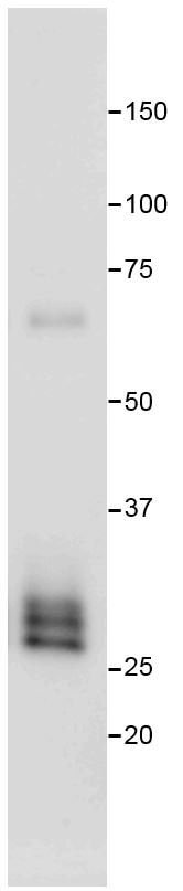
Detection of Human Neutrophil Elastase/ELA2 by Western Blot.
Western blot shows lysates of human bone marrow and human lung tissue. PVDF membrane was probed with 0.1 µg/mL of Mouse Anti-Human Neutrophil Elastase/ELA2 Monoclonal Antibody (Catalog # MAB91671) followed by HRP-conjugated Anti-Mouse IgG Secondary Antibody (Catalog # HAF018). Specific bands were detected for Neutrophil Elastase/ELA2 at approximately 25-30 kDa (as indicated). This experiment was conducted under reducing conditions and using Immunoblot Buffer Group 1.
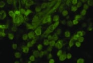
Neutrophil Elastase/ELA2 in THP‑1 Human Cell Line.
Neutrophil Elastase/ELA2 was detected in immersion fixed THP-1 human acute monocytic leukemia cell line using Mouse Anti-Human Neutrophil Elastase/ELA2 Monoclonal Antibody (Catalog # MAB91671) at 5 µg/mL for 3 hours at room temperature. Cells were stained using the NorthernLights™ 557-conjugated Anti-Mouse IgG Secondary Antibody (red; Catalog # NL007) and counterstained with DAPI (blue). Specific staining was localized to cytoplasm. View our protocol for Fluorescent ICC Staining of Non-adherent Cells.
Neutrophil Elastase/ELA2 in Human Lymphoma.
Neutrophil Elastase/ELA2 was detected in immersion fixed paraffin-embedded sections of human lymphoma using Mouse Anti-Human Neutrophil Elastase/ELA2 Monoclonal Antibody (Catalog # MAB91671) at 5 µg/mL for 1 hour at room temperature followed by incubation with the Anti-Mouse IgG VisUCyte™ HRP Polymer Antibody (Catalog # VC001). Tissue was stained using DAB (brown) and counterstained with hematoxylin (blue). Specific staining was localized to cytoplasm. View our protocol for IHC Staining with VisUCyte HRP Polymer Detection Reagents.
Detection of Neutrophil Elastase/ELA2 in THP‑1 Human Cell Line by Flow Cytometry.
THP-1 human acute monocytic leukemia cell line treated with 3 µM monensin for 3 hours was stained with Mouse Anti-Human Neutrophil Elastase/ELA2 Monoclonal Antibody (Catalog # MAB91671, filled histogram) or isotype control antibody (Catalog # MAB002, open histogram), followed by Phycoerythrin-conjugated Anti-Mouse IgG Secondary Antibody (Catalog # F0102B). To facilitate intracellular staining, cells were fixed with Flow Cytometry Fixation Buffer (Catalog # FC004) and permeabilized with Flow Cytometry Permeabilization/Wash Buffer I (Catalog # FC005). View our protocol for Staining Intracellular Molecules.Applications for Human Neutrophil Elastase/ELA2 Antibody (950317)
Application
Recommended Usage
CyTOF-ready
Ready to be labeled using established conjugation methods. No BSA or other carrier proteins that could interfere with conjugation.
Immunocytochemistry
5-25 µg/mL
Sample: Immersion fixed THP‑1 human acute monocytic leukemia cell line
Sample: Immersion fixed THP‑1 human acute monocytic leukemia cell line
Immunohistochemistry
3-25 µg/mL
Sample: Immersion fixed paraffin-embedded sections of human lymphoma
Sample: Immersion fixed paraffin-embedded sections of human lymphoma
Intracellular Staining by Flow Cytometry
0.25 µg/106 cells
Sample: THP‑1 human acute monocytic leukemia cell line treated with monensin were fixed with Flow Cytometry Fixation Buffer (Catalog # FC004) and permeabilized with Flow Cytometry Permeabilization/Wash Buffer I (Catalog # FC005)
Sample: THP‑1 human acute monocytic leukemia cell line treated with monensin were fixed with Flow Cytometry Fixation Buffer (Catalog # FC004) and permeabilized with Flow Cytometry Permeabilization/Wash Buffer I (Catalog # FC005)
Western Blot
0.1 µg/mL
Sample: Human bone marrow and human lung tissue
Sample: Human bone marrow and human lung tissue
Reviewed Applications
Read 2 reviews rated 5 using MAB91671 in the following applications:
Flow Cytometry Panel Builder
Bio-Techne Knows Flow Cytometry
Save time and reduce costly mistakes by quickly finding compatible reagents using the Panel Builder Tool.
Advanced Features
- Spectra Viewer - Custom analysis of spectra from multiple fluorochromes
- Spillover Popups - Visualize the spectra of individual fluorochromes
- Antigen Density Selector - Match fluorochrome brightness with antigen density

Formulation, Preparation, and Storage
Purification
Protein A or G purified from hybridoma culture supernatant
Reconstitution
Reconstitute at 0.5 mg/mL in sterile PBS. For liquid material, refer to CoA for concentration.
Loading...
Formulation
Lyophilized from a 0.2 μm filtered solution in PBS with Trehalose. *Small pack size (SP) is supplied either lyophilized or as a 0.2 µm filtered solution in PBS.
Shipping
Lyophilized product is shipped at ambient temperature. Liquid small pack size (-SP) is shipped with polar packs. Upon receipt, store immediately at the temperature recommended below.
Stability & Storage
Use a manual defrost freezer and avoid repeated freeze-thaw cycles.
- 12 months from date of receipt, -20 to -70 °C as supplied.
- 1 month, 2 to 8 °C under sterile conditions after reconstitution.
- 6 months, -20 to -70 °C under sterile conditions after reconstitution.
Calculators
Background: Neutrophil Elastase/ELA2
Trappin-2/Elafin (7-11). Its activity in the lung is increased by exposure to tobacco smoke which inactivates Serpin A1 through methionine oxidation (12). Mature human Neutrophil Elastase shares 73% amino acid sequence identity with mouse and rat Neutrophil Elastase (13, 14). Multiple mutations in the human ELANE gene are causative of severe congenital and cyclic neutropenias (15).
References
- Korkmaz, B. et al. (2010) Pharmacol. Rev. 62:726.
- Stein, R.L. et al. (1987) Biochemistry 26:1301.
- Bachovchin, W.W. (1986) Biochemistry 25:7751.
- Garwicz, D. et al. (2005) Haematologica 90:38.
- Owen, C.A. et al. (1995) J. Cell Biol. 131:775.
- Stephan, A. and M. Fabri (2015) Exp. Dermatol. 24:161.
- Carrell, R.W. et al. (1982) Nature 298:329.
- Rice, W.G. and S.J. Weiss (1990) Science 249:178.
- Thompson, R.C. et al. (1986) Proc. Natl. Acad. Sci. USA 83:6692.
- Cooley, J. et al. (2001) Biochemistry 40:15762.
- Wiedow, O. et al. (1990) J. Biol. Chem. 265:14791.
- Taggart, C. et al. (2000) J. Biol. Chem. 275:27258.
- Sinha, S. et al. (1987) Proc. Natl. Acad. Sci. USA 84:2228.
- Okano, K. et al. (1987) J. Biochem. 102:13.
- Makaryan, V. et al. (2015) Curr. Opin. Hematol. 22:3.
Alternate Names
ELA2, ELANE, Elastase-2, Leukocyte Elastase, Medullasin
Gene Symbol
ELANE
UniProt
Additional Neutrophil Elastase/ELA2 Products
Product Documents for Human Neutrophil Elastase/ELA2 Antibody (950317)
Certificate of Analysis
To download a Certificate of Analysis, please enter a lot or batch number in the search box below.
Note: Certificate of Analysis not available for kit components.
Product Specific Notices for Human Neutrophil Elastase/ELA2 Antibody (950317)
For research use only
Related Research Areas
Citations for Human Neutrophil Elastase/ELA2 Antibody (950317)
 Powered by Bioz
Customer Reviews for Human Neutrophil Elastase/ELA2 Antibody (950317) (2)
5 out of 5
2 Customer Ratings
Have you used Human Neutrophil Elastase/ELA2 Antibody (950317)?
Submit a review and receive an Amazon gift card!
$25/€18/£15/$25CAN/¥2500 Yen for a review with an image
$10/€7/£6/$10CAN/¥1110 Yen for a review without an image
Submit a review
Customer Images
Showing
1
-
2 of
2 reviews
Showing All
Filter By:
-
Application: Immunocytochemistry/ImmunofluorescenceSample Tested: Periapical granulomaSpecies: HumanVerified Customer | Posted 11/05/2021

-
Application: Western BlotSample Tested: Peripheral blood neutrophilsSpecies: HumanVerified Customer | Posted 06/19/2017samples: Human neutrophil lysate Block: 5% skim milk in PBS-T for 1hr at RT Dilution: 1/2000
There are no reviews that match your criteria.
Protocols
Find general support by application which include: protocols, troubleshooting, illustrated assays, videos and webinars.
- 7-Amino Actinomycin D (7-AAD) Cell Viability Flow Cytometry Protocol
- Antigen Retrieval Protocol (PIER)
- Antigen Retrieval for Frozen Sections Protocol
- Appropriate Fixation of IHC/ICC Samples
- Cellular Response to Hypoxia Protocols
- Chromogenic IHC Staining of Formalin-Fixed Paraffin-Embedded (FFPE) Tissue Protocol
- Chromogenic Immunohistochemistry Staining of Frozen Tissue
- ClariTSA™ Fluorophore Kits
- Detection & Visualization of Antibody Binding
- Extracellular Membrane Flow Cytometry Protocol
- Flow Cytometry Protocol for Cell Surface Markers
- Flow Cytometry Protocol for Staining Membrane Associated Proteins
- Flow Cytometry Staining Protocols
- Flow Cytometry Troubleshooting Guide
- Fluorescent IHC Staining of Frozen Tissue Protocol
- Graphic Protocol for Heat-induced Epitope Retrieval
- Graphic Protocol for the Preparation and Fluorescent IHC Staining of Frozen Tissue Sections
- Graphic Protocol for the Preparation and Fluorescent IHC Staining of Paraffin-embedded Tissue Sections
- Graphic Protocol for the Preparation of Gelatin-coated Slides for Histological Tissue Sections
- ICC Cell Smear Protocol for Suspension Cells
- ICC Immunocytochemistry Protocol Videos
- ICC for Adherent Cells
- IHC Sample Preparation (Frozen sections vs Paraffin)
- Immunocytochemistry (ICC) Protocol
- Immunocytochemistry Troubleshooting
- Immunofluorescence of Organoids Embedded in Cultrex Basement Membrane Extract
- Immunofluorescent IHC Staining of Formalin-Fixed Paraffin-Embedded (FFPE) Tissue Protocol
- Immunohistochemistry (IHC) and Immunocytochemistry (ICC) Protocols
- Immunohistochemistry Frozen Troubleshooting
- Immunohistochemistry Paraffin Troubleshooting
- Intracellular Flow Cytometry Protocol Using Alcohol (Methanol)
- Intracellular Flow Cytometry Protocol Using Detergents
- Intracellular Nuclear Staining Flow Cytometry Protocol Using Detergents
- Intracellular Staining Flow Cytometry Protocol Using Alcohol Permeabilization
- Intracellular Staining Flow Cytometry Protocol Using Detergents to Permeabilize Cells
- Preparing Samples for IHC/ICC Experiments
- Preventing Non-Specific Staining (Non-Specific Binding)
- Primary Antibody Selection & Optimization
- Propidium Iodide Cell Viability Flow Cytometry Protocol
- Protocol for Heat-Induced Epitope Retrieval (HIER)
- Protocol for Liperfluo
- Protocol for Making a 4% Formaldehyde Solution in PBS
- Protocol for VisUCyte™ HRP Polymer Detection Reagent
- Protocol for the Characterization of Human Th22 Cells
- Protocol for the Characterization of Human Th9 Cells
- Protocol for the Fluorescent ICC Staining of Cell Smears - Graphic
- Protocol for the Fluorescent ICC Staining of Cultured Cells on Coverslips - Graphic
- Protocol for the Preparation & Fixation of Cells on Coverslips
- Protocol for the Preparation and Chromogenic IHC Staining of Frozen Tissue Sections
- Protocol for the Preparation and Chromogenic IHC Staining of Frozen Tissue Sections - Graphic
- Protocol for the Preparation and Chromogenic IHC Staining of Paraffin-embedded Tissue Sections
- Protocol for the Preparation and Chromogenic IHC Staining of Paraffin-embedded Tissue Sections - Graphic
- Protocol for the Preparation and Fluorescent ICC Staining of Cells on Coverslips
- Protocol for the Preparation and Fluorescent ICC Staining of Non-adherent Cells
- Protocol for the Preparation and Fluorescent ICC Staining of Stem Cells on Coverslips
- Protocol for the Preparation and Fluorescent IHC Staining of Frozen Tissue Sections
- Protocol for the Preparation and Fluorescent IHC Staining of Paraffin-embedded Tissue Sections
- Protocol for the Preparation of Gelatin-coated Slides for Histological Tissue Sections
- Protocol for the Preparation of a Cell Smear for Non-adherent Cell ICC - Graphic
- Protocol: Annexin V and PI Staining by Flow Cytometry
- Protocol: Annexin V and PI Staining for Apoptosis by Flow Cytometry
- R&D Systems Quality Control Western Blot Protocol
- TUNEL and Active Caspase-3 Detection by IHC/ICC Protocol
- The Importance of IHC/ICC Controls
- Troubleshooting Guide: Fluorokine Flow Cytometry Kits
- Troubleshooting Guide: Immunohistochemistry
- Troubleshooting Guide: Western Blot Figures
- Western Blot Conditions
- Western Blot Protocol
- Western Blot Protocol for Cell Lysates
- Western Blot Troubleshooting
- Western Blot Troubleshooting Guide
- View all Protocols, Troubleshooting, Illustrated assays and Webinars
Loading...