
Human NMNAT-1 Antibody (675112)
R&D Systems | Catalog # MAB5865


Scientific images may be resized, cropped, or adjusted for brightness or contrast for presentation purposes. These updates are limited to presentation and do not affect the underlying scientific interpretation of the data. Where available, additional source data may be provided upon request.
Key Product Details
Species Reactivity
Validated:
Cited:
Applications
Validated:
Cited:
Label
Antibody Source
Product Specifications
Immunogen
Met1-Thr279
Accession # Q9HAN9
Specificity
Clonality
Host
Isotype
Scientific Data Images for Human NMNAT-1 Antibody (675112)
Detection of NMNAT‑1 in Human Kidney.
NMNAT‑1 was detected in immersion fixed paraffin-embedded sections of Human Kidney using Mouse Anti-Human NMNAT‑1 Monoclonal Antibody (Catalog # MAB5865) at 5 µg/mL for 1 hour at room temperature followed by incubation with the Anti-Mouse IgG VisUCyte™ HRP Polymer Antibody (Catalog # VC001). Before incubation with the primary antibody, tissue was subjected to heat-induced epitope retrieval using VisUCyte Antigen Retrieval Reagent-Basic (Catalog # VCTS021). Tissue was stained using DAB (brown) and counterstained with hematoxylin (blue). Specific staining was localized to cell nuclei in convoluted tubules. View our protocol for IHC Staining with VisUCyte HRP Polymer Detection Reagents.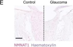
Detection of NMNAT-1 by Immunohistochemistry
NAD salvage pathway machinery are reduced in RGC relevant layers of the retina and in the optic nerve head. A NAMPT labelling remains detectable across the whole retina in glaucoma but is significantly reduced in the GCC (n = 11 control eyes, 7 glaucoma eyes). B NMNAT1 is significantly reduced in both the GCC and INL in glaucoma and remains stable in the outer retina (n = 11 control eyes, 7 glaucoma eyes). C NMNAT2 is significantly reduced in the GCC in central retina and is otherwise unchanged in the INL and outer retina (n = 11 control eyes, 7 glaucoma eyes. D–F in the ONH, NAMPT, NMNAT1, and NMNAT2 labelling is significantly reduced in the first 500 µm but remain comparable to controls in the proceeding optic nerve (NAMPT: n = 11 control eyes, 5 glaucoma eyes; NMNAT1: n = 10 control eyes, 5 glaucoma eyes; NMNAT2: n = 8 control eyes, 5 glaucoma eyes). GCC ganglion cell complex, INL inner nuclear layer, OR outer retina, ONH optic nerve head, Ctrl control, Glau glaucoma. Scale bars = 50 µm in A–C, 500 µm in D–E Image collected and cropped by CiteAb from the following open publication (https://pubmed.ncbi.nlm.nih.gov/36681854), licensed under a CC-BY license. Not internally tested by R&D Systems.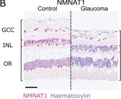
Detection of NMNAT-1 by Immunohistochemistry
NAD salvage pathway machinery are reduced in RGC relevant layers of the retina and in the optic nerve head. A NAMPT labelling remains detectable across the whole retina in glaucoma but is significantly reduced in the GCC (n = 11 control eyes, 7 glaucoma eyes). B NMNAT1 is significantly reduced in both the GCC and INL in glaucoma and remains stable in the outer retina (n = 11 control eyes, 7 glaucoma eyes). C NMNAT2 is significantly reduced in the GCC in central retina and is otherwise unchanged in the INL and outer retina (n = 11 control eyes, 7 glaucoma eyes. D–F in the ONH, NAMPT, NMNAT1, and NMNAT2 labelling is significantly reduced in the first 500 µm but remain comparable to controls in the proceeding optic nerve (NAMPT: n = 11 control eyes, 5 glaucoma eyes; NMNAT1: n = 10 control eyes, 5 glaucoma eyes; NMNAT2: n = 8 control eyes, 5 glaucoma eyes). GCC ganglion cell complex, INL inner nuclear layer, OR outer retina, ONH optic nerve head, Ctrl control, Glau glaucoma. Scale bars = 50 µm in A–C, 500 µm in D–E Image collected and cropped by CiteAb from the following open publication (https://pubmed.ncbi.nlm.nih.gov/36681854), licensed under a CC-BY license. Not internally tested by R&D Systems.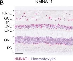
Detection of NMNAT-1 by Immunohistochemistry
Labelling of NAD salvage pathway machinery in human retina. A NAMPT labelling is detected across all retinal layers, localizing to both nuclear (haematoxylin + ve) and cytoplasmic (haematoxylin −ve) cellular compartments, with the most intense labeling in the INL (n = 11 eyes). B NMNAT1 labelling is strongest in nuclear layers (GCL, INL, ONL) localizing to nuclei (n = 11 eyes). C NMNAT2 labelling localized to cytoplasmic compartments and was detected across all retinal layers, with the greatest intensity in the GCL, reflecting high expression in RGCs (n = 11 eyes). D In the ONH, NAMPT followed the same trend as in the retina, with labelling localizing to both nuclear and cytoplasmic compartments. Labeling intensity did not vary along the length of the optic nerve (n = 11 eyes). E NMNAT1 again localized only to nuclei in the ONH (glial framework), and its intensity was consistent along the length of the optic nerve (n = 10 eyes). F NMNAT2 conversely, localized only to the axonal compartments of the optic nerve and was significantly greater in the first 500 µm of the ONH (n = 8 eyes), potentially reflecting the density of axons relative to glial framework here, or the need for more NMNAT2 at a critical metabolic portion of the axons. N.B. these data do not allow for direct comparison of abundances of these enzymes given the nature of antibody labelling (efficiency and concentration of the antibodies etc.). RNFL retinal nerve fiber layer, GCL ganglion cell layer, IPL inner plexiform layer, INL inner nuclear layer, OPL outer plexiform layer, ONL outer nuclear layer, PS photoreceptor segments. Scale bars = 50 µm in A–C, 500 µm in D–E (overview images) and 50 µm in D–E (inset images) Image collected and cropped by CiteAb from the following open publication (https://pubmed.ncbi.nlm.nih.gov/36681854), licensed under a CC-BY license. Not internally tested by R&D Systems.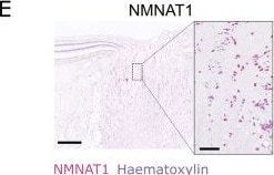
Detection of NMNAT-1 by Immunohistochemistry
Labelling of NAD salvage pathway machinery in human retina. A NAMPT labelling is detected across all retinal layers, localizing to both nuclear (haematoxylin + ve) and cytoplasmic (haematoxylin −ve) cellular compartments, with the most intense labeling in the INL (n = 11 eyes). B NMNAT1 labelling is strongest in nuclear layers (GCL, INL, ONL) localizing to nuclei (n = 11 eyes). C NMNAT2 labelling localized to cytoplasmic compartments and was detected across all retinal layers, with the greatest intensity in the GCL, reflecting high expression in RGCs (n = 11 eyes). D In the ONH, NAMPT followed the same trend as in the retina, with labelling localizing to both nuclear and cytoplasmic compartments. Labeling intensity did not vary along the length of the optic nerve (n = 11 eyes). E NMNAT1 again localized only to nuclei in the ONH (glial framework), and its intensity was consistent along the length of the optic nerve (n = 10 eyes). F NMNAT2 conversely, localized only to the axonal compartments of the optic nerve and was significantly greater in the first 500 µm of the ONH (n = 8 eyes), potentially reflecting the density of axons relative to glial framework here, or the need for more NMNAT2 at a critical metabolic portion of the axons. N.B. these data do not allow for direct comparison of abundances of these enzymes given the nature of antibody labelling (efficiency and concentration of the antibodies etc.). RNFL retinal nerve fiber layer, GCL ganglion cell layer, IPL inner plexiform layer, INL inner nuclear layer, OPL outer plexiform layer, ONL outer nuclear layer, PS photoreceptor segments. Scale bars = 50 µm in A–C, 500 µm in D–E (overview images) and 50 µm in D–E (inset images) Image collected and cropped by CiteAb from the following open publication (https://pubmed.ncbi.nlm.nih.gov/36681854), licensed under a CC-BY license. Not internally tested by R&D Systems.Applications for Human NMNAT-1 Antibody (675112)
Immunohistochemistry
Sample: Immersion fixed paraffin-embedded sections of Human Kidney
Formulation, Preparation, and Storage
Purification
Reconstitution
Sterile PBS to a final concentration of 0.5 mg/mL. For liquid material, refer to CoA for concentration.
Formulation
Shipping
Stability & Storage
- 12 months from date of receipt, -20 to -70 °C as supplied.
- 1 month, 2 to 8 °C under sterile conditions after reconstitution.
- 6 months, -20 to -70 °C under sterile conditions after reconstitution.
Calculators
Background: NMNAT-1
References
- Emanuelli, M. et al. (2001) J. Biol.Chem. 276:406.
- Schweiger, M. et al. (2001) FEBS Lett. 492:95.
- Berger, F. et al. (2007) Proc. Natl. Acad. Sci. USA 104:3765.
- Revollo, J.R. et al. (2004) J. Biol. Chem. 279:50754.
- Mack, T.G. et al. (2001) Nature Neurosci. 4:1199.
- Boulton, S. et al. (1997) Br. J. Cancer 76:845.
- Berger, F. et al. (2005) J. Biol. Chem. 280:36334.
Long Name
Alternate Names
Entrez Gene IDs
Gene Symbol
UniProt
Additional NMNAT-1 Products
Product Documents for Human NMNAT-1 Antibody (675112)
Certificate of Analysis
To download a Certificate of Analysis, please enter a lot or batch number in the search box below.
Note: Certificate of Analysis not available for kit components.
Product Specific Notices for Human NMNAT-1 Antibody (675112)
For research use only
Citations for Human NMNAT-1 Antibody (675112)
 Powered by Bioz
Customer Reviews for Human NMNAT-1 Antibody (675112)
There are currently no reviews for this product. Be the first to review Human NMNAT-1 Antibody (675112) and earn rewards!
Have you used Human NMNAT-1 Antibody (675112)?
Submit a review and receive an Amazon gift card!
$25/€18/£15/$25CAN/¥2500 for a review with an image
$10/€7/£6/$10CAN/¥1110 for a review without an image
Submit a review
Protocols
Find general support by application which include: protocols, troubleshooting, illustrated assays, videos and webinars.
- Antigen Retrieval Protocol (PIER)
- Antigen Retrieval for Frozen Sections Protocol
- Appropriate Fixation of IHC/ICC Samples
- Cellular Response to Hypoxia Protocols
- Chromogenic IHC Staining of Formalin-Fixed Paraffin-Embedded (FFPE) Tissue Protocol
- Chromogenic Immunohistochemistry Staining of Frozen Tissue
- ClariTSA™ Fluorophore Kits
- Detection & Visualization of Antibody Binding
- Fluorescent IHC Staining of Frozen Tissue Protocol
- Graphic Protocol for Heat-induced Epitope Retrieval
- Graphic Protocol for the Preparation and Fluorescent IHC Staining of Frozen Tissue Sections
- Graphic Protocol for the Preparation and Fluorescent IHC Staining of Paraffin-embedded Tissue Sections
- Graphic Protocol for the Preparation of Gelatin-coated Slides for Histological Tissue Sections
- IHC Sample Preparation (Frozen sections vs Paraffin)
- Immunofluorescent IHC Staining of Formalin-Fixed Paraffin-Embedded (FFPE) Tissue Protocol
- Immunohistochemistry (IHC) and Immunocytochemistry (ICC) Protocols
- Immunohistochemistry Frozen Troubleshooting
- Immunohistochemistry Paraffin Troubleshooting
- Preparing Samples for IHC/ICC Experiments
- Preventing Non-Specific Staining (Non-Specific Binding)
- Primary Antibody Selection & Optimization
- Protocol for Heat-Induced Epitope Retrieval (HIER)
- Protocol for Making a 4% Formaldehyde Solution in PBS
- Protocol for VisUCyte™ HRP Polymer Detection Reagent
- Protocol for the Preparation & Fixation of Cells on Coverslips
- Protocol for the Preparation and Chromogenic IHC Staining of Frozen Tissue Sections
- Protocol for the Preparation and Chromogenic IHC Staining of Frozen Tissue Sections - Graphic
- Protocol for the Preparation and Chromogenic IHC Staining of Paraffin-embedded Tissue Sections
- Protocol for the Preparation and Chromogenic IHC Staining of Paraffin-embedded Tissue Sections - Graphic
- Protocol for the Preparation and Fluorescent IHC Staining of Frozen Tissue Sections
- Protocol for the Preparation and Fluorescent IHC Staining of Paraffin-embedded Tissue Sections
- Protocol for the Preparation of Gelatin-coated Slides for Histological Tissue Sections
- TUNEL and Active Caspase-3 Detection by IHC/ICC Protocol
- The Importance of IHC/ICC Controls
- Troubleshooting Guide: Immunohistochemistry
- View all Protocols, Troubleshooting, Illustrated assays and Webinars