Best Seller
Human Otx2 Antibody
R&D Systems | Catalog # AF1979

Key Product Details
Species Reactivity
Validated:
Human
Cited:
Human, Mouse, Rat, Porcine, Avian - Chicken, Chicken, Fish - Danio rerio (Zebrafish), Fish - Oryzias latipes (Medaka or Japanese Rice Fish), Primate - Callithrix jacchus (Common Marmoset), Primate - Macaca fascicularis (Crab-eating Monkey or Cynomolgus Macaque), Primate - Pan troglodytes (Chimpanzee), Transgenic Mouse
Applications
Validated:
Immunohistochemistry, Western Blot, Immunocytochemistry, Simple Western
Cited:
Immunohistochemistry, Immunohistochemistry-Paraffin, Immunohistochemistry-Frozen, Western Blot, Flow Cytometry, Immunofluorescence, Immunocytochemistry, Bioassay, Cell Culture, Differentiation
Label
Unconjugated
Antibody Source
Polyclonal Goat IgG
Loading...
Product Specifications
Immunogen
E. coli-derived recombinant human Otx2
Met1-Leu289
Accession # P32243
Met1-Leu289
Accession # P32243
Specificity
Detects human Otx2 in direct ELISAs and Western blots.
Clonality
Polyclonal
Host
Goat
Isotype
IgG
Scientific Data Images for Human Otx2 Antibody
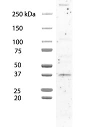
Detection of Human Otx2 by Western Blot.
Western blot shows lysates of IMR-32 human neuroblastoma cell line. PVDF membrane was probed with 1 µg/mL of Goat Anti-Human Otx2 Antigen Affinity-purified Polyclonal Antibody (Catalog # AF1979) followed by HRP-conjugated Anti-Goat IgG Secondary Antibody (Catalog # HAF017). A specific band was detected for Otx2 at approximately 37 kDa (as indicated). This experiment was conducted under reducing conditions and using Immunoblot Buffer Group 1.
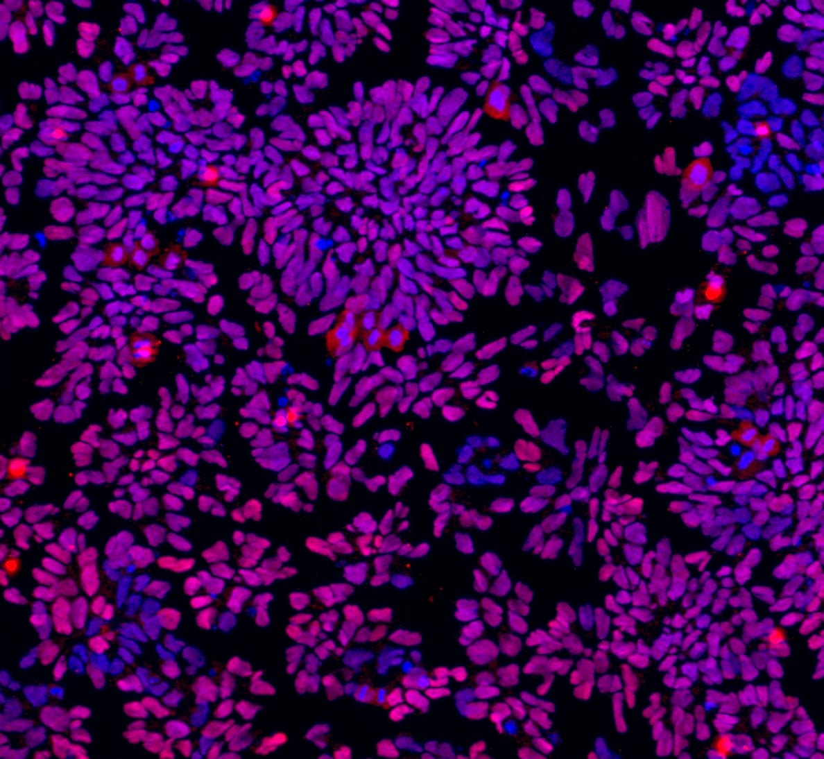
Otx2 in NTera‑2 Human Cell Line.
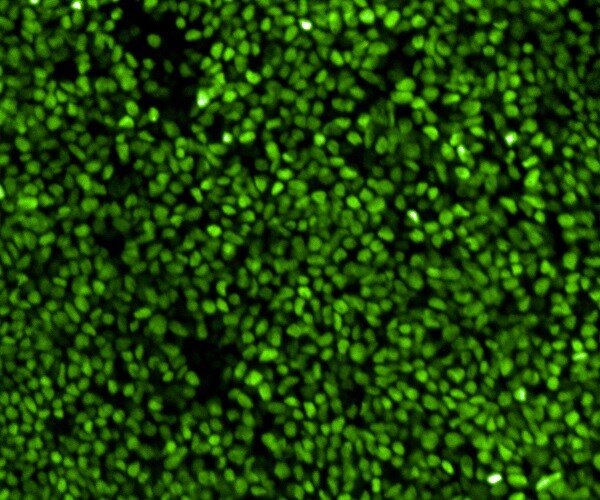
Otx2 was detected in immersion fixed NTera-2 human testicular embryonic carcinoma cell line using Goat Anti-Human Otx2 Antigen Affinity-purified Polyclonal Antibody (Catalog # AF1979) at 10 µg/mL for 3 hours at room temperature. Cells were stained using the NorthernLights™ 557-conjugated Anti-Goat IgG Secondary Antibody (yellow, upper panel; Catalog # NL001) and counterstained with DAPI (blue, lower panel). View our protocol for Fluorescent ICC Staining of Cells on Coverslips.
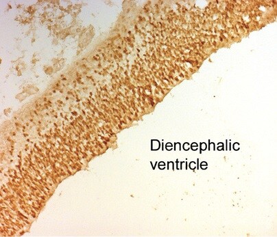
Otx2 in Mouse Embryo.
Otx2 was detected in immersion fixed paraffin-embedded sections of mouse embryo (14 d.p.c.) using Goat Anti-Human Otx2 Antigen Affinity-purified Polyclonal Antibody (Catalog # AF1979) at 1 µg/mL for 1 hour at room temperature followed by incubation with the Anti-Goat IgG VisUCyte™ HRP Polymer Antibody (Catalog # VC004). Before incubation with the primary antibody, tissue was subjected to heat-induced epitope retrieval using Antigen Retrieval Reagent-Basic (Catalog # CTS013). Tissue was stained using DAB (brown) and counterstained with hematoxylin (blue). Specific staining was localized to developing nervous system. View our protocol for IHC Staining with VisUCyte HRP Polymer Detection Reagents.
Detection of Human Otx2 by Simple WesternTM.
Simple Western lane view shows lysates of IMR-32 human neuroblastoma cell line, loaded at 0.2 mg/mL. A specific band was detected for Otx2 at approximately 48 kDa (as indicated) using 10 µg/mL of Goat Anti-Human Otx2 Antigen Affinity-purified Polyclonal Antibody (Catalog # AF1979) followed by 1:50 dilution of HRP-conjugated Anti-Goat IgG Secondary Antibody (Catalog # HAF109). This experiment was conducted under reducing conditions and using the 12-230 kDa separation system.
Detection of Human Otx2 by Immunocytochemistry/Immunofluorescence
Generation and characterization of systemic lupus erythematosus (SLE)‐specific human‐induced pluripotent stem cells (hiPSCs). (A) Dermal fibroblasts derived from patient with SLE were reprogrammed into iPSCs using Sendai virus vectors and three clones (#1, #2 and #3) were characterized. RT‐PCR confirms the loss of transgenes in hiPSCs‐SLE (lanes 1, 2 and 3), presence (lane 4) in infected fibroblasts (ipF‐SLE) and absence of Sendai viral transgenes in parental fibroblasts (pF‐SLE) (lane 5). Full‐length gels are presented in File S2. (B) The hiPSCs‐SLE colonies expressed alkaline phosphatase. Scale bar, 500 μm. (C) RT‐qPCR analysis of pluripotency genes OCT4, NANOG, SOX2 and REX1 was performed in fibroblasts and in hiPSCs derived from patient with SLE. All expression values are normalized to GAPDH and relative donor fibroblasts. Data are mean ± SEM and all statistical analysis was made between hiPSCs‐SLE clones and relative fibroblasts by Student's t test showing P‐values ≤ .05 in each comparison. (D) PluriTest assays combines novelty score (blue) on x‐axis and pluripotency score (red) on y‐axis. hiPSCs‐SLE localize in the red cloud suggesting the empirical distribution of pluripotent cells compared to non‐pluripotent blue cloud. (E) Representative images of M‐FISH staining show normal karyotypes of hiPSCs‐SLE clones. (F) Immunofluorescence analysis of pluripotent stem cell markers Nanog (green), Oct4 (red) and co‐staining with DAPI (blue) in hiPSCs‐SLE. Scale bar, 50 μm. (G) Representative images of floating and adherent EBs derived from hiPSCs‐SLE at differentiation day 8 and 18, respectively. Scale bar, 500 μm. (H) RT‐qPCR results confirm the capability of hiPSCs‐SLE to differentiate into all three germ layers. The expression levels of GATA4, HAND1 and PAX6 in EBs are relative to undifferentiated hiPSCs. All expression values are normalized to GAPDH and relative hiPSCs. Data are mean ± SEM and all statistical analysis was made between EBs‐SLE and relative hiPSCs‐SL
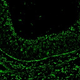
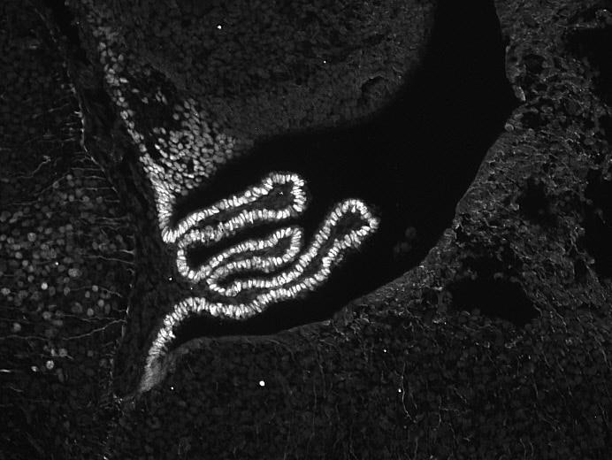
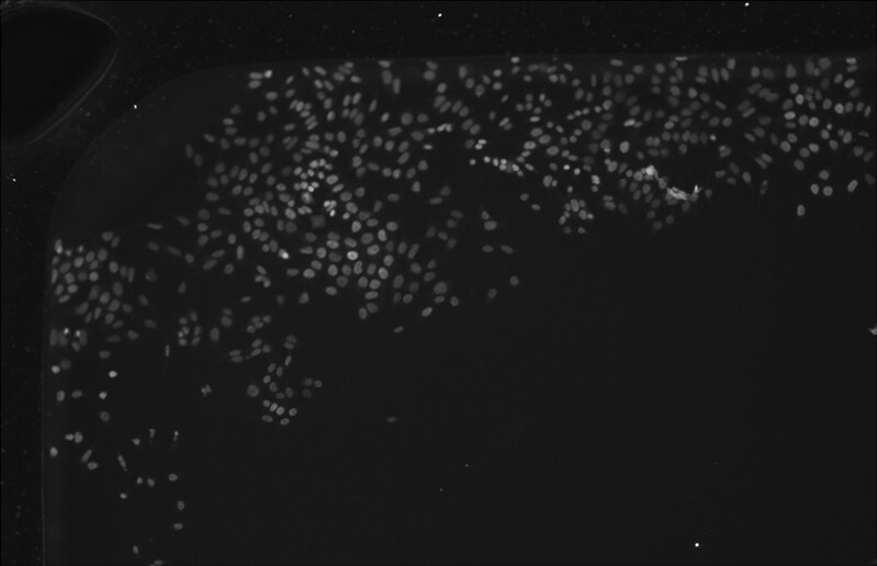
Detection of Zebrafish Otx2 by Immunohistochemistry
Medaka-derived organoids show onset of retinal differentiation. (d) Optical sections showing expression of HuC/D (amacrine&ganglion cells), Otx2 (bipolar&photoreceptor cells),&Prox1 (horizontal cells) in day 4 organoid. Image collected & cropped by CiteAb from the following open publication (https://pubmed.ncbi.nlm.nih.gov/34252023), licensed under a CC-BY license. Not internally tested by R&D Systems.
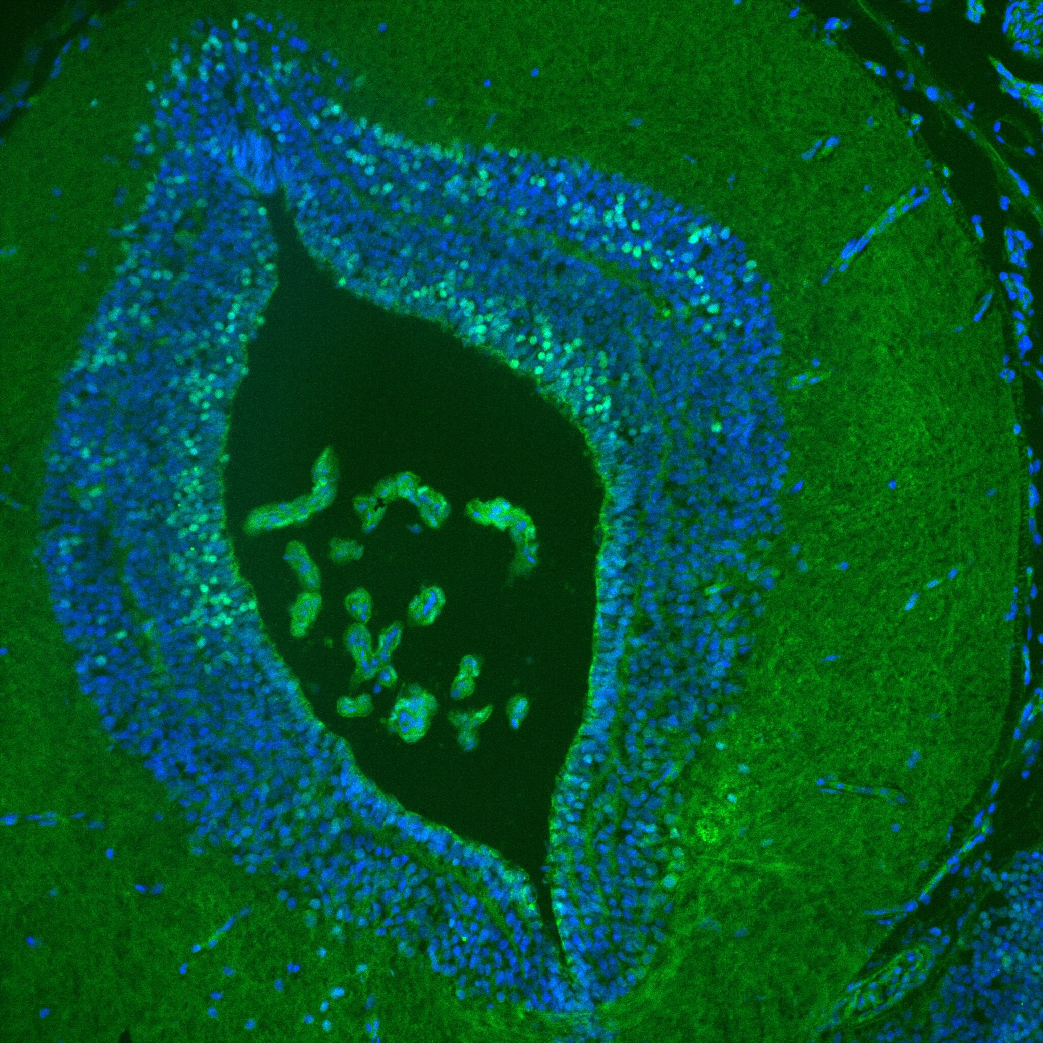
Detection of Chicken Otx2 by Immunohistochemistry
The short-term effects of Notch inhibition on RPC reporters. (d) Quantification of the %age of VSX2, Otx2&double marker-positive cells within the electroporated cell population from cell counting. Sectioned chick retinas electroporated ex vivo at E5 with the co-electroporation control CAG::Nuc beta gal with or without CAG::MAML-DN. The retinas cultured for 20 h&immunostained with VSX2 or Otx2 antibodies. The Shapiro–Wilk normality test was used to confirm the normal distribution. * signifies p < 0.05, *** signifies p < 0.001 with a two-tailed student’s t-test. Each point represents one biological replicate. The columns represent mean,&the error bars represent standard deviation. Image collected & cropped by CiteAb from the following open publication (https://pubmed.ncbi.nlm.nih.gov/34267251), licensed under a CC-BY license. Not internally tested by R&D Systems.
Detection of Chicken Otx2 by Flow Cytometry
Targeted expression of MAML-DN to cone/HC restricted RPCs does not alter cone formation. (d) Flow cytometry quantification of the %age of Otx2, Lim1, AP2 alpha, OC1ECR22&ThrbCRM2 reporter-positive cells within the ThrbCRM1 lineage traced cell population. Dissociated chick retinal cells electroporated ex vivo at E5 with the co-electroporation control CAG::TdTomato, ThrbCRM1::PhiC31&its responder plasmid,&with or without CAG::MAML-DN. The retinas cultured for 2 days post-electroporation. The Shapiro–Wilk normality test was used to confirm the normal distribution. Mann–Whitney test was used to test significance in ThrbCRM2 quantification. A two-tailed student’s t-test was used to test significance in the rest of the quantifications. * signifies p < 0.05, ** signifies p < 0.01. Each point represents one biological replicate. Image collected & cropped by CiteAb from the following open publication (https://pubmed.ncbi.nlm.nih.gov/34267251), licensed under a CC-BY license. Not internally tested by R&D Systems.
Detection of Zebrafish Otx2 by Immunohistochemistry
Medaka primary embryonic pluripotent cells form optic vesicle-like structures. (f) Maximal projection of day 2 organoids&1 dpf embryo generated from Rx3::H2B-GFP transgenic or wild-type blastulae&stained with neural tissue-specific anti-Sox2 (n = 12/12)&anti-Otx2 (n = 10/10) antibodies, co-stained with anti-Rx2&DAPI nuclear stain. hpa, hours post-aggregation; dpf, days post-fertilization. Scale bars: 100 μm.Overview of complexity&variability of organoid morphology generated within one experiment.Overlay of fluorescence&bright-field images of retinal organoids on day 2 generated from the Rx3::H2B-GFP reporter line formed by aggregation of <1000 primary embryonic pluripotent cells stained with anti-GFP antibody. Scale bar: 100 μm.Examples of organoids generated by aggregation of <1000 cells forming one, two,&three optic vesicle-like structures.Fluorescence images, bright-field images,&their overlay of retinal organoids on day 2 generated from the Rx3::H2B-GFP reporter line formed by aggregation of <1000 primary embryonic pluripotent cells stained with anti-GFP antibody. Scale bar: 100 μm. Image collected & cropped by CiteAb from the following open publication (https://pubmed.ncbi.nlm.nih.gov/34252023), licensed under a CC-BY license. Not internally tested by R&D Systems.
Detection of Zebrafish Otx2 by Immunohistochemistry
Examples of organoids generated by aggregation of <1000 cells forming one, two, and three optic vesicle-like structures.Fluorescence images, bright-field images, and their overlay of retinal organoids on day 2 generated from the Rx3::H2B-GFP reporter line formed by aggregation of <1000 primary embryonic pluripotent cells stained with anti-GFP antibody. Scale bar: 100 μm. Image collected and cropped by CiteAb from the following open publication (https://pubmed.ncbi.nlm.nih.gov/34252023), licensed under a CC-BY license. Not internally tested by R&D Systems.
Detection of Zebrafish Otx2 by Immunohistochemistry
Medaka primary embryonic pluripotent cells form optic vesicle-like structures. (e) Bright-field&fluorescent images of day 2 Rx3::H2B-GFP organoids stained with anti-GFP antibody. Optical sections of an organoid (day 2) (n = 9/10)&an embryo (1 dpf) stained with anti-Rx2&anti-Otx2 antibodies. Image collected & cropped by CiteAb from the following open publication (https://pubmed.ncbi.nlm.nih.gov/34252023), licensed under a CC-BY license. Not internally tested by R&D Systems.Applications for Human Otx2 Antibody
Application
Recommended Usage
Immunocytochemistry
5-15 µg/mL
Sample: Immersion fixed NTera-2 human testicular embryonic carcinoma cell line
Sample: Immersion fixed NTera-2 human testicular embryonic carcinoma cell line
Immunohistochemistry
1-15 µg/mL
Sample: Immersion fixed paraffin-embedded sections of mouse embryo (14 d.p.c.)
Sample: Immersion fixed paraffin-embedded sections of mouse embryo (14 d.p.c.)
Simple Western
10 µg/mL
Sample: IMR‑32 human neuroblastoma cell line
Sample: IMR‑32 human neuroblastoma cell line
Western Blot
1 µg/mL
Sample: IMR‑32 human neuroblastoma cell line
Sample: IMR‑32 human neuroblastoma cell line
Reviewed Applications
Read 11 reviews rated 4.8 using AF1979 in the following applications:
Formulation, Preparation, and Storage
Purification
Antigen Affinity-purified
Reconstitution
Reconstitute at 0.2 mg/mL in sterile PBS. For liquid material, refer to CoA for concentration.
Loading...
Formulation
Lyophilized from a 0.2 μm filtered solution in PBS with Trehalose. *Small pack size (SP) is supplied either lyophilized or as a 0.2 µm filtered solution in PBS.
Shipping
Lyophilized product is shipped at ambient temperature. Liquid small pack size (-SP) is shipped with polar packs. Upon receipt, store immediately at the temperature recommended below.
Stability & Storage
Use a manual defrost freezer and avoid repeated freeze-thaw cycles.
- 12 months from date of receipt, -20 to -70 °C as supplied.
- 1 month, 2 to 8 °C under sterile conditions after reconstitution.
- 6 months, -20 to -70 °C under sterile conditions after reconstitution.
Calculators
Background: Otx2
References
- Nagao, T. et al. (1998) Proc. Natl. Acad. Sci. USA 95:3737.
Long Name
Orthodenticle Homolog 2
Alternate Names
homeobox protein OTX2, MCOPS5, MGC45000, orthodenticle homeobox 2, Orthodenticle homolog 2, orthodenticle homolog 2 (Drosophila), OTX2
Entrez Gene IDs
5015 (Human)
Gene Symbol
OTX2
UniProt
Additional Otx2 Products
Product Documents for Human Otx2 Antibody
Certificate of Analysis
To download a Certificate of Analysis, please enter a lot or batch number in the search box below.
Note: Certificate of Analysis not available for kit components.
Product Specific Notices for Human Otx2 Antibody
For research use only
Related Research Areas
Citations for Human Otx2 Antibody
 Powered by Bioz
Customer Reviews for Human Otx2 Antibody (11)
4.8 out of 5
11 Customer Ratings
Have you used Human Otx2 Antibody?
Submit a review and receive an Amazon gift card!
$25/€18/£15/$25CAN/¥2500 Yen for a review with an image
$10/€7/£6/$10CAN/¥1110 Yen for a review without an image
Submit a review
Customer Images
Showing
1
-
5 of
11 reviews
Showing All
Filter By:
-
Application: Western BlotSample Tested: HCT-116 human colorectal carcinoma cell lineSpecies: HumanVerified Customer | Posted 01/01/2026Used at 1 ug/mL

-
Application: Western BlotSample Tested: AdipocytesSpecies: HumanVerified Customer | Posted 07/17/2025
-
Application: Immunocytochemistry/ImmunofluorescenceSample Tested: iPS2 human induced pluripotent stem cellsSpecies: HumanVerified Customer | Posted 09/21/2023
-
Application: Immunocytochemistry/ImmunofluorescenceSample Tested: neuron cells and BrainSpecies: HumanVerified Customer | Posted 07/12/2021
-
Application: ImmunohistochemistrySample Tested: Adult brainSpecies: HumanVerified Customer | Posted 06/29/2021
-
Application: ImmunohistochemistrySample Tested: Fetal brainSpecies: HumanVerified Customer | Posted 12/21/2020
-
Application: ImmunocytochemistrySample Tested: brainSpecies: pleurodeles waltlVerified Customer | Posted 03/30/2020In the image, the green 488 is otx2 staining and the blue is DAPI.
-
Application: Immunocytochemistry/ImmunofluorescenceSample Tested: Embryonic brainSpecies: MouseVerified Customer | Posted 10/04/2018This antibody works very well at 1:1000 in immunofluorescence staining of mouse paraffin-embedded sections. Epitope retrieval: Citrate buffer.
-
Application: Immunocytochemistry/ImmunofluorescenceSample Tested: Undifferentiated neural progenitor cellsSpecies: HumanVerified Customer | Posted 08/22/2016ICC on hPSC derived neurl progenitors
-
Application: ImmunocytochemistrySample Tested: R1 wild type Mouse embryonic Stem CellsSpecies: MouseVerified Customer | Posted 08/04/2016Otx2 in R1 wild type mouse embryonic stem cells
-
Application: ImmunohistochemistrySample Tested: Embryonic retinaSpecies: MouseVerified Customer | Posted 06/29/2016Otx2 staining on frozen mouse embryonic day 17 eyes sections using AF1979 The sections were blocked using 0.01% Triton-X, 2% donkey serum in 1X PBS 1 hour at room temperature. OTX2 was diluted 1:500 using blocking buffer and incubated overnight at 4°C. The secondary antibody used was donkey ant goat conjugated to Alexa Fluor 488 (1:2000).
There are no reviews that match your criteria.
Protocols
Find general support by application which include: protocols, troubleshooting, illustrated assays, videos and webinars.
- Antigen Retrieval Protocol (PIER)
- Antigen Retrieval for Frozen Sections Protocol
- Appropriate Fixation of IHC/ICC Samples
- Cellular Response to Hypoxia Protocols
- Chromogenic IHC Staining of Formalin-Fixed Paraffin-Embedded (FFPE) Tissue Protocol
- Chromogenic Immunohistochemistry Staining of Frozen Tissue
- ClariTSA™ Fluorophore Kits
- Detection & Visualization of Antibody Binding
- Fluorescent IHC Staining of Frozen Tissue Protocol
- Graphic Protocol for Heat-induced Epitope Retrieval
- Graphic Protocol for the Preparation and Fluorescent IHC Staining of Frozen Tissue Sections
- Graphic Protocol for the Preparation and Fluorescent IHC Staining of Paraffin-embedded Tissue Sections
- Graphic Protocol for the Preparation of Gelatin-coated Slides for Histological Tissue Sections
- ICC Cell Smear Protocol for Suspension Cells
- ICC Immunocytochemistry Protocol Videos
- ICC for Adherent Cells
- IHC Sample Preparation (Frozen sections vs Paraffin)
- Immunocytochemistry (ICC) Protocol
- Immunocytochemistry Troubleshooting
- Immunofluorescence of Organoids Embedded in Cultrex Basement Membrane Extract
- Immunofluorescent IHC Staining of Formalin-Fixed Paraffin-Embedded (FFPE) Tissue Protocol
- Immunohistochemistry (IHC) and Immunocytochemistry (ICC) Protocols
- Immunohistochemistry Frozen Troubleshooting
- Immunohistochemistry Paraffin Troubleshooting
- Preparing Samples for IHC/ICC Experiments
- Preventing Non-Specific Staining (Non-Specific Binding)
- Primary Antibody Selection & Optimization
- Protocol for Heat-Induced Epitope Retrieval (HIER)
- Protocol for Making a 4% Formaldehyde Solution in PBS
- Protocol for VisUCyte™ HRP Polymer Detection Reagent
- Protocol for the Fluorescent ICC Staining of Cell Smears - Graphic
- Protocol for the Fluorescent ICC Staining of Cultured Cells on Coverslips - Graphic
- Protocol for the Preparation & Fixation of Cells on Coverslips
- Protocol for the Preparation and Chromogenic IHC Staining of Frozen Tissue Sections
- Protocol for the Preparation and Chromogenic IHC Staining of Frozen Tissue Sections - Graphic
- Protocol for the Preparation and Chromogenic IHC Staining of Paraffin-embedded Tissue Sections
- Protocol for the Preparation and Chromogenic IHC Staining of Paraffin-embedded Tissue Sections - Graphic
- Protocol for the Preparation and Fluorescent ICC Staining of Cells on Coverslips
- Protocol for the Preparation and Fluorescent ICC Staining of Non-adherent Cells
- Protocol for the Preparation and Fluorescent ICC Staining of Stem Cells on Coverslips
- Protocol for the Preparation and Fluorescent IHC Staining of Frozen Tissue Sections
- Protocol for the Preparation and Fluorescent IHC Staining of Paraffin-embedded Tissue Sections
- Protocol for the Preparation of Gelatin-coated Slides for Histological Tissue Sections
- Protocol for the Preparation of a Cell Smear for Non-adherent Cell ICC - Graphic
- R&D Systems Quality Control Western Blot Protocol
- TUNEL and Active Caspase-3 Detection by IHC/ICC Protocol
- The Importance of IHC/ICC Controls
- Troubleshooting Guide: Immunohistochemistry
- Troubleshooting Guide: Western Blot Figures
- Western Blot Conditions
- Western Blot Protocol
- Western Blot Protocol for Cell Lysates
- Western Blot Troubleshooting
- Western Blot Troubleshooting Guide
- View all Protocols, Troubleshooting, Illustrated assays and Webinars
Loading...
Associated Pathways
