
Key Product Details
Validated by
Biological Validation
Species Reactivity
Validated:
Human
Cited:
Human
Applications
Validated:
Immunohistochemistry, Western Blot, Intracellular Staining by Flow Cytometry, Immunocytochemistry, Simple Western, Immunoprecipitation, CyTOF-ready
Cited:
Western Blot, Immunocytochemistry
Label
Unconjugated
Antibody Source
Polyclonal Goat IgG
Loading...
Product Specifications
Immunogen
E. coli-derived recombinant human p21
Ser2-Pro164
Accession # P38936
Ser2-Pro164
Accession # P38936
Specificity
Detects human p21 in Western blots.
Clonality
Polyclonal
Host
Goat
Isotype
IgG
Scientific Data Images for Human p21/CIP1/CDKN1A Antibody
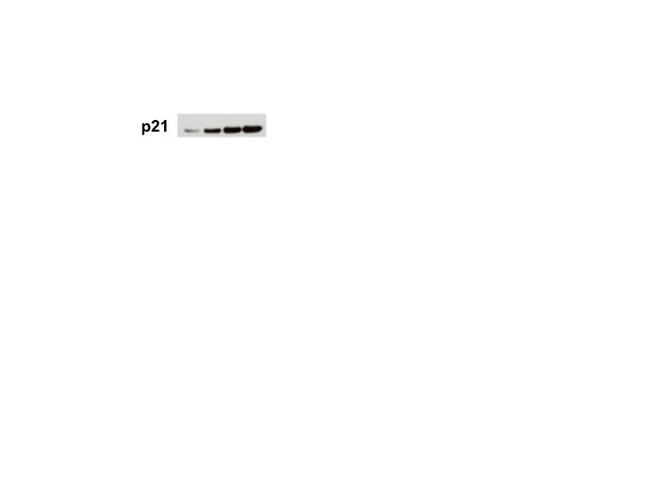
Detection of Human p21/CIP1/CDKN1A by Western Blot.
Western blot shows lysates of MCF-7 human breast cancer cell line untreated (-) or treated (+) with 1 µM camptothecin (CPT) for 16 hours. PVDF membrane was probed with 0.5 µg/mL of Goat Anti-Human p21/CIP1/CDKN1A Antigen Affinity-purified Polyclonal Antibody (Catalog # AF1047), followed by HRP-conjugated Anti-Goat IgG Secondary Antibody (Catalog # HAF017). A specific band was detected for p21/CIP1/CDKN1A at approximately 21 kDa (as indicated). This experiment was conducted under reducing conditions and using Immunoblot Buffer Group 1.
p21/CIP1/CDKN1A in MCF‑7 Human Cell Line.
p21/CIP1/CDKN1A was detected in immersion fixed MCF-7 human breast cancer cell line treated with (left panel) or without (right panel) camptothecin using Goat Anti-Human p21/CIP1/CDKN1A Antigen Affinity-purified Polyclonal Antibody (Catalog # AF1047) at 10 µg/mL for 3 hours at room temperature. Cells were stained using the NorthernLights™ 557-conjugated Anti-Goat IgG Secondary Antibody (red; Catalog # NL001) and counterstained with DAPI (blue). Specific staining was localized to nuclei. View our protocol for Fluorescent ICC Staining of Cells on Coverslips.
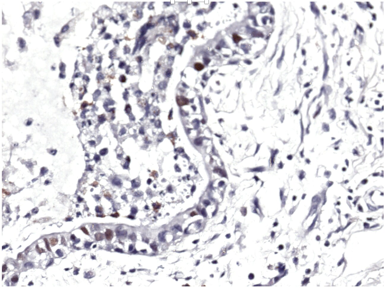
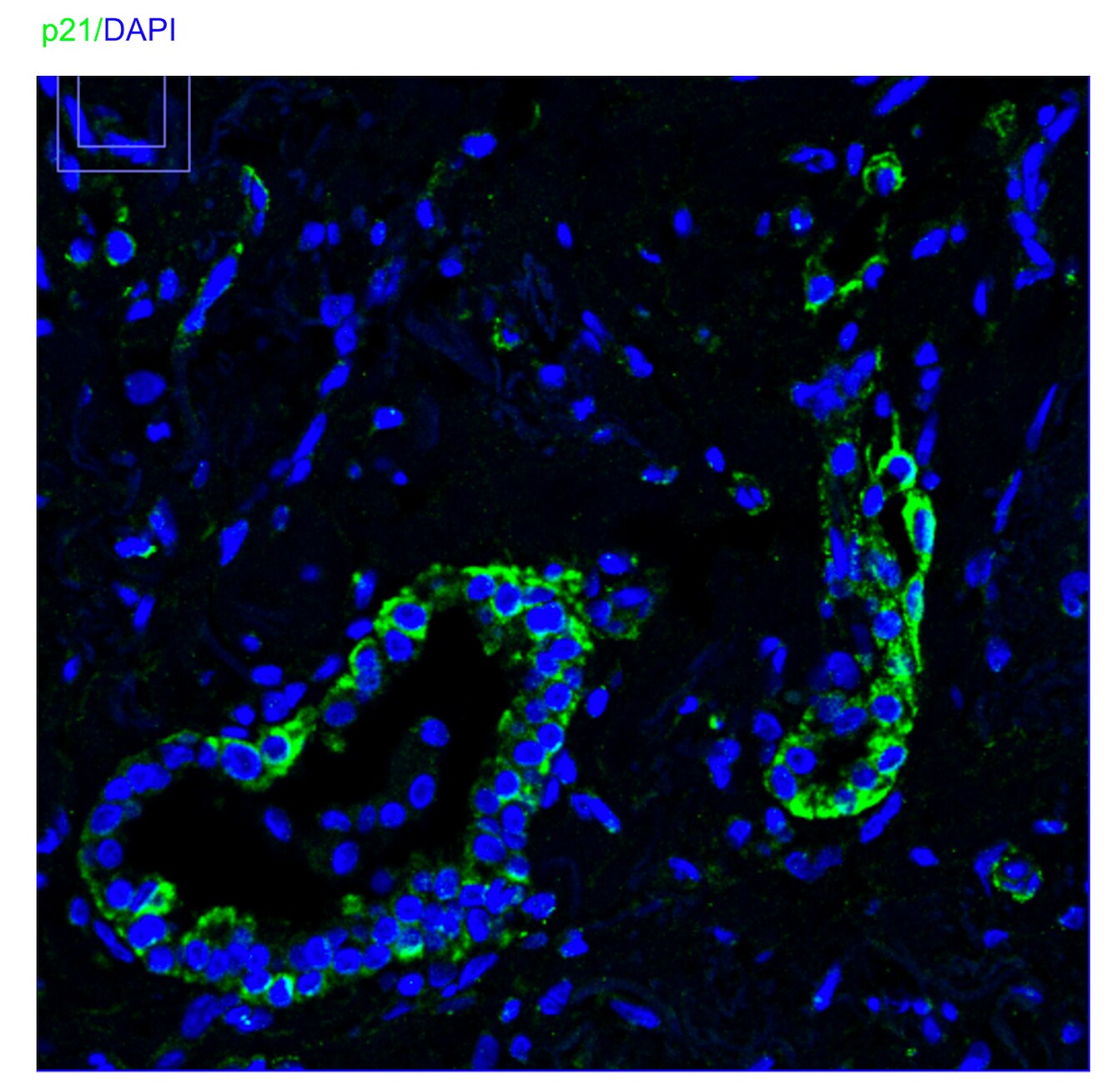
p21/CIP1/CDKN1A in Human Breast Cancer Tissue.
p21/CIP1/CDKN1A was detected in immersion fixed paraffin-embedded sections of human breast cancer tissue using 1.7 µg/mL Goat Anti-Human p21/CIP1/CDKN1A Antigen Affinity-purified Polyclonal Antibody (Catalog # AF1047) overnight at 4 °C. Tissue was stained with the Anti-Goat HRP-DAB Cell & Tissue Staining Kit (brown; Catalog # CTS008) and counterstained with hematoxylin (blue). View our protocol for Chromogenic IHC Staining of Paraffin-embedded Tissue Sections.
Detection of p21/CIP1/CDKN1A in MCF‑7 Human Cell Line by Flow Cytometry.
MCF-7 human breast cancer cell line was unstimulated (light orange filled histogram) or treated with 1 µM camphtothecin for 16 hours, then stained with Goat Anti-Human p21/CIP1/CDKN1A Antigen Affinity-purified Polyclonal Antibody (Catalog # AF1047, dark orange filled histogram) or isotype control antibody (Catalog # AB-108-C, open histogram), followed by Allophycocyanin-conjugated Anti-Goat IgG Secondary Antibody (Catalog # F0108). To facilitate intracellular staining, cells were fixed with paraformaldehyde and permeabilized with methanol.
Detection of Human p21/CIP1/CDKN1A by Simple WesternTM.
Simple Western lane view shows lysates of MCF-7 human breast cancer cell line untreated (-) or treated (+) with 1 µM Camptothecin (CPT) for 16 hours, loaded at 0.2 mg/mL. A specific band was detected for p21/CIP1/CDKN1A at approximately 30 kDa (as indicated) using 5 µg/mL of Goat Anti-Human p21/CIP1/CDKN1A Antigen Affinity-purified Polyclonal Antibody (Catalog # AF1047) followed by 1:50 dilution of HRP-conjugated Anti-Goat IgG Secondary Antibody (Catalog # HAF109). This experiment was conducted under reducing conditions and using the 12-230 kDa separation system.Applications for Human p21/CIP1/CDKN1A Antibody
Application
Recommended Usage
CyTOF-ready
Ready to be labeled using established conjugation methods. No BSA or other carrier proteins that could interfere with conjugation.
Immunocytochemistry
5-15 µg/mL
Sample: Immersion fixed MCF-7 human breast cancer cell line treated with Camptothecin
Sample: Immersion fixed MCF-7 human breast cancer cell line treated with Camptothecin
Immunohistochemistry
5-15 µg/mL
Sample: Immersion fixed paraffin-embedded sections of human breast cancer tissue
Sample: Immersion fixed paraffin-embedded sections of human breast cancer tissue
Immunoprecipitation
1-2 µg/500 µg cell lysate
Sample: MCF‑7 human breast cancer cell line, see our available Western blot detection antibodies
Sample: MCF‑7 human breast cancer cell line, see our available Western blot detection antibodies
Intracellular Staining by Flow Cytometry
0.25 µg/106 cells
Sample: MCF‑7 human breast cancer cell line fixed with paraformaldehyde and permeabilized with methanol
Sample: MCF‑7 human breast cancer cell line fixed with paraformaldehyde and permeabilized with methanol
Simple Western
5 µg/mL
Sample: MCF‑7 human breast cancer cell line treated with Camptothecin (CPT)
Sample: MCF‑7 human breast cancer cell line treated with Camptothecin (CPT)
Western Blot
0.5 µg/mL
Sample: Camptothecin-treated MCF‑7 human breast cancer cell line
Sample: Camptothecin-treated MCF‑7 human breast cancer cell line
Reviewed Applications
Read 4 reviews rated 4.3 using AF1047 in the following applications:
Flow Cytometry Panel Builder
Bio-Techne Knows Flow Cytometry
Save time and reduce costly mistakes by quickly finding compatible reagents using the Panel Builder Tool.
Advanced Features
- Spectra Viewer - Custom analysis of spectra from multiple fluorochromes
- Spillover Popups - Visualize the spectra of individual fluorochromes
- Antigen Density Selector - Match fluorochrome brightness with antigen density

Formulation, Preparation, and Storage
Purification
Antigen Affinity-purified
Reconstitution
Reconstitute at 0.2 mg/mL in sterile PBS. For liquid material, refer to CoA for concentration.
Loading...
Formulation
Lyophilized from a 0.2 μm filtered solution in PBS with Trehalose. *Small pack size (SP) is supplied either lyophilized or as a 0.2 µm filtered solution in PBS.
Shipping
Lyophilized product is shipped at ambient temperature. Liquid small pack size (-SP) is shipped with polar packs. Upon receipt, store immediately at the temperature recommended below.
Stability & Storage
Use a manual defrost freezer and avoid repeated freeze-thaw cycles.
- 12 months from date of receipt, -20 to -70 °C as supplied.
- 1 month, 2 to 8 °C under sterile conditions after reconstitution.
- 6 months, -20 to -70 °C under sterile conditions after reconstitution.
Calculators
Background: p21/CIP1/CDKN1A
Long Name
p21 Cyclin Dependent Kinase 4 Inhibitor 1A
Alternate Names
CDKN1A, CIP1
Gene Symbol
CDKN1A
UniProt
Additional p21/CIP1/CDKN1A Products
Product Documents for Human p21/CIP1/CDKN1A Antibody
Certificate of Analysis
To download a Certificate of Analysis, please enter a lot or batch number in the search box below.
Note: Certificate of Analysis not available for kit components.
Product Specific Notices for Human p21/CIP1/CDKN1A Antibody
For research use only
Related Research Areas
Citations for Human p21/CIP1/CDKN1A Antibody
 Powered by Bioz
Customer Reviews for Human p21/CIP1/CDKN1A Antibody (4)
4.3 out of 5
4 Customer Ratings
Have you used Human p21/CIP1/CDKN1A Antibody?
Submit a review and receive an Amazon gift card!
$25/€18/£15/$25CAN/¥2500 Yen for a review with an image
$10/€7/£6/$10CAN/¥1110 Yen for a review without an image
Submit a review
Customer Images

Showing
1
-
4 of
4 reviews
Showing All
Filter By:
-
Application: Western BlotSample Tested: Prostate cancerSpecies: HumanVerified Customer | Posted 01/25/2018

-
Application: Western BlotSample Tested: MDA-MB-231 human breast cancer cell lineSpecies: HumanVerified Customer | Posted 01/17/2018
-
Application: ImmunohistochemistrySample Tested: Adult lungSpecies: HumanVerified Customer | Posted 11/04/2016
-
Application: Immunocytochemistry/ImmunofluorescenceSample Tested: Adult lungSpecies: HumanVerified Customer | Posted 10/18/2016
There are no reviews that match your criteria.
Protocols
Find general support by application which include: protocols, troubleshooting, illustrated assays, videos and webinars.
- 7-Amino Actinomycin D (7-AAD) Cell Viability Flow Cytometry Protocol
- Antigen Retrieval Protocol (PIER)
- Antigen Retrieval for Frozen Sections Protocol
- Appropriate Fixation of IHC/ICC Samples
- Cellular Response to Hypoxia Protocols
- Chromogenic IHC Staining of Formalin-Fixed Paraffin-Embedded (FFPE) Tissue Protocol
- Chromogenic Immunohistochemistry Staining of Frozen Tissue
- ClariTSA™ Fluorophore Kits
- Detection & Visualization of Antibody Binding
- Extracellular Membrane Flow Cytometry Protocol
- Flow Cytometry Protocol for Cell Surface Markers
- Flow Cytometry Protocol for Staining Membrane Associated Proteins
- Flow Cytometry Staining Protocols
- Flow Cytometry Troubleshooting Guide
- Fluorescent IHC Staining of Frozen Tissue Protocol
- Graphic Protocol for Heat-induced Epitope Retrieval
- Graphic Protocol for the Preparation and Fluorescent IHC Staining of Frozen Tissue Sections
- Graphic Protocol for the Preparation and Fluorescent IHC Staining of Paraffin-embedded Tissue Sections
- Graphic Protocol for the Preparation of Gelatin-coated Slides for Histological Tissue Sections
- ICC Cell Smear Protocol for Suspension Cells
- ICC Immunocytochemistry Protocol Videos
- ICC for Adherent Cells
- IHC Sample Preparation (Frozen sections vs Paraffin)
- Immunocytochemistry (ICC) Protocol
- Immunocytochemistry Troubleshooting
- Immunofluorescence of Organoids Embedded in Cultrex Basement Membrane Extract
- Immunofluorescent IHC Staining of Formalin-Fixed Paraffin-Embedded (FFPE) Tissue Protocol
- Immunohistochemistry (IHC) and Immunocytochemistry (ICC) Protocols
- Immunohistochemistry Frozen Troubleshooting
- Immunohistochemistry Paraffin Troubleshooting
- Immunoprecipitation Protocol
- Intracellular Flow Cytometry Protocol Using Alcohol (Methanol)
- Intracellular Flow Cytometry Protocol Using Detergents
- Intracellular Nuclear Staining Flow Cytometry Protocol Using Detergents
- Intracellular Staining Flow Cytometry Protocol Using Alcohol Permeabilization
- Intracellular Staining Flow Cytometry Protocol Using Detergents to Permeabilize Cells
- Preparing Samples for IHC/ICC Experiments
- Preventing Non-Specific Staining (Non-Specific Binding)
- Primary Antibody Selection & Optimization
- Propidium Iodide Cell Viability Flow Cytometry Protocol
- Protocol for Heat-Induced Epitope Retrieval (HIER)
- Protocol for Liperfluo
- Protocol for Making a 4% Formaldehyde Solution in PBS
- Protocol for VisUCyte™ HRP Polymer Detection Reagent
- Protocol for the Characterization of Human Th22 Cells
- Protocol for the Characterization of Human Th9 Cells
- Protocol for the Fluorescent ICC Staining of Cell Smears - Graphic
- Protocol for the Fluorescent ICC Staining of Cultured Cells on Coverslips - Graphic
- Protocol for the Preparation & Fixation of Cells on Coverslips
- Protocol for the Preparation and Chromogenic IHC Staining of Frozen Tissue Sections
- Protocol for the Preparation and Chromogenic IHC Staining of Frozen Tissue Sections - Graphic
- Protocol for the Preparation and Chromogenic IHC Staining of Paraffin-embedded Tissue Sections
- Protocol for the Preparation and Chromogenic IHC Staining of Paraffin-embedded Tissue Sections - Graphic
- Protocol for the Preparation and Fluorescent ICC Staining of Cells on Coverslips
- Protocol for the Preparation and Fluorescent ICC Staining of Non-adherent Cells
- Protocol for the Preparation and Fluorescent ICC Staining of Stem Cells on Coverslips
- Protocol for the Preparation and Fluorescent IHC Staining of Frozen Tissue Sections
- Protocol for the Preparation and Fluorescent IHC Staining of Paraffin-embedded Tissue Sections
- Protocol for the Preparation of Gelatin-coated Slides for Histological Tissue Sections
- Protocol for the Preparation of a Cell Smear for Non-adherent Cell ICC - Graphic
- Protocol: Annexin V and PI Staining by Flow Cytometry
- Protocol: Annexin V and PI Staining for Apoptosis by Flow Cytometry
- R&D Systems Quality Control Western Blot Protocol
- TUNEL and Active Caspase-3 Detection by IHC/ICC Protocol
- The Importance of IHC/ICC Controls
- Troubleshooting Guide: Fluorokine Flow Cytometry Kits
- Troubleshooting Guide: Immunohistochemistry
- Troubleshooting Guide: Western Blot Figures
- Western Blot Conditions
- Western Blot Protocol
- Western Blot Protocol for Cell Lysates
- Western Blot Troubleshooting
- Western Blot Troubleshooting Guide
- View all Protocols, Troubleshooting, Illustrated assays and Webinars
Loading...
Associated Pathways

