
Key Product Details
Species Reactivity
Validated:
Human
Cited:
Human, Mouse, Primate - Macaca mulatta (Rhesus Macaque)
Applications
Validated:
Western Blot, Intracellular Staining by Flow Cytometry, Immunocytochemistry, Simple Western
Cited:
Immunohistochemistry, Western Blot, Immunocytochemistry
Label
Unconjugated
Antibody Source
Polyclonal Sheep IgG
Loading...
Product Specifications
Immunogen
E. coli-derived recombinant human Pax6
Met1-Arg272
Accession # P26367
Met1-Arg272
Accession # P26367
Specificity
Detects human Pax6 in direct ELISAs.
Clonality
Polyclonal
Host
Sheep
Isotype
IgG
Scientific Data Images for Human Pax6 Antibody
Detection of Human and Rat Pax6 by Western Blot.
Western blot shows lysates of HeLa human cervical epithelial carcinoma cell line and rat cortical stem cells. PVDF membrane was probed with 0.5 µg/mL of Sheep Anti-Human Pax6 Antigen Affinity-purified Polyclonal Antibody (Catalog # AF8150) followed by HRP-conjugated Anti-Sheep IgG Secondary Antibody (Catalog # HAF016). A specific band was detected for Pax6 at approximately 48-50 kDa (as indicated). This experiment was conducted under reducing conditions and using Immunoblot Buffer Group 1.
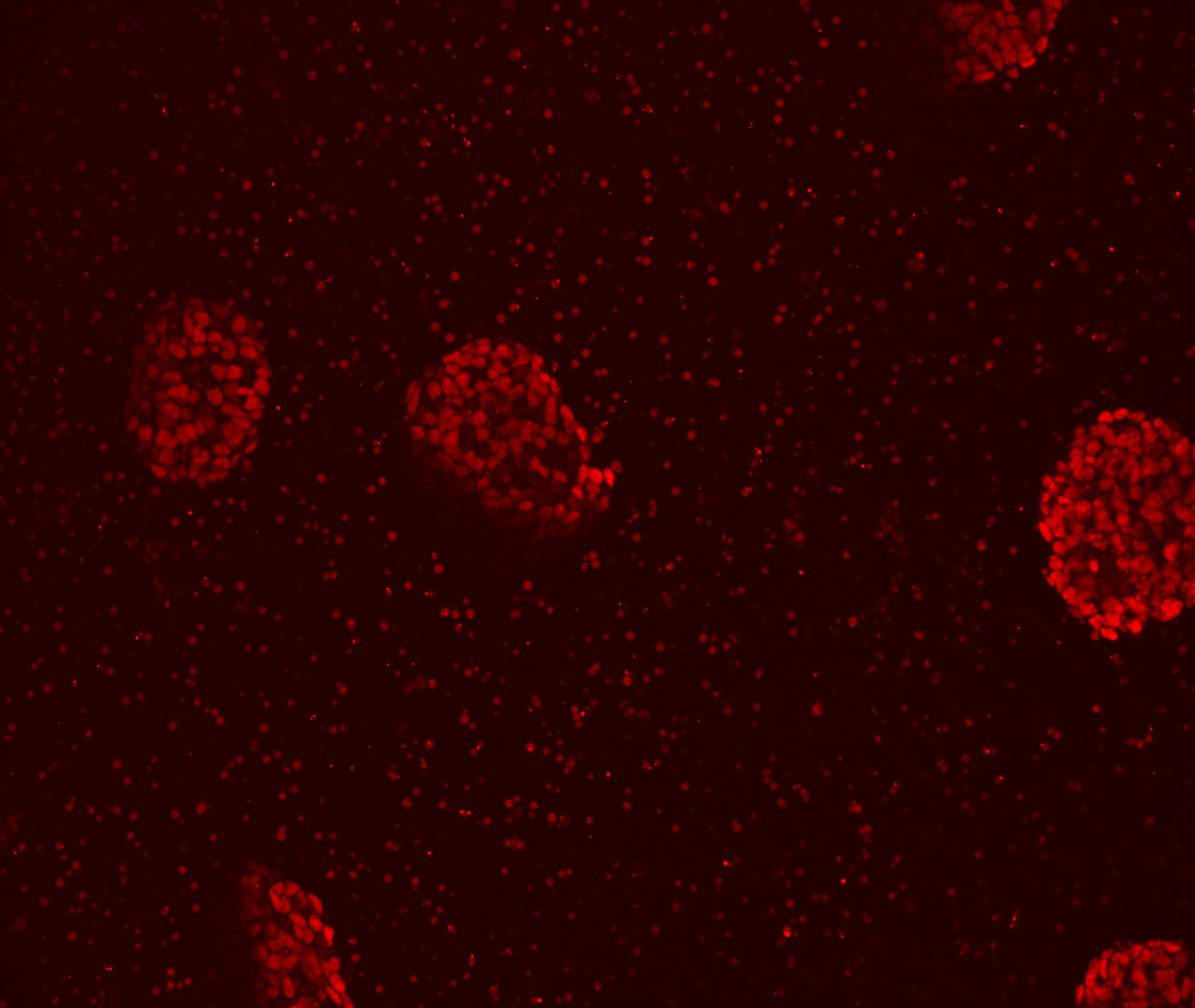
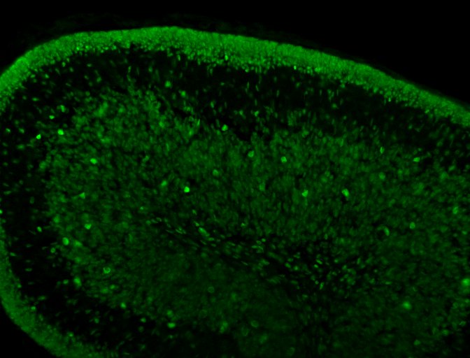
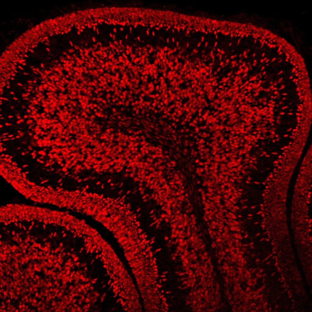
Pax6 in SA01 Human Embryonic Stem Cells.
Immersion fixed SA01 human embryonic stem cells were differentiated for 6 days with Recombinant Human Noggin (Catalog # 6057-NG) and SB431542 (upper panel) or differentiated for 6 days with Recombinant Human BMP-4 (negative control, lower panel; Catalog # 314-BP). Pax6 was detected using Sheep Anti-Human Pax6 Antigen Affinity-purified Polyclonal Antibody (Catalog # AF8150) at 5 µg/mL. Cells were stained using an Alexa Fluor 488-conjugated Anti-Sheep IgG Secondary Antibody (green) and counterstained with DAPI (blue). Specific staining was localized to nuclei.Images courtesy of Dr. Ron McKay, Leiber Institute for Brain Development, Baltimore, Maryland, USA.
Detection of Human Pax6 by Simple WesternTM.
Simple Western lane view shows lysates of HeLa human cervical epithelial carcinoma cell line, loaded at 0.2 mg/mL. A specific band was detected for Pax6 at approximately 59 kDa (as indicated) using 5 µg/mL of Sheep Anti-Human Pax6 Antigen Affinity-purified Polyclonal Antibody (Catalog # AF8150) followed by 1:50 dilution of HRP-conjugated Anti-Sheep IgG Secondary Antibody (Catalog # HAF016). This experiment was conducted under reducing conditions and using the 12-230 kDa separation system.
Detection of Pax6 in Jurkat cells by Flow Cytometry.
Jurkat cells were stained with Sheep Anti-Human Pax6 Antigen Affinity-purified Polyclonal Antibody (Catalog # AF8150, filled histogram) or isotype control antibody (Catalog # 5-001-A, open histogram), followed by Phycoerythrin-conjugated Anti-Sheep IgG Secondary Antibody (Catalog # F0126). To facilitate intracellular staining, cells were fixed and permeabilized with FlowX FoxP3 Fixation & Permeabilization Buffer Kit (Catalog # FC012). View our protocol for Staining Intracellular Molecules.
Detection of Pax6 by Flow Cytometry
BAF250a was dispensable for neuroectoderm differentiation. (A) FACS analysis of differentiation of WT and BAF250a KO hESC to neuroectoderm using PAX6 antibody at differentiation day 12. (B) Relative mRNA expression of TFAP2A and PAX6 at day 12 differentiation. (C) Immunoprecipitation of day 2 differentiation cells after LDN193189 and SB43142 treatment using anti-OCT4 antibody, followed by Western blotting to detect beta -CATENIN and active beta -CATENIN. Five percent of cell lysates were used as input controls. (D) Relative band intensity of precipitated beta -CATENIN and active beta -CATENIN in WT and BAF250 KO cells. ChIP assays with (E) anti-OCT4 and (F) anti-beta -CATENIN antibodies using day 2 differentiated cells after LDN193189 and SB43142 treatment at TFAP2A and PAX6 promoters. n = 3. Image collected and cropped by CiteAb from the following open publication (https://pubmed.ncbi.nlm.nih.gov/32039194), licensed under a CC-BY license. Not internally tested by R&D Systems.Applications for Human Pax6 Antibody
Application
Recommended Usage
Immunocytochemistry
5-15 µg/mL
Sample: Immersion fixed SA01 human embryonic stem cells
Sample: Immersion fixed SA01 human embryonic stem cells
Intracellular Staining by Flow Cytometry
0.25 µg/106 cells
Sample: Jurkat human acute T cell leukemia cell line
Sample: Jurkat human acute T cell leukemia cell line
Simple Western
5 µg/mL
Sample: HeLa human cervical epithelial carcinoma cell line
Sample: HeLa human cervical epithelial carcinoma cell line
Western Blot
0.5 µg/mL
Sample: HeLa human cervical epithelial carcinoma cell line and rat cortical stem cells
Sample: HeLa human cervical epithelial carcinoma cell line and rat cortical stem cells
Reviewed Applications
Read 3 reviews rated 4.3 using AF8150 in the following applications:
Flow Cytometry Panel Builder
Bio-Techne Knows Flow Cytometry
Save time and reduce costly mistakes by quickly finding compatible reagents using the Panel Builder Tool.
Advanced Features
- Spectra Viewer - Custom analysis of spectra from multiple fluorochromes
- Spillover Popups - Visualize the spectra of individual fluorochromes
- Antigen Density Selector - Match fluorochrome brightness with antigen density

Formulation, Preparation, and Storage
Purification
Antigen Affinity-purified
Reconstitution
Reconstitute at 0.2 mg/mL in sterile PBS. For liquid material, refer to CoA for concentration.
Loading...
Formulation
Lyophilized from a 0.2 μm filtered solution in PBS with Trehalose. *Small pack size (SP) is supplied either lyophilized or as a 0.2 µm filtered solution in PBS.
Shipping
Lyophilized product is shipped at ambient temperature. Liquid small pack size (-SP) is shipped with polar packs. Upon receipt, store immediately at the temperature recommended below.
Stability & Storage
Use a manual defrost freezer and avoid repeated freeze-thaw cycles.
- 12 months from date of receipt, -20 to -70 °C as supplied.
- 1 month, 2 to 8 °C under sterile conditions after reconstitution.
- 6 months, -20 to -70 °C under sterile conditions after reconstitution.
Calculators
Background: Pax6
Long Name
Paired Box Gene 6
Alternate Names
keratitis), MGC17209, Oculorhombin, paired box 6, paired box protein Pax-6
Gene Symbol
PAX6
UniProt
Additional Pax6 Products
Product Documents for Human Pax6 Antibody
Certificate of Analysis
To download a Certificate of Analysis, please enter a lot or batch number in the search box below.
Note: Certificate of Analysis not available for kit components.
Product Specific Notices for Human Pax6 Antibody
For research use only
Citations for Human Pax6 Antibody
 Powered by Bioz
Customer Reviews for Human Pax6 Antibody (3)
4.3 out of 5
3 Customer Ratings
Have you used Human Pax6 Antibody?
Submit a review and receive an Amazon gift card!
$25/€18/£15/$25CAN/¥2500 Yen for a review with an image
$10/€7/£6/$10CAN/¥1110 Yen for a review without an image
Submit a review
Customer Images
Showing
1
-
3 of
3 reviews
Showing All
Filter By:
-
Application: Immunocytochemistry/ImmunofluorescenceSample Tested: Neural progenitor cellsSpecies: HumanVerified Customer | Posted 09/26/2018Pax6 was detected human embryonic stem cells-derived neural stem cells using Sheep Anti-Human PAX6 Antigen Affinity-purified Polyclonal Antibody (Catalog # AF8150) at 1 µg/mL for overnight at 4°C. Cells were stained using the Donkey anti-sheep IgG (H+L) Cross-Adsorbed, Alexa Fluor® 568.

-
Application: ImmunohistochemistrySample Tested: Cerebellum tissueSpecies: MouseVerified Customer | Posted 11/02/2017Works fine at 1/1000 dilution. O.N. incubation at 4C.
-
Application: ImmunohistochemistrySample Tested: Cerebellum tissueSpecies: MouseVerified Customer | Posted 07/13/2016
There are no reviews that match your criteria.
Protocols
Find general support by application which include: protocols, troubleshooting, illustrated assays, videos and webinars.
- 7-Amino Actinomycin D (7-AAD) Cell Viability Flow Cytometry Protocol
- Appropriate Fixation of IHC/ICC Samples
- Cellular Response to Hypoxia Protocols
- ClariTSA™ Fluorophore Kits
- Detection & Visualization of Antibody Binding
- Extracellular Membrane Flow Cytometry Protocol
- Flow Cytometry Protocol for Cell Surface Markers
- Flow Cytometry Protocol for Staining Membrane Associated Proteins
- Flow Cytometry Staining Protocols
- Flow Cytometry Troubleshooting Guide
- ICC Cell Smear Protocol for Suspension Cells
- ICC Immunocytochemistry Protocol Videos
- ICC for Adherent Cells
- Immunocytochemistry (ICC) Protocol
- Immunocytochemistry Troubleshooting
- Immunofluorescence of Organoids Embedded in Cultrex Basement Membrane Extract
- Immunohistochemistry (IHC) and Immunocytochemistry (ICC) Protocols
- Intracellular Flow Cytometry Protocol Using Alcohol (Methanol)
- Intracellular Flow Cytometry Protocol Using Detergents
- Intracellular Nuclear Staining Flow Cytometry Protocol Using Detergents
- Intracellular Staining Flow Cytometry Protocol Using Alcohol Permeabilization
- Intracellular Staining Flow Cytometry Protocol Using Detergents to Permeabilize Cells
- Preparing Samples for IHC/ICC Experiments
- Preventing Non-Specific Staining (Non-Specific Binding)
- Primary Antibody Selection & Optimization
- Propidium Iodide Cell Viability Flow Cytometry Protocol
- Protocol for Liperfluo
- Protocol for VisUCyte™ HRP Polymer Detection Reagent
- Protocol for the Characterization of Human Th22 Cells
- Protocol for the Characterization of Human Th9 Cells
- Protocol for the Fluorescent ICC Staining of Cell Smears - Graphic
- Protocol for the Fluorescent ICC Staining of Cultured Cells on Coverslips - Graphic
- Protocol for the Preparation and Fluorescent ICC Staining of Cells on Coverslips
- Protocol for the Preparation and Fluorescent ICC Staining of Non-adherent Cells
- Protocol for the Preparation and Fluorescent ICC Staining of Stem Cells on Coverslips
- Protocol for the Preparation of a Cell Smear for Non-adherent Cell ICC - Graphic
- Protocol: Annexin V and PI Staining by Flow Cytometry
- Protocol: Annexin V and PI Staining for Apoptosis by Flow Cytometry
- R&D Systems Quality Control Western Blot Protocol
- TUNEL and Active Caspase-3 Detection by IHC/ICC Protocol
- The Importance of IHC/ICC Controls
- Troubleshooting Guide: Fluorokine Flow Cytometry Kits
- Troubleshooting Guide: Western Blot Figures
- Western Blot Conditions
- Western Blot Protocol
- Western Blot Protocol for Cell Lysates
- Western Blot Troubleshooting
- Western Blot Troubleshooting Guide
- View all Protocols, Troubleshooting, Illustrated assays and Webinars

