PDGF is a major serum mitogen that can exist as a homo- or heterodimeric protein consisting of disulfide-linked PDGF-A and PDGF-B chains. The PDGF-AA, PDGF‑BB and PDGF-AB isoforms have been shown to bind to two distinct cell surface PDGF receptors with different affinities. Whereas PDGF R alpha binds all three PDGF isoforms with high affinity, PDGF R beta binds PDGF‑BB and AB, but not PDGF-AA. Both PDGF R alpha and PDGF R beta are members of the class III subfamily of receptor tyrosine kinases (RTK) that also includes the receptors for M-CSF, SCF and Flt3 ligand. All class III RTKs are characterized by the presence of five immunoglobulin-like domains in their extracellular region and a split kinase domain in their intracellular region. PDGF binding induces receptor homo-and heterodimerization and signal transduction. The expression of the alpha and beta receptors is independently regulated in various cell types. Only PDGF R alpha is expressed in oligodendrocyte progenitor cells, mesothelial cell and liver endothelial cells. Soluble PDGF-R alpha has been detected in cell conditioned medium and human plasma. Recombinant soluble PDGF R alpha binds PDGF with high affinity and is a potent PDGF antagonist (1).
Human PDGF R alpha Antibody (35248)
R&D Systems | Catalog # MAB322

Key Product Details
Species Reactivity
Validated:
Human
Cited:
Human, Mouse, Rat
Applications
Validated:
Immunohistochemistry, Western Blot, Neutralization
Cited:
Immunohistochemistry, Immunohistochemistry-Paraffin, Western Blot, Neutralization, Flow Cytometry, Immunocytochemistry
Label
Unconjugated
Antibody Source
Monoclonal Mouse IgG1 Clone # 35248
Loading...
Product Specifications
Immunogen
S. frugiperda insect ovarian cell line Sf 21-derived recombinant human PDGF R alpha
Specificity
Detects human PDGF R alpha in direct ELISAs and Western blots. In direct ELISAs and Western blots, no cross-reactivity with recombinant human PDGF R beta or recombinant mouse PDGF R alpha is observed.
Clonality
Monoclonal
Host
Mouse
Isotype
IgG1
Endotoxin Level
<0.10 EU per 1 μg of the antibody by the LAL method.
Scientific Data Images for Human PDGF R alpha Antibody (35248)
Neutralization by Human PDGF R alpha Antibody
In a functional ELISA, Human PDGF R alpha Antibody (Catalog # MAB322) blocks the binding of Recombinant Human PDGF R alpha Fc Chimera Protein (6765-PR) to Recombinant Human PDGF AA (221-AA). The Neutralization Dose (ND50) for this effect is typically 40.0-400 ng/mL.
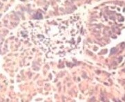
PDGF R alpha in Human Breast Cancer Tissue.
PDGF Ra was detected in immersion fixed paraffin-embedded sections of human breast cancer tissue using Mouse Anti-Human PDGF Ra Monoclonal Antibody (Catalog # MAB322) at 25 µg/mL overnight at 4 °C. Tissue was stained using the Anti-Mouse HRP-DAB Cell & Tissue Staining Kit (brown; Catalog # CTS002) and counterstained with hematoxylin (blue). Specific staining was localized to epithelial cells. View our protocol for Chromogenic IHC Staining of Paraffin-embedded Tissue Sections.Applications for Human PDGF R alpha Antibody (35248)
Application
Recommended Usage
Immunohistochemistry
8-25 µg/mL
Sample: Immersion fixed paraffin-embedded sections of human breast cancer tissue
Sample: Immersion fixed paraffin-embedded sections of human breast cancer tissue
Western Blot
1 µg/mL
Sample: Recombinant Human PDGF R alpha (Catalog # 322-PR) under non-reducing conditions only
Sample: Recombinant Human PDGF R alpha (Catalog # 322-PR) under non-reducing conditions only
Neutralization
Reviewed Applications
Read 3 reviews rated 4.3 using MAB322 in the following applications:
Formulation, Preparation, and Storage
Purification
Protein A or G purified from hybridoma culture supernatant
Reconstitution
Reconstitute at 0.5 mg/mL in sterile PBS. For liquid material, refer to CoA for concentration.
Loading...
Formulation
Lyophilized from a 0.2 μm filtered solution in PBS with Trehalose. See Certificate of Analysis for details.
*Small pack size (-SP) is supplied either lyophilized or as a 0.2 µm filtered solution in PBS.
*Small pack size (-SP) is supplied either lyophilized or as a 0.2 µm filtered solution in PBS.
Shipping
Lyophilized product is shipped at ambient temperature. Liquid small pack size (-SP) is shipped with polar packs. Upon receipt, store immediately at the temperature recommended below.
Stability & Storage
Use a manual defrost freezer and avoid repeated freeze-thaw cycles.
- 12 months from date of receipt, -20 to -70 °C as supplied.
- 1 month, 2 to 8 °C under sterile conditions after reconstitution.
- 6 months, -20 to -70 °C under sterile conditions after reconstitution.
Calculators
Background: PDGF R alpha
References
- Heldin, C.H. and L. Claesson-Welsh (1994) Guidebook to Cytokines and Their Receptors, Nicola, N.A. (ed) Oxford University Press, New York, NY p. 202.
Long Name
Platelet-derived Growth Factor Receptor alpha
Alternate Names
CD140a, PDGFR alpha, PDGFRA
Gene Symbol
PDGFRA
Additional PDGF R alpha Products
Product Documents for Human PDGF R alpha Antibody (35248)
Certificate of Analysis
To download a Certificate of Analysis, please enter a lot or batch number in the search box below.
Note: Certificate of Analysis not available for kit components.
Product Specific Notices for Human PDGF R alpha Antibody (35248)
For research use only
Citations for Human PDGF R alpha Antibody (35248)
 Powered by Bioz
Customer Reviews for Human PDGF R alpha Antibody (35248) (3)
4.3 out of 5
3 Customer Ratings
Have you used Human PDGF R alpha Antibody (35248)?
Submit a review and receive an Amazon gift card!
$25/€18/£15/$25CAN/¥2500 Yen for a review with an image
$10/€7/£6/$10CAN/¥1110 Yen for a review without an image
Submit a review
Customer Images
Showing
1
-
3 of
3 reviews
Showing All
Filter By:
-
Application: ImmunohistochemistrySample Tested: Ovary tissueSpecies: HumanVerified Customer | Posted 09/13/2021

-
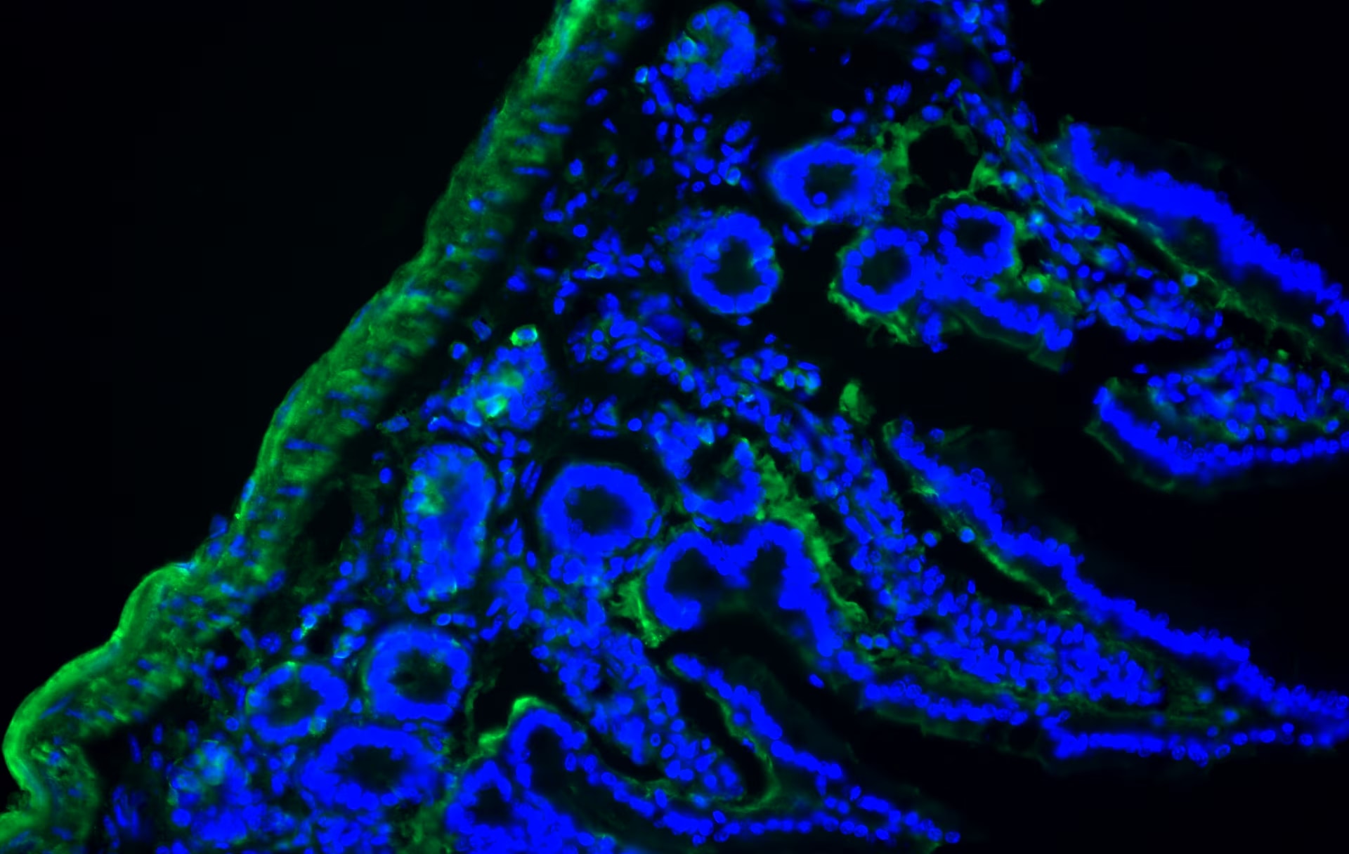
Application: Immunohistochemistry-FrozenSample Tested: Adult small intestineSpecies: CanineVerified Customer | Posted 03/19/2018Diffuse MAB322 antibody reactivity was visualized with green signal (Alexa 488 conjugated anti-mouse antibody). Blue=DAPI.
-
Application: ELISASample Tested: Purified StandardSpecies: HumanVerified Customer | Posted 12/01/2017
There are no reviews that match your criteria.
Protocols
Find general support by application which include: protocols, troubleshooting, illustrated assays, videos and webinars.
- Antigen Retrieval Protocol (PIER)
- Antigen Retrieval for Frozen Sections Protocol
- Appropriate Fixation of IHC/ICC Samples
- Cellular Response to Hypoxia Protocols
- Chromogenic IHC Staining of Formalin-Fixed Paraffin-Embedded (FFPE) Tissue Protocol
- Chromogenic Immunohistochemistry Staining of Frozen Tissue
- ClariTSA™ Fluorophore Kits
- Detection & Visualization of Antibody Binding
- Fluorescent IHC Staining of Frozen Tissue Protocol
- Graphic Protocol for Heat-induced Epitope Retrieval
- Graphic Protocol for the Preparation and Fluorescent IHC Staining of Frozen Tissue Sections
- Graphic Protocol for the Preparation and Fluorescent IHC Staining of Paraffin-embedded Tissue Sections
- Graphic Protocol for the Preparation of Gelatin-coated Slides for Histological Tissue Sections
- IHC Sample Preparation (Frozen sections vs Paraffin)
- Immunofluorescent IHC Staining of Formalin-Fixed Paraffin-Embedded (FFPE) Tissue Protocol
- Immunohistochemistry (IHC) and Immunocytochemistry (ICC) Protocols
- Immunohistochemistry Frozen Troubleshooting
- Immunohistochemistry Paraffin Troubleshooting
- Preparing Samples for IHC/ICC Experiments
- Preventing Non-Specific Staining (Non-Specific Binding)
- Primary Antibody Selection & Optimization
- Protocol for Heat-Induced Epitope Retrieval (HIER)
- Protocol for Making a 4% Formaldehyde Solution in PBS
- Protocol for VisUCyte™ HRP Polymer Detection Reagent
- Protocol for the Preparation & Fixation of Cells on Coverslips
- Protocol for the Preparation and Chromogenic IHC Staining of Frozen Tissue Sections
- Protocol for the Preparation and Chromogenic IHC Staining of Frozen Tissue Sections - Graphic
- Protocol for the Preparation and Chromogenic IHC Staining of Paraffin-embedded Tissue Sections
- Protocol for the Preparation and Chromogenic IHC Staining of Paraffin-embedded Tissue Sections - Graphic
- Protocol for the Preparation and Fluorescent IHC Staining of Frozen Tissue Sections
- Protocol for the Preparation and Fluorescent IHC Staining of Paraffin-embedded Tissue Sections
- Protocol for the Preparation of Gelatin-coated Slides for Histological Tissue Sections
- R&D Systems Quality Control Western Blot Protocol
- TUNEL and Active Caspase-3 Detection by IHC/ICC Protocol
- The Importance of IHC/ICC Controls
- Troubleshooting Guide: Immunohistochemistry
- Troubleshooting Guide: Western Blot Figures
- Western Blot Conditions
- Western Blot Protocol
- Western Blot Protocol for Cell Lysates
- Western Blot Troubleshooting
- Western Blot Troubleshooting Guide
- View all Protocols, Troubleshooting, Illustrated assays and Webinars


