PDGF is a major serum mitogen that can exist as a homo or hetero-dimeric protein consisting of disulfide-linked PDGF-A and PDGF‑B chains. The PDGF-AA, PDGF‑BB and PDGF-AB isoforms have been shown to bind to two distinct cell surface PDGF receptors with different affinities. Where as PDGF R alpha binds all three PDGF isoforms with high affinity, PDGF R beta binds PDGF-BB only with high-affinity. Both PDGF R alpha and PDGF R beta are members of the class III subfamily of receptor tyrosine kinases (RTK) that also includes the receptors for M-CSF, SCF and Flt3 ligand. All class III RTKs are characterized by the presence of five immuno‑globulinlike domains in their extracellular region and a split kinase domain in their intracellular region. PDGF binding induces receptor homo-and hetero‑dimerization and signal transduction. The expression of the alpha and beta receptors is independently regulated in various cell types. Recombinant soluble PDGF R beta binds PDGF with high affinity and is potent PDGF antagonist (4).
Human PDGF R beta Antibody (PR7212)
R&D Systems | Catalog # MAB1263

Key Product Details
Species Reactivity
Validated:
Human
Cited:
Human, Mouse
Applications
Validated:
Western Blot, Flow Cytometry, Immunoprecipitation
Cited:
Immunohistochemistry, Western Blot, Flow Cytometry, Immunocytochemistry, Immunoprecipitation
Label
Unconjugated
Antibody Source
Monoclonal Mouse IgG1 Clone # PR7212
Loading...
Product Specifications
Immunogen
Human skin fibroblast membrane extracts
Specificity
Detects human PDGF R beta in Western blots. Specificity has been confirmed in binding studies using several different cell lines (1, 2) and by its ability to immunoprecipitate PDGF receptor beta ‑subunit complexed with
Clonality
Monoclonal
Host
Mouse
Isotype
IgG1
Scientific Data Images for Human PDGF R beta Antibody (PR7212)
Detection of PDGF R beta in MG-63 cells by Flow Cytometry
MG-63 cells were stained with Mouse Anti-Human PDGF R beta Monoclonal Antibody (Catalog # MAB1263, filled histogram) or isotype control antibody (Catalog # MAB002, open histogram) followed by Allophycocyanin-conjugated Anti-Mouse IgG Secondary Antibody (Catalog # F0101B). View our protocol for Staining Membrane-associated Proteins.
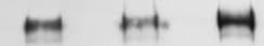
Detection of PDGF R beta by Western Blot
Immunoblot image are shown (A), along with densitometric analysis of (B) PDGFR beta, (C) Cyr61, (D) CD97, (E) Glypican-1, (F) MUC18/CD146 and (G) uPAR. (H) A summary of proteins detected by mass spectrometry and immunoblot. Graphs represent mean ± SEM of n = 3 independent biological experiments, shown as 1,2,3. Unpaired t-test was used as statistical test. *P < 0.05, **P < 0.01, ***P < 0.001 Image collected and cropped by CiteAb from the following open publication (https://pubmed.ncbi.nlm.nih.gov/36171564), licensed under a CC-BY license. Not internally tested by R&D Systems.
Detection of PDGF R beta by Western Blot
Immunoblot image are shown (A), along with densitometric analysis of (B) PDGFR beta, (C) Cyr61, (D) CD97, (E) Glypican-1, (F) MUC18/CD146 and (G) uPAR. (H) A summary of proteins detected by mass spectrometry and immunoblot. Graphs represent mean ± SEM of n = 3 independent biological experiments, shown as 1,2,3. Unpaired t-test was used as statistical test. *P < 0.05, **P < 0.01, ***P < 0.001 Image collected and cropped by CiteAb from the following open publication (https://pubmed.ncbi.nlm.nih.gov/36171564), licensed under a CC-BY license. Not internally tested by R&D Systems.Applications for Human PDGF R beta Antibody (PR7212)
Application
Recommended Usage
Flow Cytometry
0.25 µg/106 cells
Sample: MG-63 human osteosarcoma cell line
Sample: MG-63 human osteosarcoma cell line
Immunoprecipitation
Gronwald et al. (1988) Proc. Natl. Acad. Sci. 85:3435.
Western Blot
1 µg/mL
Sample: Recombinant Human PDGF R beta Fc Chimera (Catalog # 385-PR)
Sample: Recombinant Human PDGF R beta Fc Chimera (Catalog # 385-PR)
Reviewed Applications
Read 4 reviews rated 4 using MAB1263 in the following applications:
Flow Cytometry Panel Builder
Bio-Techne Knows Flow Cytometry
Save time and reduce costly mistakes by quickly finding compatible reagents using the Panel Builder Tool.
Advanced Features
- Spectra Viewer - Custom analysis of spectra from multiple fluorochromes
- Spillover Popups - Visualize the spectra of individual fluorochromes
- Antigen Density Selector - Match fluorochrome brightness with antigen density

Formulation, Preparation, and Storage
Purification
Protein A or G purified from ascites
Reconstitution
For liquid material, refer to CoA for concentration.
Formulation
Supplied as a 0.2 μm filtered solution in PBS. *Small pack size (SP) is supplied either lyophilized or as a 0.2 µm filtered solution in PBS.
Shipping
Lyophilized product is shipped at ambient temperature. Liquid small pack size (-SP) is shipped with polar packs. Upon receipt, store immediately at the temperature recommended below.
Stability & Storage
Use a manual defrost freezer and avoid repeated freeze-thaw cycles.
- 12 months from date of receipt, -20 to -70 °C, as supplied.
- 1 month, 2 to 8 °C under sterile conditions after opening.
- 6 months, -20 to -70 °C under sterile conditions after opening.
Calculators
Background: PDGF R beta
References
- Hart et al. (1987) J. Biol. Chem. 262:10780.
- Gronwald et al. (1988) Proc. Natl. Acad. Sci. 85:3435.
- Seifert et al. (1989) J. Biol. Chem. 264:8771.
- Heldin, C.H. and L. Claesson-Welsh (1994) in Guidebook to Cytokines and Their Receptors, Nicola, N.A. ed. Oxford University Press, New York, p. 202.
Long Name
Platelet-derived Growth Factor Receptor beta
Alternate Names
CD140b, PDGFRB
Gene Symbol
PDGFRB
Additional PDGF R beta Products
Product Documents for Human PDGF R beta Antibody (PR7212)
Certificate of Analysis
To download a Certificate of Analysis, please enter a lot or batch number in the search box below.
Note: Certificate of Analysis not available for kit components.
Product Specific Notices for Human PDGF R beta Antibody (PR7212)
For research use only
Related Research Areas
Citations for Human PDGF R beta Antibody (PR7212)
 Powered by Bioz
Customer Reviews for Human PDGF R beta Antibody (PR7212) (4)
4 out of 5
4 Customer Ratings
Have you used Human PDGF R beta Antibody (PR7212)?
Submit a review and receive an Amazon gift card!
$25/€18/£15/$25CAN/¥2500 Yen for a review with an image
$10/€7/£6/$10CAN/¥1110 Yen for a review without an image
Submit a review
Customer Images
Showing
1
-
4 of
4 reviews
Showing All
Filter By:
-
Application: Western BlotSample Tested: Breast CancerSpecies: HumanVerified Customer | Posted 09/06/2021

-
Application: MicroarraysSample Tested: EDTA PlasmaSpecies: HumanVerified Customer | Posted 06/10/2020
-
Application: MicroarraysSample Tested: EDTA PlasmaSpecies: HumanVerified Customer | Posted 11/07/2018
-
Application: MicroarraySample Tested: EDTA PlasmaSpecies: HumanVerified Customer | Posted 11/02/2018
There are no reviews that match your criteria.
Protocols
Find general support by application which include: protocols, troubleshooting, illustrated assays, videos and webinars.
- 7-Amino Actinomycin D (7-AAD) Cell Viability Flow Cytometry Protocol
- Cellular Response to Hypoxia Protocols
- Extracellular Membrane Flow Cytometry Protocol
- Flow Cytometry Protocol for Cell Surface Markers
- Flow Cytometry Protocol for Staining Membrane Associated Proteins
- Flow Cytometry Staining Protocols
- Flow Cytometry Troubleshooting Guide
- Immunoprecipitation Protocol
- Intracellular Flow Cytometry Protocol Using Alcohol (Methanol)
- Intracellular Flow Cytometry Protocol Using Detergents
- Intracellular Nuclear Staining Flow Cytometry Protocol Using Detergents
- Intracellular Staining Flow Cytometry Protocol Using Alcohol Permeabilization
- Intracellular Staining Flow Cytometry Protocol Using Detergents to Permeabilize Cells
- Propidium Iodide Cell Viability Flow Cytometry Protocol
- Protocol for Liperfluo
- Protocol for the Characterization of Human Th22 Cells
- Protocol for the Characterization of Human Th9 Cells
- Protocol: Annexin V and PI Staining by Flow Cytometry
- Protocol: Annexin V and PI Staining for Apoptosis by Flow Cytometry
- R&D Systems Quality Control Western Blot Protocol
- Troubleshooting Guide: Fluorokine Flow Cytometry Kits
- Troubleshooting Guide: Western Blot Figures
- Western Blot Conditions
- Western Blot Protocol
- Western Blot Protocol for Cell Lysates
- Western Blot Troubleshooting
- Western Blot Troubleshooting Guide
- View all Protocols, Troubleshooting, Illustrated assays and Webinars


