Platelet endothelial aggregation receptor 1 (PEAR1) is a 150 kDa type I transmembrane protein and member of the MEGF family of proteins (1). Human PEAR1 is synthesized as a 1037 amino acid (aa) precursor that contains a 20 aa signal sequence, a 735 aa extracellular domain (ECD), a 21 aa transmembrane region, and a 261 aa cytoplasmic region. The ECD consists of 15 EGF-like repeats that vary in length from 39 to 42 aa and contain a consensus sequence of CX1-2GX2GX2-4CX3CX1-3CX1-2GX1-2CX4GX1CX1CX2GX2GX2C (1). The consensus repeat contains six conserved glycine residues and eight conserved cysteine residues, suggesting four disulfide-bonded cysteine pairs in each EGF repeat (1). Within the ECD, there are also five potential sites for N-linked glycosylation. The cytoplasmic region contains five potential Src homology 3-binding, proline-rich domains (1). Mature human PEAR1 is 84% aa identical to mature mouse PEAR1. PEAR1 is most highly expressed in platelets and endothelial cells (1). Functionally, PEAR1 is a receptor for a yet undetermined ligand that signals upon the formation of platelet‑platelet contacts induced both by platelet aggregations and by platelet centrifugation (1). The signaling enhances and stabilizes platelet thrombi (2). Upon aggregation, the surface-expressed protein is tyrosine-phosphorylated (1). This phosphorylation event is inhibited by the alpha IIb beta 3 antagonist eptifibatide, thus demonstrating that PEAR1 tyrosine phosphorylation is dependent on surface contacts between activated platelets (1). PEAR1 can be phosphorylated in an alpha IIb beta 3 integrin-dependent manner on tyrosine (Tyr925) and serine residues (Ser593 and Ser1029) and, potentially, at Tyr804, Tyr943, and Tyr979 (1). Inherited PEAR1 variations that alter expression or function of the platelet signaling molecule could modify agonist-induced aggregation in native platelets (2). In addition, a genetic variant in PEAR1 could be an important determinant of residual platelet function during aspirin treatment, because the COX1/thromboxane A2 pathway will be strongly inhibited by aspirin, and maximal aggregation will then be dependent on other secondary signaling pathways (2).

Key Product Details
Species Reactivity
Validated:
Human
Cited:
Human
Applications
Validated:
Western Blot, Flow Cytometry, CyTOF-ready
Cited:
Immunoprecipitation
Label
Unconjugated
Antibody Source
Monoclonal Mouse IgG2A Clone # 492621
Loading...
Product Specifications
Immunogen
Chinese hamster ovary cell line CHO-derived recombinant human PEAR1
Leu21-Ser754
Accession # Q5VY43
Leu21-Ser754
Accession # Q5VY43
Specificity
Detects human PEAR1 in direct ELISAs and Western blots. In Western blots, no cross-reactivity with recombinant human Tenascin R or recombinant mouse Tenascin C is observed.
Clonality
Monoclonal
Host
Mouse
Isotype
IgG2A
Scientific Data Images for Human PEAR1 Antibody (492621)
Detection of PEAR1 in Human Peripheral Blood Platelets by Flow Cytometry
Human peripheral blood platelets were stained with Mouse Anti-Human PEAR1 Monoclonal Antibody (Catalog # MAB4527) followed by Allophycocyanin-conjugated Anti-Mouse IgG F(ab')2Secondary Antibody (Catalog # F0101B) and Integrin a 2b/CD41 Fluorescein-conjugated Monoclonal Antibody. Quadrant markers were set based on isotype control antibody staining (Catalog # MAB002).Applications for Human PEAR1 Antibody (492621)
Application
Recommended Usage
CyTOF-ready
Ready to be labeled using established conjugation methods. No BSA or other carrier proteins that could interfere with conjugation.
Flow Cytometry
2.5 µg/106 cells
Sample: Human Peripheral Blood Platelets
Sample: Human Peripheral Blood Platelets
Western Blot
1 µg/mL
Sample: Recombinant Human PEAR1 (Catalog # 4527-PR)
Sample: Recombinant Human PEAR1 (Catalog # 4527-PR)
Reviewed Applications
Read 3 reviews rated 3.7 using MAB4527 in the following applications:
Flow Cytometry Panel Builder
Bio-Techne Knows Flow Cytometry
Save time and reduce costly mistakes by quickly finding compatible reagents using the Panel Builder Tool.
Advanced Features
- Spectra Viewer - Custom analysis of spectra from multiple fluorochromes
- Spillover Popups - Visualize the spectra of individual fluorochromes
- Antigen Density Selector - Match fluorochrome brightness with antigen density

Formulation, Preparation, and Storage
Purification
Protein A or G purified from hybridoma culture supernatant
Reconstitution
Reconstitute at 0.5 mg/mL in sterile PBS. For liquid material, refer to CoA for concentration.
Loading...
Formulation
Lyophilized from a 0.2 μm filtered solution in PBS with Trehalose. *Small pack size (SP) is supplied either lyophilized or as a 0.2 µm filtered solution in PBS.
Shipping
Lyophilized product is shipped at ambient temperature. Liquid small pack size (-SP) is shipped with polar packs. Upon receipt, store immediately at the temperature recommended below.
Stability & Storage
Use a manual defrost freezer and avoid repeated freeze-thaw cycles.
- 12 months from date of receipt, -20 to -70 °C as supplied.
- 1 month, 2 to 8 °C under sterile conditions after reconstitution.
- 6 months, -20 to -70 °C under sterile conditions after reconstitution.
Calculators
Background: PEAR1
References
- Nanda, N. et al. (2005) J. Biol. Chem. 280:24680.
- Herrera-Galeano, J.E. et al. (2008) Arterioscler. Thromb. Vasc. Biol. 28:1484.
Long Name
Platelet endothelial aggregation receptor 1
Alternate Names
Jedi
Gene Symbol
PEAR1
UniProt
Additional PEAR1 Products
Product Documents for Human PEAR1 Antibody (492621)
Certificate of Analysis
To download a Certificate of Analysis, please enter a lot or batch number in the search box below.
Note: Certificate of Analysis not available for kit components.
Product Specific Notices for Human PEAR1 Antibody (492621)
For research use only
Related Research Areas
Citations for Human PEAR1 Antibody (492621)
 Powered by Bioz
Customer Reviews for Human PEAR1 Antibody (492621) (3)
3.7 out of 5
3 Customer Ratings
Have you used Human PEAR1 Antibody (492621)?
Submit a review and receive an Amazon gift card!
$25/€18/£15/$25CAN/¥2500 Yen for a review with an image
$10/€7/£6/$10CAN/¥1110 Yen for a review without an image
Submit a review
Customer Images
Showing
1
-
3 of
3 reviews
Showing All
Filter By:
-
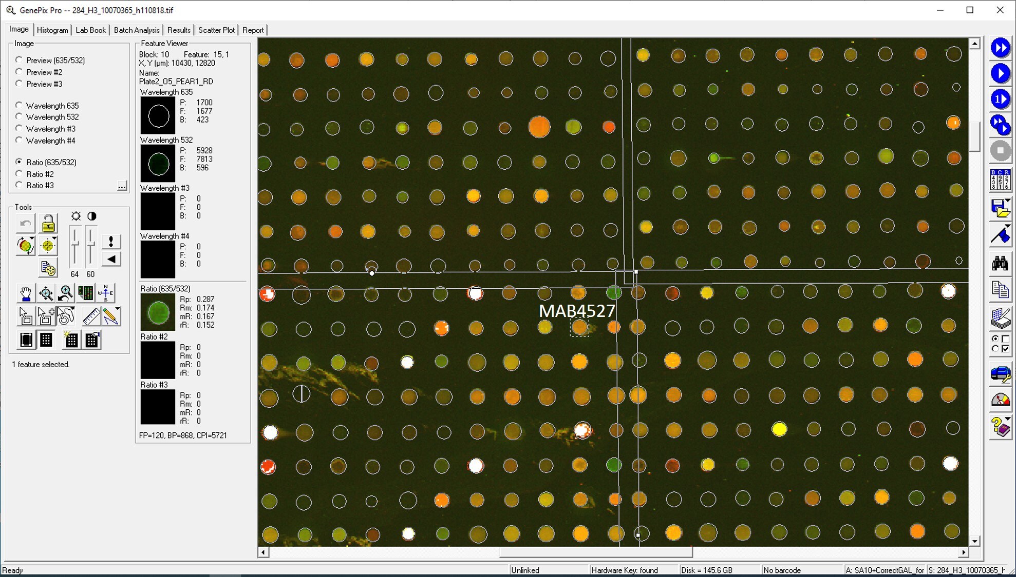
Application: MicroarraysSample Tested: EDTA PlasmaSpecies: HumanVerified Customer | Posted 01/14/2021

-
Application: MicroarraysSample Tested: EDTA PlasmaSpecies: HumanVerified Customer | Posted 11/14/2018
-
Application: MicroarraySample Tested: EDTA PlasmaSpecies: HumanVerified Customer | Posted 11/09/2018
There are no reviews that match your criteria.
Protocols
Find general support by application which include: protocols, troubleshooting, illustrated assays, videos and webinars.
- 7-Amino Actinomycin D (7-AAD) Cell Viability Flow Cytometry Protocol
- Cellular Response to Hypoxia Protocols
- Extracellular Membrane Flow Cytometry Protocol
- Flow Cytometry Protocol for Cell Surface Markers
- Flow Cytometry Protocol for Staining Membrane Associated Proteins
- Flow Cytometry Staining Protocols
- Flow Cytometry Troubleshooting Guide
- Intracellular Flow Cytometry Protocol Using Alcohol (Methanol)
- Intracellular Flow Cytometry Protocol Using Detergents
- Intracellular Nuclear Staining Flow Cytometry Protocol Using Detergents
- Intracellular Staining Flow Cytometry Protocol Using Alcohol Permeabilization
- Intracellular Staining Flow Cytometry Protocol Using Detergents to Permeabilize Cells
- Propidium Iodide Cell Viability Flow Cytometry Protocol
- Protocol for Liperfluo
- Protocol for the Characterization of Human Th22 Cells
- Protocol for the Characterization of Human Th9 Cells
- Protocol: Annexin V and PI Staining by Flow Cytometry
- Protocol: Annexin V and PI Staining for Apoptosis by Flow Cytometry
- R&D Systems Quality Control Western Blot Protocol
- Troubleshooting Guide: Fluorokine Flow Cytometry Kits
- Troubleshooting Guide: Western Blot Figures
- Western Blot Conditions
- Western Blot Protocol
- Western Blot Protocol for Cell Lysates
- Western Blot Troubleshooting
- Western Blot Troubleshooting Guide
- View all Protocols, Troubleshooting, Illustrated assays and Webinars
Loading...
Associated Pathways
